Introduction
The effects of cadmium (Cd) on biological systems
have been extensively studied; however, they remain relatively
enigmatic. Cd has an exceptionally long half-life in biological
systems (years) which may underlie the physiological changes
associated with exposure to Cd. Cd is found widely throughout the
environment and is one of the more abundant toxicants to which
humans are exposed in both occupational and environmental settings
(1-3). Cadmium exerts toxic effects on many
organs and systems of organs (4,5).
Similarly, its endocrine disruptive activities have been described
(6-9), as well as its hormetic effects
(10). Cadmium, as well as
Cd-containing compounds, have been classified as carcinogenic by
the International Agency for Research on Cancer (11,12) based on epidemiological studies
showing a causal connection with the development of lung cancer.
Epidemiological studies have also implicated exposure to Cd to be
involved in the development of other types of cancer, such as
kidney cancer (13-15), prostate cancer (14,16,17), cancer of the testis (17), bladder cancer (18), breast cancer (19) and overall cancer mortality
(20).
The latest report on the global burden of cancer
worldwide using GLOBOCAN 2018 estimated pancreatic cancer (PC) as
the seventh leading cause of cancer-related mortality among both
males and females with the projection that in the 28 countries of
the European Union, this type of cancer will surpass breast cancer
as the third leading cause of cancer-related mortality in the
future (21). The prognosis of
this type of cancer is very poor, mainly due to late-stage
diagnosis. Hence, the elucidation of the etiology of this type of
cancer and the identification of prognostic factors for it are of
utmost importance. The quest for a legitimate biomarker to identify
PC in the early stages is ongoing (22,23). A recent review article suggested
the possible role of exposure to Cd in the development of PC
(24). Some of the mechanisms
involved in the carcinogenetic effects of Cd on the pancreas may be
the induction of oxidative stress and interaction with bioelements,
which have been observed and discussed in a number of studies
(25-32). The development of PC is likely to
be associated with the silencing of certain tumor suppressor genes
and/or the activation of oncogenes, although the underlying
molecular mechanisms remain undefined (22,23,33,34). However, interactions between
certain environmental chemicals, with Cd being one of them, and
these genes can be expected. Furthermore, the micro-RNA-mediated
regulation of pancreatic cell proliferation has been demonstrated
by Basu et al (35).
Subsequently, microRNAs (miRNAs or miRs) were found to be present
specifically in the mitochondria, originating from mitochondrial
DNA and regulating genes coding for mitochondrial proteins in a
direct manner, and consequently mitochondrial function (36). Concerning the role of Cd as a
mitochondrial toxin, it has recently been studied, but with mixed
results, suggesting that Cd may exert direct and indirect effects
on the mitochondria (37-40). This ability of Cd to exert toxic
effects on the mitochondria could certainly be one of the
mechanisms of its carcinogenic effects on cells.
The nature of Cd-related mitochondrial toxicity has
led to speculation that the toxicity observed can be mediated
either by direct or indirect mechanisms. The aim of this study was
to enhance our understanding of the effects of Cd on the
mitochondria and to determine whether Cd is a direct or an indirect
mitochondrial toxin. Current literature has been unclear and at
times, conflicting (37). One
concept that is becoming more accepted is that there is an
organ-specificity regarding Cd toxicity. There is overlap between
some of the major organs examined, although there are also
differences in Cd-mediated responses. In the kidneys, there is
speculation that Cd interferes with normal mitochondrial function,
leading to increased oxidative stress and apoptosis (40), which is further supported by in
vitro studies utilizing human kidney cell lines showing that Cd
directly alters mitochondrial membrane permeability and potential
(39). Li et al extended
these findings to isolated and purified mitochondria (38). In hepatic cells, exposure to Cd
has been shown to decrease mitochondrial membrane permeability, but
also to directly alter apoptotic pathways by increasing the
activity of both caspase-3 and caspase-9, as well as increasing the
activity of p53 (41). Recently,
the effects of CdCl2 on the pancreas and pancreatic
cells have been further described (24). In pancreatic β-cells (RIN-m5F
cells), exposure to CdCl2 has been shown to result in
the death of β-cells following increased oxidative stress followed
by a downstream-mediated activation of apoptotic pathways regulated
by the mitochondria (42). Some
researchers have postulated that environmental or occupational
exposure to Cd, and the resulting Cd-mediated death or damage to
β-cells is an underlying cause of diabetes and obesity (43).
In this study, we examined the effects of cadmium
chloride (CdCl2) on mitochondrial function due to the
involvement of mitochondria in the 'Warburg effect'. The Warburg
effect describes how tumor cells bypass oxidative phosphorylation
[responsible for the generation of 38 adenosine triphosphates
(ATPs)] and instead utilize aerobic glycolysis [generating only 2
ATPs, but other metabolites which could be useful in cellular
proliferation (44)]. An
advancement in our understanding of the peculiar phenomenon that
was identified by Otto Warburg almost 100 years ago has progressed
very little until the last 5-10 years when a significant surge in
interest has taken place (45,46). Although one single theory has not
come to the forefront, numerous hypotheses have been presented,
suggesting that the switch from oxidative phosphorylation to
aerobic glycolysis signals the transition from a normal cell to a
tumor cell (46). Another
hypothesis is that switching to aerobic glycolysis can protect the
tumor cell from sugar-induced death when glucose uptake is high,
and by changing to a high production of lactate, pro-proliferative
intermediates can be produced (47). Compounds, such as Cd, that damage
the mitochondria promote a shift towards aerobic glycolysis, thus
leading to tumor formation. Researchers that have examined toxicant
[1-methyl-4-phenyl-1,2,3,6-tetrahydro-pyridine (MPTP)] damage to
the mitochondria have reported that oxidative phosphorylation is
decreased, that apoptotic pathways are altered and there is a
bioenergetic shift in glucose utilization (48). Indirect genetic damage to the
mitochondria that will result in epigenetic alterations has also
been demonstrated in tumor cells, as well as inherited genetic
abnormalities that predispose or facilitate the progression of the
Warburg effect in tumor cells (49). The apparent importance of the
mitochondria and the Warburg effect has led researchers to
postulate that either 'turning' oxidative phosphorylation 'back on'
or shunting away from lactate production/utilization may be an
effective means of controlling tumor growth (45,50).
Recently, we performed a literature search for
'cadmium', 'Warburg Effect', or 'Crabtree Effect' and the
references that we obtained were more specific for the effects of
Cd on sugar utilization in yeast, which is what the Crabtree effect
initially described (51,52). The effect of Cd on cellular energy
utilization is an area of research that needs to be addressed for a
better understanding of the effects of Cd on the pancreas and on
the development of PC, diabetes, obesity and other pancreatic
dysfunctions. In this study, we designed a series of assays that
would systematically address the Cd-mediated effects on pancreatic
cells using a control epithelial cell line (HPNE) and the
pancreatic tumor cell line, AsPC-1. The goals of these experiments
were to first determine the level of toxicity following the
exposure of both cell lines to Cd, and to then use subtoxic
concentrations to determine mitochondrial toxicity. Our initial
concentrations were determined using Cd concentrations obtained
from pancreatic tissue, from a pancreatic tumor, the area
surrounding the tumor and from 'healthy' pancreatic tissue
(53). After determining the
threshold for Cd-mediated toxicity, we examined the production of
free radicals following exposure to Cd and then used the
glucose-galactose assay to assess the toxic potential of Cd on the
mitochondria by measuring the shift in the toxicity curve and
change in IC50 values comparing the glucose curve to the
galactose curve. From these experiments, we aim to improve our
understanding of Cd-mediated intracellular changes in pancreatic
cells.
Materials and methods
Cell lines and cell culture
maintenance
All cell culture experiments were performed using
standard Biosafety Level 2 (BSL-2) guidelines. The study was
approved by the Oklahoma State University Center for Health Science
Institutional Biosafety committee in compliance with all National
Institutes of Health regulations. Cell lines purchased from the
American Type Culture Collection (ATCC). Pancreatic hTERT-HPNE
('human pancreatic Nestin-expressing' cells; ATCC®
CRL-4023™, immortalized pancreatic control cells, referred to as
'HPNE') and AsPC-1 (ATCC® CRL-1682™, pancreatic tumor
cells) cells were grown and maintained as described in the
ATCC-suggested protocols. HPNE cell growth media consisted of DMEM
base media with 2 mM glutamine, and 1.5 g/l sodium bicarbonate
supplemented with M3 Base media supplement (M300F-500; Incell
Corp.) at a ratio of 3:1. To produce complete growth media, 5%
fetal bovine serum, 10 ng/ml human epidermal growth factor, 1 mg/ml
glucose and 750 ng/ml Puromycin (Sigma-Aldrich and Corning) were
added to the DMEM:M3 base. Base AsPC-1 media was RPMI-1640 with 2
mM glutamine, and complete growth media consisted of
supplementation with 10% fetal bovine serum, and 1%
penicillin/streptomycin (Sigma-Aldrich and Corning). Assay media
used in the cytotoxicity assays was a base MEM supplemented with 2
mM glutamine and 1% fetal bovine serum. Assay media was phenol-free
and low serum to minimize the confounding factors associated with
the influence of protective growth factors in serum. Cells were
allowed to adhere for at least 24 h before exposure. Following
adherence, growth media were removed and replaced with assay media
containing the appropriate treatment(s).
Cell viability and cell growth
assays
All lactate dehydrogenase (LDH) assays (Cytotox-ONE™
kit; G7891, Promega) were performed essentially as previously
described (54). The HPNE and
AsPC-1 cell lines (20-50,000 cells/well) were plated and allowed to
adhere for at least 24 h prior to the assay. LDH assays determined
i) the number of viable cells within the culture; ii) the
percentage of the total number of cells that were viable. Initial
serum-comparison assays experiments examined the effects of various
serum concentrations on cell growth and viability. Following the
removal of the growth media, MEM supplemented with 0% (control), or
1, 2.5, 5 and 10% fetal bovine serum was added to appropriate wells
followed by incubation at 37°C in 5% CO2 for 48 h.
Following exposure, the LDH assay was carried out according to the
manufacturer's instructions. The ability to quantify dead and live
cells is described in the following formula: {[(total RFU) - (Media
RFU)] ÷ (total RFU)} ×100. The amount of fluorescence produced was
quantified using a Biotek plate reader (Biotek Instruments) set to
an excitation wavelength of 485 nm and an emission wavelength of
530 nm.
Once we determined whether or not changing the serum
concentration would affect cell viability and cell number, we
exposed both the HPNE and AsPC-1 cells to 2 ppm (18 μM), 6
ppm (54 μM) and 14 ppm (126 μM) CdCl2 for
48 h. We have previously analyzed human pancreatic tissue and
measured the content of Cd (53).
The highest concentration was found within the tumor, the
intermediate concentration was in the region immediately
surrounding the tumor zone, and the lowest concentration was found
at a site distant from the tumor. We used the molecular
contribution of Cd to CdCl2 and calculated the
concentration of Cd alone. This assay was performed to aid in the
identification of the threshold of toxicity associated with
exposure to CdCl2. Following exposure for 48 h, both
cell viability and the total number of cells were determined as
described in the serum-comparison assays described above. The
rationale for using CdCl2 as our form of Cd is based on
existing literature and published studies using CdCl2
both in in vitro and in vivo studies (24,25,27,31,53). The use of CdCl2 will
strengthen conclusions comparing our findings to the existing
literature using CdCl2.
Measurement of oxidative stress
Both cell lines were grown in 25 cm2
flasks and seeded into a 96-well (black/clear bottom) plate at an
initial density of 10−5 cells/ml (or 10−4
cells/well). The cells were allowed to adhere for 24 h and to
initiate the assay, the growth media was removed followed by
washing the cells (twice) with warmed PBS. The cells were then
loaded with 100 μM DCFH-diacetate (DCFH-DA) for 30 min at
37°C in 5% CO2, while being protected from light.
Following the removal and washing with PBS, the cells were exposed
to one of three treatments; control (PBS only; 0 μM
CdCl2), 50 μM CdCl2 or 100 μM
CdCl2. Baseline fluorescence was determined in the
PBS-only group prior to the addition of CdCl2. The cells
were returned to the incubator to allow for the continuation of the
reaction. The plates were removed after 30, 60, 90 and 120 min and
the fluorescence resulting from the conversion of
dichlorofluorescin (DCFH) to dichlorofluorescein (DCF) was measured
using a Biotek plate reader set to an excitation wavelength of 485
nm and an emission wavelength of 530 nm. The amount of fluorescence
detected is directly correlated to the amount of oxidative stress
occurring within the cell. Data were then expressed as the means ±
SEM of n=8 assayed in duplicate.
Determination of lethal concentration 50%
(LC50) values for CdCl2
The HPNE and AsPC-1 cell lines were maintained as
described above. The cells were plated at a final density of
approximately 105 cells/ml in clear 96-well plates. The
cells were allowed to adhere for at least 24 h and the media were
carefully removed and assay media with the appropriate
concentrations of CdCl2 (0-10−3 M) were added
to the wells. Following 48 h of exposure, 12 mM stock (1.2 mM final
concentration) MTT (Molecular Probes) was added to each well and
the plates were returned to the incubator (37°C and 5%
CO2) for 4 h followed by the addition of detergent for
another 3-4 h. The amount of insoluble formazan was determined by
measuring the absorbance at 565 nm using the BioTek plate reader.
Data were then expressed as the means ± SEM of n=6 in
duplicate.
Mitochondrial toxicity assays
HPNE and AsPC-1 cell lines were plated at a final
density of 20,000-50,000 cells/well and allowed to adhere for at
least 24 h prior to the assay. The initiation of toxicant exposure
begins by removing the growth media and replacing with DMEM
supplemented with either 25 mM glucose or 10 mM galactose. This
method of determining mitochondrial toxicity is similar to that
described in the study by Sanuki et al (55) and Dott et al (56). The treatment groups consisted of
cells exposed to Cd (0.1-100 μM) or control (media only) for
4 h followed by the addition of 12 mM MTT dye (1.2 mM final
concentration). The absorbance was measured at 565 nm, and the data
were then expressed as the means ± SEM of n=12 in duplicate.
To establish the association between cell death and
the loss of ATP, we used a luciferin-based detection system
(Mitochondrial ToxGlo™; Promega) (57). The principle of the mitochondrial
toxicity tests is that substituting 10 mM galactose for 25 mM
glucose will increase susceptibility to mitochondrial toxins
(58-60). The cells were exposed to
CdCl2 (1 or 50 μM) for 48 h prior to the
initiation of the assay. These two concentrations were selected
based on 50 μM approximating 6 ppm, and 1 μM was
significantly lower than our threshold of 10 μM. Following
30 min of incubation at 37°C in a CO2 incubator,
viability was determined by measuring the fluorescence (485
nmex/525 nmem) emitted. The second step in
the multiplex was to quantify the amount of ATP present by directly
adding the luciferin-based detection system into the wells that
have already been quantified for cell viability with the amount of
luminescence being directly proportional to the amount of ATP
present. Collectively, by comparing the cellular responses in the
two datasets, a better understanding of CdCl2 can be
obtained as a cellular toxicant that results in mitochondrial
dysfunction or non-mitochondrial related cytotoxic mechanisms.
Effect of CdCl2 on
mitochondrial permeability
To assess cell health, we utilized a dye that is
permeable to the mitochondrial and will fluoresce red or green
depending on the membrane potential of the mitochondria. A high
mitochondrial membrane potential (ΔψM) is representative
of a healthy cell, whereas a low ΔψM is indicative of an
unhealthy cell. The higher the ratio, the greater the polarization
of the mitochondrial membrane. We utilized the JC-1 kit (Cayman
Chemical Co.) for this purpose. The cells were plated at
approximately 105 cells/well and allowed to adhere for
24 h. Exposure was initiated by removing the growth media from each
well and adding the treatment media with CdCl2 (1
μM), or the vehicle (water). The cells were returned to the
incubator for 48 h at 37°C and 5% CO2. Following
exposure, the cells were prepared according to manufacturer
instructions; staining was initiated by the addition of 10 μ
JC-1 dye and the cells were returned to the incubator for 20 min.
The fluorescence intensity was measured using the BioTek plate
reader at 535ex/595em to measure JC-1 aggregates (healthy cells;
green) and then a second reading using 485ex/535em which measures
JC-1 monomers (apoptotic/unhealthy cells; red).
Statistical analysis: All data were
analyzed using GraphPad Prism software (version 7.04)
Data from initial CdCl2 exposure assays
were analyzed by one-way ANOVA followed by Dunnett's post hoc
comparison to control values. In the oxidative stress and LDH
assays, data were analyzed by two-way ANOVA (2×3 or cell line x
concentration) using Sidak's or Tukey's test for multiple
comparisons, respectively. All data are expressed as the means ±
SEM of n=4 (ATP assay), n=6 (MTT LC50 assay), n=8 (DCFH
oxidative stress assay) or n=12 (mitochondrial toxicity assay)
performed in either duplicate or triplicate. In the LC50
and mitochondrial toxicity assays, the data were first fit by
non-linear single-site nonlinear regression inhibition curve
(single-site) to determine the IC50 value for
CdCl2 in each media. A ≥3-fold leftward shift in
LC50 or IC50 values in the galactose group
was indicative of mitochondrial toxicity. When curve-fitting, we
utilized the D'Agostino and Pearson Test for Normality (omnibus
K2) to assure that the curves followed a Gaussian
distribution. One-way analysis of variance was performed with
Dunnett's test for post hoc comparison to control LC50
values. For individual comparisons of LC50 values, a
Student's t-test was used to compared cell lines and treatments.
For analyzing the mitochondrial toxicity data using dual-labeling,
we utilized a three-way ANOVA (2×2×2; cell type, concentration,
media) design with Sidak's test for post hoc multiple comparisons.
Data are presented first as the result of the ANOVA where
appropriate (F-value) representing whether there was a significant
association within the group, such as a concentration effect.
Post-hoc multiple comparisons were then performed, usually
comparing to the control value; however, the value obtained from
the post-hoc comparison is labeled in the figures by a symbol. A
significance level was set at P<0.05.
Results
Cell viability: Effect of exposure to Cd
and low serum concentration: There is a need to perform assays in
reduced serum
Fetal bovine serum contains multiple factors that
have been known to scavenge free radicals and afford protection to
the cells when exposed to a toxic insult (61-63). These assays were performed to
assess changes in cell viability in the presence of reducing
concentrations of serum (fetal bovine serum), as well as a change
in basal media from the standard optimized growth media to MEM with
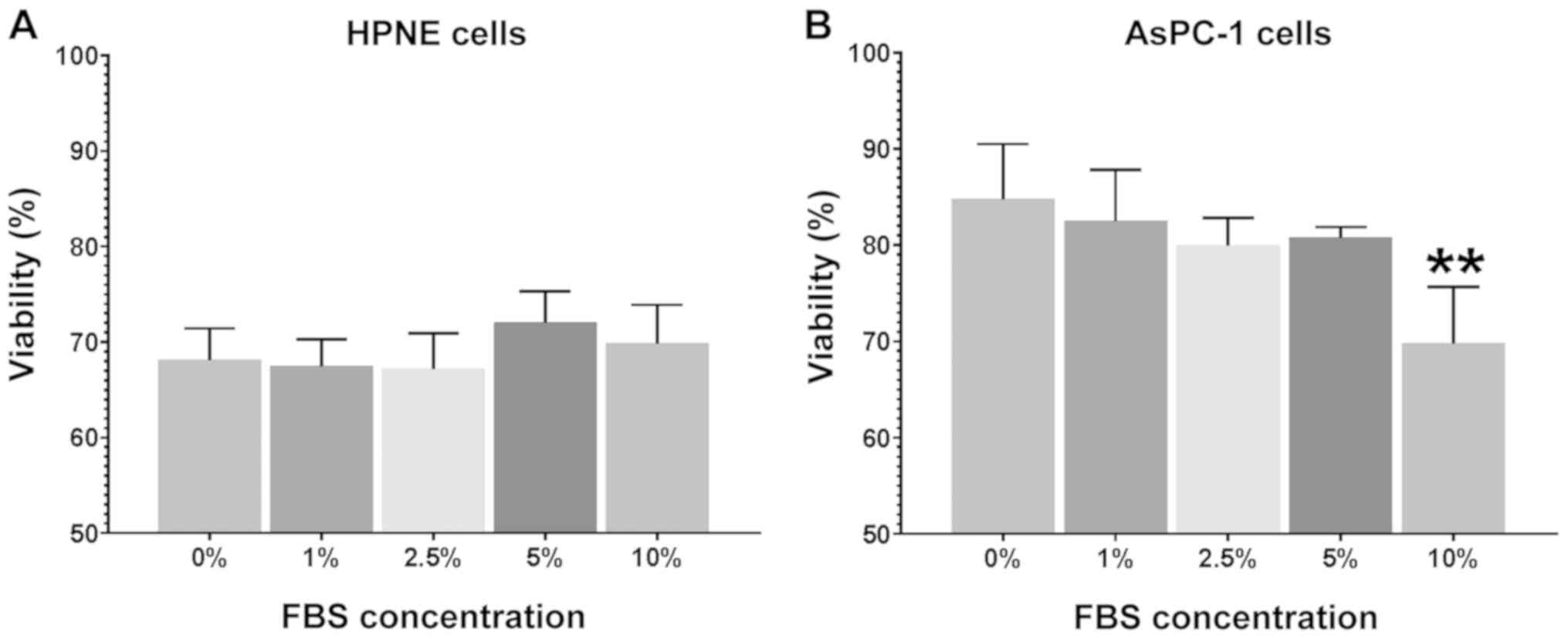
minimal supplementation. In the HPNE cells (Fig. 1A), no marked differences (P=0.61)
were observed in either viability between the serum groups or
between the growth media and corresponding MEM. In the AsPC-1
cells, there was a robust increase (F5,41=27.34;
P<0.0001) in viability in the presence of reduced serum
(Fig. 1B). From 0 to 5% the
AsPC-1 cells exhibited a 20-25% greater viability compared to the
growth media. It was noteworthy that reduced serum improved the
viability of AsPC-1 cells. Previous studies have suggested that
reducing the serum concentration will reduce viability and cellular
proliferation in a variety of cell lines (64,65). This study, at least to the best of
our knowledge, is the first to examine the effects of a reduced
serum concentration on AsPC-1 viability and growth.
Initial experiments examined the effects of
CdCl2 on the viability of HPNE and AsPC-1 cells by
exposing the cells to CdCl2 at final concentrations of
0, 2, 6 and 14 ppm (Table I).
These concentrations were selected based on data from human studies
showing 14 ppm CdCl2 in the core of a pancreatic tumor,
with 6 ppm on the tumor margin and 2 ppm associated with 'healthy'
tissue distant from the tumor center (53). As evidenced by the data presented
in Table I, we observed only
minimal changes in viability or cell number in cells exposed to
CdCl2. In the HPNE cells, we observed an effect of
treatment, (approximately 5%) in both cell number and viability
compared to the control values (F4,15=37.59;
P<0.0001). Post hoc comparisons revealed no differences between
the treatment and control groups. In the AsPC-1 cells, exposure to
CdCl2 (treatment effect) resulted in a significant
change in both viability (F4,15=66.69; P<0.0001) and
in cell number (F4,15=10.91; P=0.0002). Dunnett's post
hoc analysis was used to compare changes in the treatment groups to
the control values. Only exposure to the highest concentration of
CdCl2, 14 ppm, elicited significant reductions in both
cellular viability (51%) and cell number (54%). Collectively, these
data suggest that the control HPNE cells are relatively resistant
following changes in serum concentration, as well as
CdCl2 exposure. We observed minimal changes in both cell
viability, as well as in cell number which would be indicative of
proliferation/growth changes. The AsPC-1 cells, on the other hand,
were more sensitive to the changes in serum concentration and
exposure to CdCl2. Generally, only the highest
concentration of CdCl2 elicited significant reductions
in both cell viability and number. Subsequent experiments assessed
CdCl2-mediated changes in oxidative stress and
mitochondrial toxicity as a secondary means for CdCl2
exposure resulting in cellular damage.
 | Table IEffects of exposure to
CdCl2 for 48 h on the live cell number and viability of
HPNE and AsPC-1 cells. |
Table I
Effects of exposure to
CdCl2 for 48 h on the live cell number and viability of
HPNE and AsPC-1 cells.
| | | CdCl2
concentration
|
|---|
| Growth media | Low-serum MEM | 2 ppm | 6 ppm | 14 ppm |
|---|
| HPNE | | | | | |
| Live cell no.
(RFU) | 3,711±100 | 3,591±71 | 3,478±70 | 3,585±52 | 3,518±87 |
| Viability (%) | 84.2±0.3 | 81.9±0.2 | 80.7±0.4 | 80.6±0.2 | 78.6±0.5 |
| AsPC-1 | | | | | |
| Live cell no.
(RFU) | 15,501±1,518 | 15,247±1,206 | 12,318±231 | 12,048±1,033 | 7,054±678a |
| Viability (%) | 62.4±2.3 | 71.1±1.1 | 58.9±1.4 | 56.9±6.6 | 30.7±4.1a |
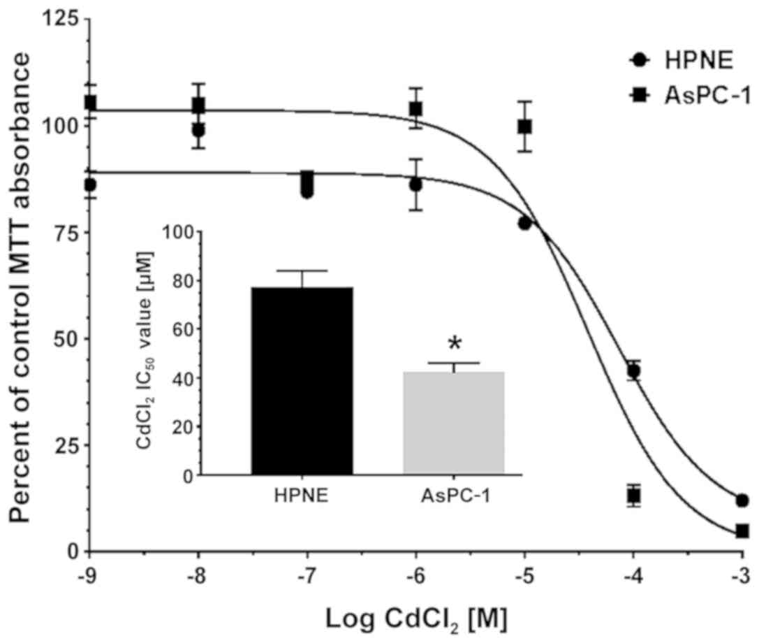
To determine the LC50 value of
CdCl2 in the HPNE and AsPC-1 cells, each cell line was
incubated with 7 concentrations of CdCl2 (1 to 1 mM) for
48 h, and cell viability was determined by MTT staining (n=6).
Curves for both cell lines were best fit to a single-site model
(Fig. 2). Individual response
curves were analyzed to assess the LC50 for
CdCl2 in each assay run. LC50 data were
compared for the HPNE (77.3±6.8 μM) and AsPC-1 (42.1±4.0
μM) cells by Student's t-test and we observed a significant
(P=0.0013) 45.5% reduction in LC50 values in the AsPC-1
group (Fig. 2, inset). We also
compared our means at each concentration to their corresponding
control values to determine the threshold at which we observed a
significant decrease in cell viability. This threshold provides a
value where CdCl2 effects are significantly different
from the control and compliments our LC50 values. Using
a one-way ANOVA with Dunnett's test to compare treatment groups to
their corresponding control, we were able to identify the threshold
at which the concentration yielded a significant reduction in cell
number. The viability of both the HPNE (F6,35=92.27;
P<0.0001) and AsPC-1 (F6,35=115; P<0.0001) cells
was significantly affected by the increasing concentrations of
CdCl2. The maximum concentration where we observed no
reduction in viability was 10 μM in both cell lines.
Therefore, although there was a slight rightward shift in
LC50 values in the AsPC-1 group, the threshold for
viability reduction was unaltered between the cell lines.
Cd-mediated induction of oxidative
stress
The precise role of CdCl2 in the
generation of free radicals has been a topic of considerable
discussion. It is clear that transition metals have been implicated
in the generation of free radicals (66,67). There is also evidence to suggest
that oxidative stress and associated inflammation are underlying
causes of cancer (68,69). The role of CdCl2 is not
as clear as with other metals such as iron, lead, mercury. In this
study, the ability of 50 or 100 μM CdCl2 to
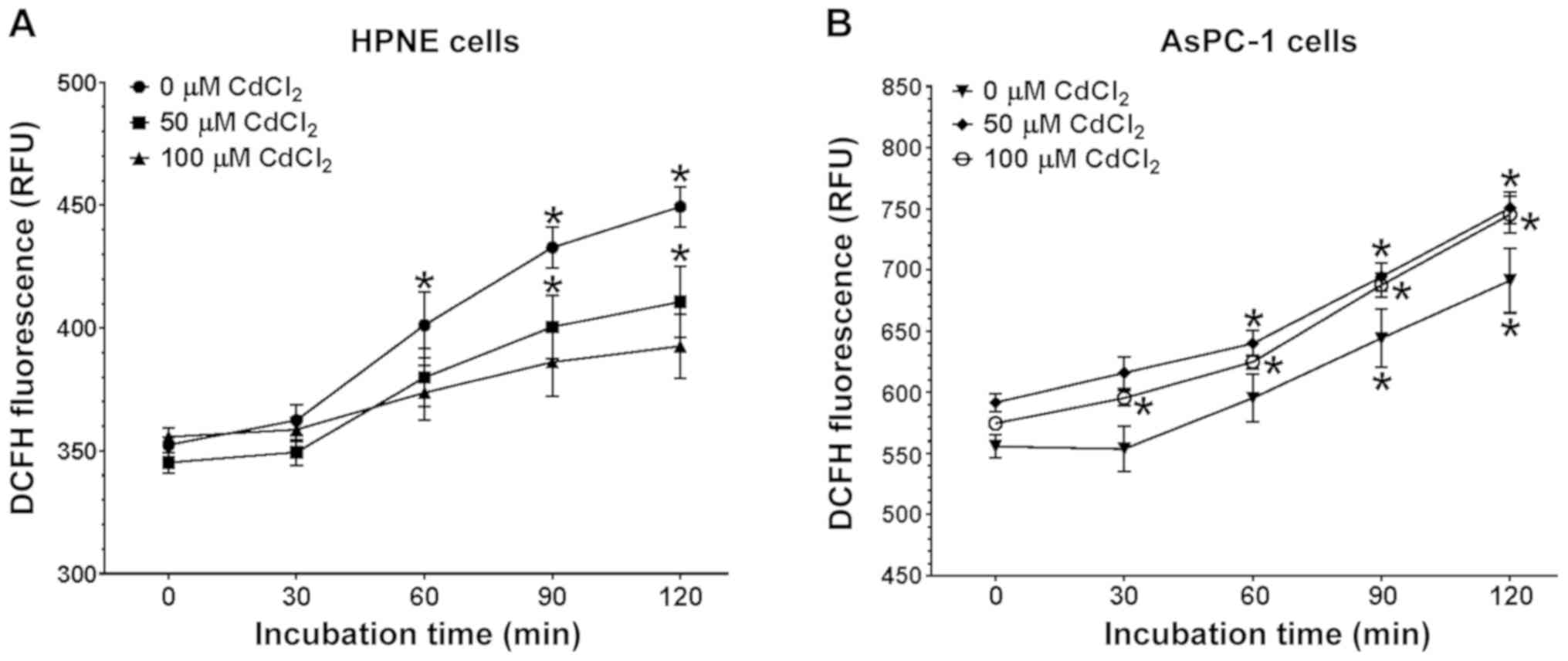
increase oxidative stress in the HPNE and AsPC-1 cells (Fig. 3A and B, respectively) was assessed
by measuring DCF fluorescence. Prior to expsoure to
CdCl2, basal DCF fluorescence was measured to yield a
baseline to which subsequent treatment values would be compared.
The basal levels of DCF fluorescence were 57.7% higher in the
AsPC-1 cells compared to the HPNE values (555.8 and 352.4 RFU
respectively; P<0.0001) at the initial 0-time-point. When
comparing the means between the AsPC-1 and HPNE cells at 60 min of
incubation, the trend was similar with the AsPC-1 cells,
demonstrating a 48.4% increase in oxidative stress (595.2 vs. 401.2
RFU respectively; P<0.0001). Data were analyzed using a
three-way ANOVA (cell type x time x treatment) and there was a
clear association regarding cell type (F1,56=531.2;
P<0.0001) and time (F4,105=162.6; P<0.0001) responses to
CdCl2. In both cell lines, increasing the time of
exposure resulted in an increased fluorescence. The trend for these
increases was similar between the HPNE (F4,105=26.75; P<0.0001)
and AsPC-1 (F4,105=59.71; P<0.0001) cells with no significant
association between time and CdCl2 concentration in
either the HPNE (F8,105=1.766; P=0.09) or AsPC-1 (F8,105=0.2748;
P=0.92) cells.
Mitochondrial toxicity elicited by
exposure to Cd
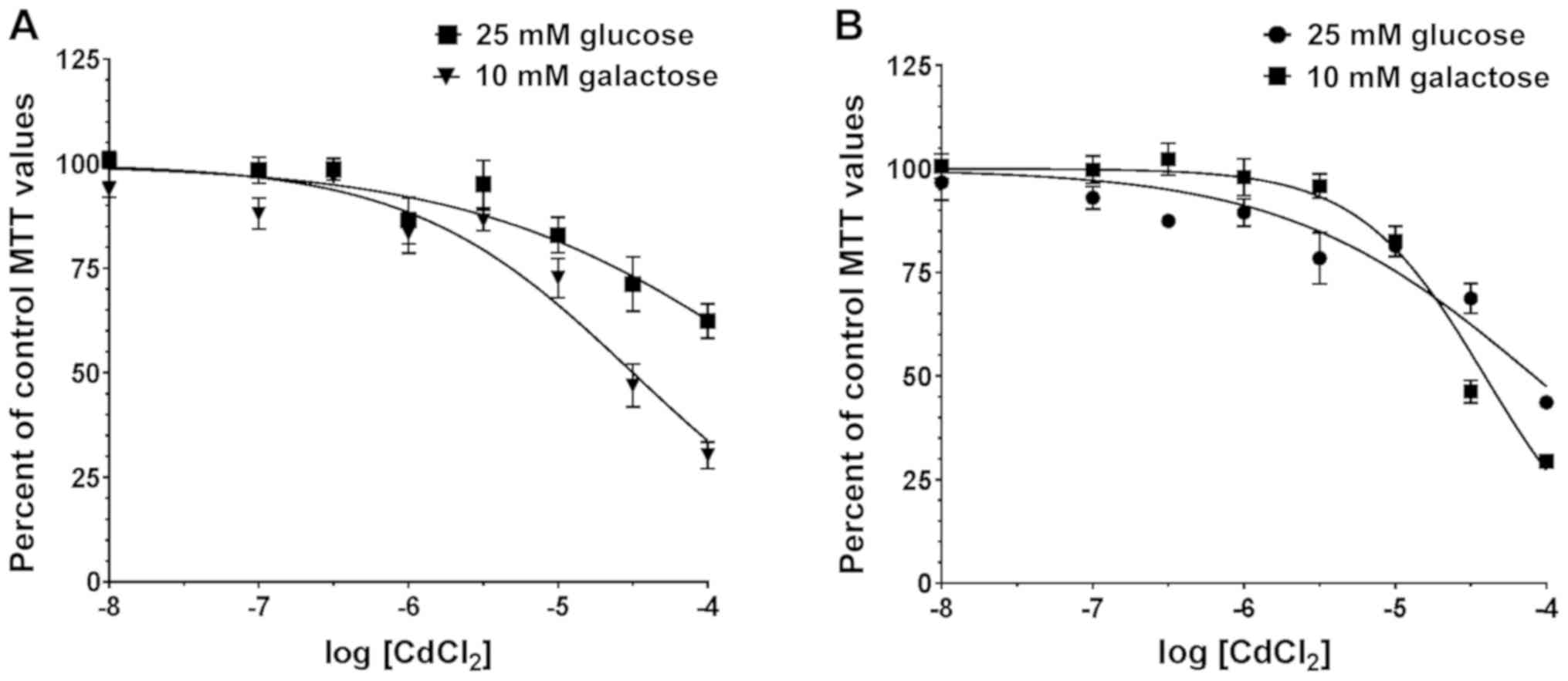
The next series of experiments examined the effects
of increasing concentrations of CdCl2 on cellular
toxicity in the HPNE (Fig. 4A)
and AsPC-1 (Fig. 4B) cells.
Following 4 h of exposure to CdCl2, cellular viability
was measured by MTT assay. This assay examined the shift in
IC50 value for CdCl2 following exposure to
the metal in the presence of 25 mM glucose or 10 mM
galactose-containing media. A rightward shift in IC50
for the galactose curve of 2.5-3.0-fold would be indicative of a
mitochondrial toxin (55,56). All curves where fit to a
single-site model with a r2 values of 0.72, 0.73, 0.75
and 0.86 for HPNE-glucose, HPNE-galactose, AsPC-1-glucose and
AsPC-1-galactose, respectively. After assessing the normality
values, all curves passed as fitting normal Gaussian distribution
(P-values of 0.07, 0.56, 0.17 and 0.92, respectively).
Individual IC50 values were obtained for
each sample using non-linear curve fitting and graphically
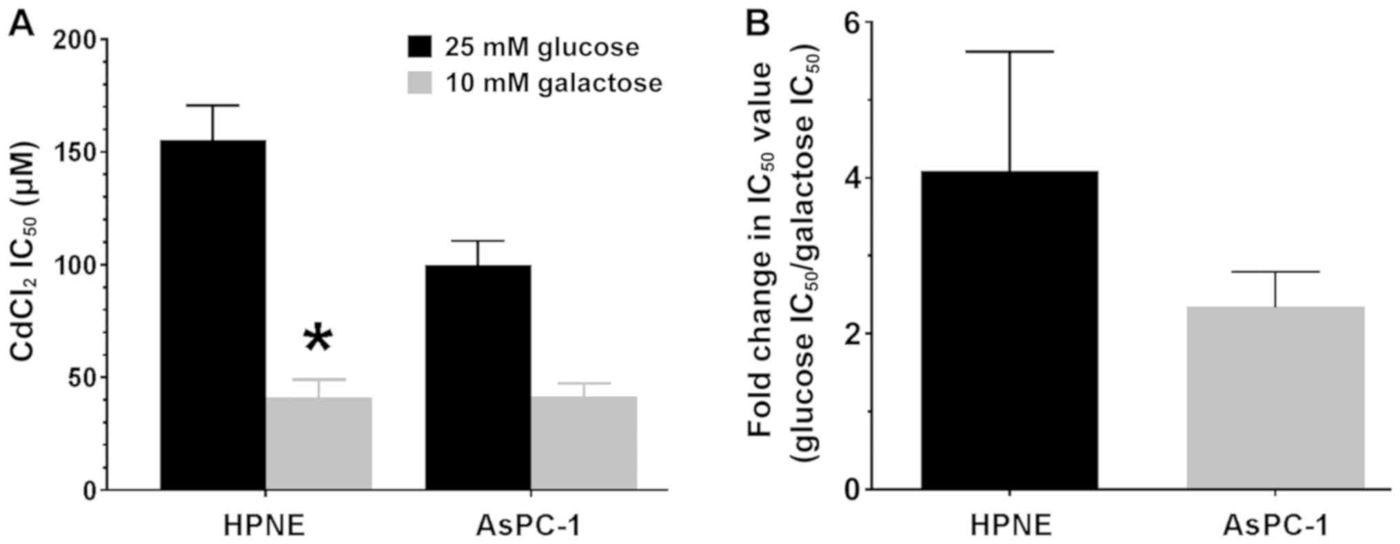
represented as the means ± SEM for each group (Fig. 5A). There was a significant effect
of media on the IC50 values (glucose vs. galactose;
F1,44=25.38; P<0.0001) that was observed in both cell
lines. Sidak post hoc analysis revealed that only the
IC50 value in the HPNE-galactose group was significantly
(P<0.01) lower than that in the HPNE-glucose group (Fig. 5A). When examining the shift in
IC50 values (Fig. 5B),
we observed that there was almost a 2- to 4-fold rightward shift in
IC50 values in both cell lines. The IC50
shift suggests that CdCl2 does act as a mitochondrial
toxin in the HPNE and AsPC-1 cells.
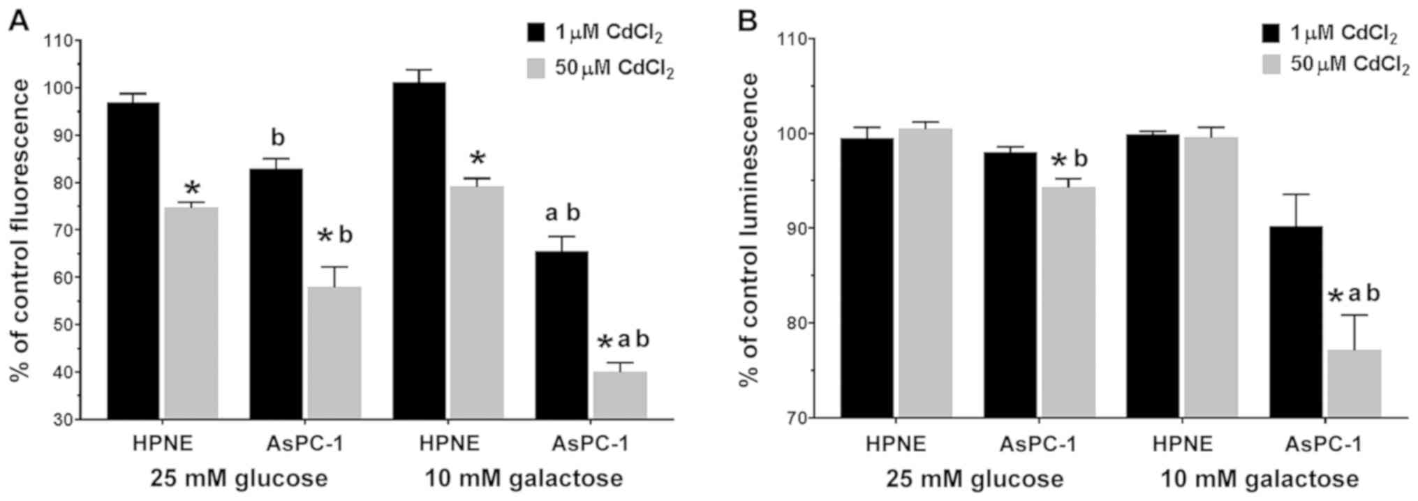
Using a dual-labeling assay kit, we measured both
cell viability (Fig. 6A) and ATP
release from the mitochondria, which is indicative of mitochondrial
toxicity (Fig. 6B). The use of
the more sensitive dual-labeling system revealed a complex response
pattern to the action of CdCl2 in both cell lines. The
four possible outcomes from this assay are: i) No change in ATP
signal + no change in membrane integrity = not a mitochondrial
toxicant; ii) reduced ATP + increased membrane integrity = primary
necrosis; iii) reduced ATP + no change in membrane integrity =
mitochondrial toxin; and iv) very low ATP at all concentrations +
discordant change in membrane integrity = mitochondrial toxicant.
Exposure to CdCl2 significantly (F1,24=214.5;
P<0.0001) affected membrane integrity (Fig. 6A). The cytotoxicity response was
also dependent on the type of media, glucose vs. galactose
(F1,24=13.54; P=0.0012) and cell type, HPNE vs. AsPC-1
(F1,24=173.4; P<0.0001). There was a significant
(F1,24=37.11; P<0.0001) association between the media
and CdCl2 concentration. In each group, exposure to 50
μM CdCl2 resulted in a significant reduction
(P<0.05) in viability compared to the corresponding 1 μM
CdCl2 values in each of the groups. Viability in both
the 1 and 50 μM CdCl2 groups in the
galactose/AsPC-1 group was significantly (P<0.05) lower than the
corresponding 25 mM glucose viability values. Across the
CdCl2 concentrations and glucose/galactose
supplementation, the viability values were significantly
(P<0.05) reduced in the AsPC-1 cells compared to the
corresponding viability values in the HPNE cells.
When examining the levels of ATP, we observed a
less robust change across he treatment groups (Fig. 6B), yet the trends were similar
compared to the data observed from the cytotoxicity experiments.
Exposure to CdCl2 significantly (F1,24=55.58;
P<0.0001) affected membrane integrity (Fig. 6A). The cytotoxicity response was
also dependent on the type of media, glucose vs. galactose
(F1,24=22.67; P<0.0001) and cell type, HPNE vs.
AsPC-1 (F1,24=9.10; P=0.006). There were two
interactions that were significantly associated and these were
media type x CdCl2 concentration
(F1,24=21.03; P=0.0001) and cell type x CdCl2
concentration (F1,24=10.53; P=0.0034), suggesting that
the CdCl2 concentration had a significant impact on both
membrane integrity and ATP quantity, depending on the media
(glucose vs. galactose) type or cell type (HPNE vs. AsPC-1). The
AsPC-1 cells exhibited a slightly higher sensitivity to the effects
of CdCl2, as indicated in Fig. 6B. The sensitivity of the AsPC-1
cells was particularly visible at the higher concentration of 50
μM of CdCl2, with the reduction in ATP was more
pronounced in the 10 mM galactose group. If there is a change in
membrane integrity associated with the changes in ATP, then that
would be indicative of a necrotic toxin, leading to a reduction in
ATP. We observed two associated interactions, with both the media
and the cell type being significantly associated with the
concentration of CdCl2.
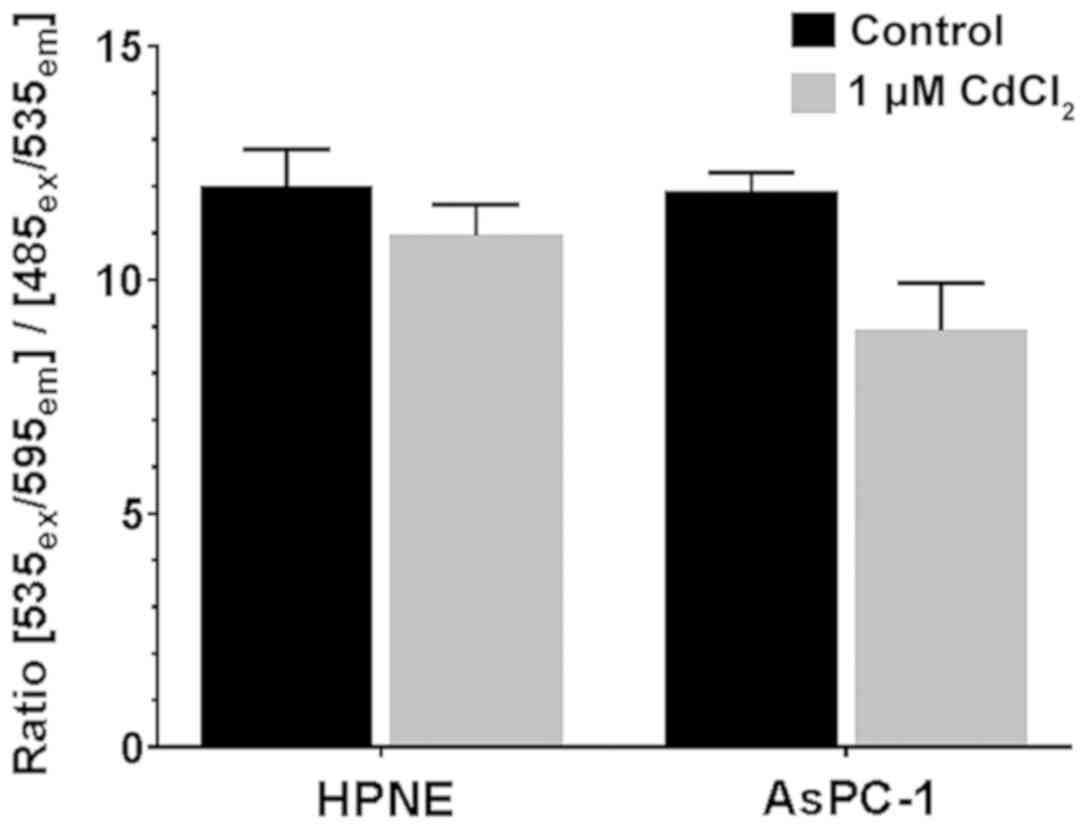
An additional measure of mitochondrial function is
the measure of mitochondrial permeability (referred to as
ΔψM). We used the JC-1 dye due to its ability to
permeate the mitochondria, and to either form aggregates (healthy
cells) or monomers (unhealthy cells) which fluoresce at different
wavelengths. We observed that CdCl2 (1 μM) had a
significant effect on the health of the cells
(F1,16=7.35; P=0.0184), slightly reducing the ratio of
health/unhealthy cells in both cell lines (Fig. 7). Neither treatment group differed
significantly from its corresponding control value.
Discussion
On the periodic table, some of the most toxic
metals reside in the d-block or transition metal block. Cd is part
of this group; however, Cd is somewhat different in that it is a
fifth-period element, unlike chromium, manganese, iron, and cobalt
which are fourth-period elements. In addition, Cd is at the end of
the transitional metal fifth-period row which makes it a
'post-translational element'. Therefore, there are similarities
between Cd and other metals that are known toxicants, carcinogens,
and free radical generators, such as mercury, iron, chromium,
cobalt, nickel and manganese, although additional studies are
required to improve our understanding of the cellular and
intracellular actions of Cd. In this study, we aimed to work
towards that end. Initial experiments examined the effects of
CdCl2 on viability and cell number in media containing
decreasing concentrations of fetal bovine serum. We did this to
confirm the lack of serum effect for short exposure times. Other
studies have reported that increasing the serum concentration up to
10% can affect the response of different cell types to toxicants
(64,70). In this study, we reported that
following exposure conducted in a low-serum environment, neither
the growth or viability of neither cell line was significantly
affected. The lack of effect aids in further in vitro
studies and we can control the influence and impact that serum has
on toxicity studies as a possible protectant. The lack of
cytotoxicity observed following the exposure of HPNE cells to
CdCl2 is not surprising since, control cells should have
better defenses to protect against CdCl2-mediated
damage. In addition, the 48-h exposure period may not be long
enough for overt cytotoxic damage to be observed. Assay condition
parameters are a consideration for further studies to avoid
obscuring potential necrotic or apoptotic changes. Subsequent
assays used a reduced serum to remove as many confounds as possible
from additional toxicology testing. One variable we did not address
in our studies is the possibility that low-level CdCl2
exposure may alter cellular responses, such as apoptosis.
The ability of elemental Cd to bioaccumulate in
humans and concentrate in organs, such as the liver and kidneys can
increase Cd-mediated toxicity over time (71). Exposure to CdCl2 can
result in apoptotic, as well as necrotic changes (72-74). The results of this study indicated
that exposure to only the higher concentration of CdCl2
resulted in a reduction in cell viability and cell number. This
reduction was only observed in the AsPC-1 cells. This study
intended to establish 'threshold' levels of exposure (concentration
and duration) for use in subsequent mitochondrial toxicity
experiments. Since we were attempting to find a threshold
concentration for toxicity, it was not unexpected that we observed
a lack of robust changes in cell viability, proliferation or cell
number after a 48-h exposure. Our understanding of Cd as an
environmental toxicant is expanding, and its role in the
development of cancer has become widely accepted (24,75-77). Health organizations have
classified Cd as a carcinogen (76). However, the exact mechanism(s)
through which Cd exerts these carcinogenic effects remain largely
unknown. Questions have been put forth as to whether it is a
'direct vs. indirect' effect, or potentially an 'upstream vs.
downstream' effect. Further studies are required to describe the
intracellular effects of Cd. In this study, we chose to target the
mitochondria as an intracellular organelle that needs to function
normally to aid the cell in providing the necessary energy
requirements. When the mitochondria fail to function normally,
tumor formation can follow (46).
The determination of a CdCl2
LC50 value in cell cultures is hard to find. It has
previously been shown that the LC50 for CdCl2
is approximately 70-80 μM in various aquatic organisms
(77). Other researchers have
indicated that the lethality of CdCl2 is dependent on
both the concentration and the duration of the exposure (78). In hepatic and pituitary cultures,
the reported LC50 values for CdCl2 range from
40-50 μM (78,79). In this study, our findings of
40-70 μM fall within the LC50 range that has been
reported elsewhere. The concentrations selected in the present
study should represent concentrations that are: i) Below; ii)
above; and iii) at the LC50 value. Caution was needed
not to use concentrations that were too high which would prove
lethal to a large percentage number of cells. That would skew our
findings and conclusions since we would primarily be examining a
subpopulation of cells that was resilient enough to guard against
CdCl2 toxicity. Yet a concentration that was too low
would not elicit a change in cellular function, and a change in
apoptotic function in particular.
The secondary aim of this study was to establish
and utilize 'subtoxic' concentrations of CdCl2 to assess
oxidative stress elicited by exposure to CdCl2. Using
approximately the mid-concentration, 6 ppm or 54 μM, of
CdCl2, we can see that CdCl2 was only weakly
a pro-oxidant, leading to a 15-20% increase in free radicals over
120 min of exposure. Measuring DCF fluorescence relies on the
direct generation of free radicals by CdCl2. In
vivo studies have shown that the administration of
CdCl2 does increase biomarker expression commonly
associated with increased oxidative stress (80,81). Therefore, changes in the oxidative
status of the cell alone cannot be sufficient alone to elicit
intracellular changes leading to apoptotic changes (82). In the kidneys, researchers have
postulated that Cd interferes with normal mitochondrial function,
leading to increased oxidative stress and apoptosis (38-40). In hepatic cells, CdCl2
exposure has been shown to decrease ΔψM, but also to
directly alter apoptotic pathways by increasing the activity of
both caspase-3 and caspase-9, as well as increasing the activity of
p53 (41). In the pancreas,
CdCl2 exposure has been shown to result in the death of
β-cells following increased oxidative stress followed by a
downstream-mediated activation of apoptotic pathways regulated by
mitochondria (42). Other
researchers have postulated that environment or occupational
exposure to Cd, and the resulting Cd-mediated death or damage to
β-cells is an underlying cause of diabetes and obesity (43). It is apparent that the Cd-mediated
effects in the pancreas can alter β-cell function, hormonal
responses, and may lead to the development of PC.
Healthy or 'normal' cells prefer oxidative
phosphorylation (generation of 38 ATPs) to derive their energy
(Crabtree), but immortalized cells lines may be adapted to using
high glucose, generating ATP via glycolysis (only generates 2
ATPs), which will decrease the sensitivity of the cells to
mitochondrial toxins (44). In
this study, we examined the effects of CdCl2 on
mitochondrial function and the Warburg effect, which involves the
differential production of ATP in media containing glucose or
galactose. Subsequently, by comparing the toxicity of a compound in
the presence of glucose media, or the presence of galactose media,
we can determine direct mitochondrial toxicity compared to other
indirect mechanisms (55,57,58). The use of galactose-containing (10
mM) media will force the cell to increase their reliance on
oxidative phosphorylation to generate ATP. The direct assessment of
CdCl2 as a mitochondrial toxin suggested that
CdCl2 is a weak mitochondrial toxicant. Comparisons of
the glucose/galactose MTT curves in the presence of increasing
concentrations of CdCl2 revealed an almost 3-fold shift
in IC50 values with glucose IC50 values >
galactose IC50 values.
In support of this finding was our observation that
ATP levels were reduced, primarily in AsPC-1 cells, with little
concomitant changes in viability, suggestive of mitochondrial
toxicity (57,59). These data suggest that the
CdCl2 concentration has a significant impact on both
membrane integrity and ATP quantity depending on media (glucose vs.
galactose) type or cell type (HPNE vs. AsPC-1). When examining the
data to determine whether CdCl2 is a mitochondrial
toxin, or whether it elicits toxicity through another mechanism,
the amount of ATP present needs to be quantified. We would have
expected a reduction in ATP in the presence of CdCl2
compared to control values suggesting a mitochondrial toxin. The
cytotoxic response must then be examined, with no change in
membrane integrity + reduction in ATP being indicative of being a
mitochondrial toxin.
Following the addition of the JC-1 dye data, we
observed only a small decrease in the ratio of healthy to unhealthy
cells in both cell lines. A caveat of these findings is that we
used 1 μM CdCl2. The rationale for this
concentration was that our threshold was approximately 10 μM
CdCl2 when we began to see a reduction in cellular
viability. We decided that a 10-fold lower concentration would be
sufficient to induce changes in cellular function without resulting
in a loss of viability, which would make our data difficult to
interpret since the mitochondrial information would be from cells
that were able to survive CdCl2 exposure. From our data,
it can be hypothesized that CdCl2 is a weak
mitochondrial toxin, with adverse effects evident at the highest
CdCl2 concentrations of 10 μM or higher.
Although one single theory has not come to the
forefront, numerous hypotheses have been presented suggesting that
the switch from oxidative phosphorylation to aerobic glycolysis
signals the transition from a normal cell to a tumor cell (46). Another hypothesis is that
switching to aerobic glycolysis can protect the tumor cell from a
sugar-induced death when glucose uptake is high, and by switching
to a high production of lactate, pro-proliferative intermediates
can be produced (47). Compounds,
such as CdCl2, that damage the mitochondria promote a
shift towards aerobic glycolysis, thus leading to tumor formation.
Studies that have examined toxicant (MPTP) damage to the
mitochondria have reported that oxidative phosphorylation is
decreased, that apoptotic pathways are altered and there is a
bioenergetic shift in glucose utilization (48). Indirect genetic damage to the
mitochondria that will result in epigenetic alterations have has
also been demonstrated in tumor cells, as well as inherited genetic
abnormalities that predispose or facilitate the progression of the
Warburg effect in tumor cells (49). The apparent importance of the
mitochondria and the Warburg effect has led researchers to
postulate that either 'turning' oxidative phosphorylation 'back on'
or shunting away from lactate production/utilization may be an
effective means of controlling tumor growth (45,50). In this study, we demonstrated a
rightward shift in IC50 values for both the HPNE and
AsPC-1 cells, suggesting that exposure to CdCl2 plays a
role in mitochondrial dysfunction. Additional experiments
supporting the finding of CdCl2 as a weak mitochondrial
toxicant, include the investigation of the aspects of mitochondrial
toxicity, such as changes in mitochondrial respiration, transition
pore permeability, and the activation of apoptotic peptides. The
ability of Cd to accumulate within mammalian tissues is a variable
that needs to be addressed due to the long biological half-life.
This characteristic of Cd may result in toxicity when exposure
concentrations are low, but exposure duration is extended.
Additional studies are underway to examine the effects of
CdCl2 exposure on intracellular function when subtoxic
concentrations of CdCl2 are added to the growth media
for extended periods of time (weeks). Exposure in this manner would
better represent long-term chronic exposure that many individuals
are subjected to in the environment.
Collectively, these data enhance our understanding
of Cd-mediated intracellular toxicity. The results also lead us to
additional questions. Unlike other well-studied metals, such as
mercury, iron, nickel and manganese, CdCl2 appears to
exert multiple weak effects, such as on oxidative stress,
viability, ATP loss and oxidative phosphorylation as an energy
source. CdCl2 does behave as a weak mitochondrial
toxicant as indicated by the 2- to 4-fold shift in IC50
values comparing the glucose and galactose curves. Since we
observed only modest changes in viability, cell number and ATP
release, it can be hypothesized that changes following low
concentrations of CdCl2 are most likely due to a complex
and cumulative set of small changes as opposed to substantial
changes. The assessment of the direct vs. indirect effects of
CdCl2 on cellular and mitochondrial function is
essential. Potential CdCl2-mediated changes which occur
upstream or downstream from the mitochondria will be pivotal in
determining where and how CdCl2 is eliciting its
cellular effects. Additional studies are underway to examine the
molecular alterations in intracellular organelles associated with
long-term CdCl2 exposure addressing our larger question
regarding CdCl2 as a mitochondrial toxicant.
Funding
The authors received support for this research in
part from OSU intramural funding [#154333-7] and Ministry of
Education, Science and Technological Development of Serbia (Project
III 46009). The funders had no role in the design of the study, or
in the collection, analyses, or interpretation of the data, in the
writing of the manuscript, and in the decision to publish the
results.
Availability of data and materials
All data generated or analyzed during this study
are included in this published article or are available from the
corresponding author on reasonable request.
Authors' contributions
DRW and ABD were responsible for the
conceptualization, methodology, formal data analysis and
interpretation, data curation, writing/original draft preparation,
writing-review and editing, project administration, and funding
acquisition. DAS and AT were involved in consultation regarding the
conceptualization and interpretation of the data, data curation,
composition of the draft manuscript, as well as the final
manuscript and post-review editing. VD was responsible for earlier
clinical studies that established the pancreatic concentrations of
Cd and working to quantify in vivo Cd concentrations. VD and
AB aided in the design of the experiments and the establishment of
appropriate/relevant concentrations of Cd to be used in the study.
AB worked with the overall design and interpretation of the data
and assisted in the writing of the final manuscript. AS worked with
the design of the viability studies, performance of viability
assays and the corresponding data acquisition and analysis for
those assays. All authors have read and approved the final
manuscript.
Ethics approval and consent to
participate
Not applicable.
Patient consent for publication
Not applicable.
Competing interests
DAS is the Editor-in-Chief for the journal, but had
no personal involvement in the reviewing process, or any influence
in terms of adjudicating on the final decision, for this article.
The other authors declare that they have no competing interests
regarding this work.
Acknowledgments
Not applicable.
References
|
1
|
Faroon O, Ashizawa A, Wright S, Tucker P,
Jenkins K, Ingerman L and Rudisill C: Toxicological Profile for
Cadmium. Agency for Toxic Substances and Disease Registry. 1–487.
2012.
|
|
2
|
Renieri E, Alegakis A, Kiriakakis M,
Vinceti M, Ozcagli E, Wilks M and Tsatsakis A: Cd, Pb and Hg
Biomonitoring in Fish of the Mediterranean Region and Risk
Estimations on Fish Consumption. Toxics. 2:417–442. 2014.
View Article : Google Scholar
|
|
3
|
Renieri EA, Safenkova IV, Alegakis AK,
Slutskaya ES, Kokaraki V, Kentouri M, Dzantiev BB and Tsatsakis AM:
Cadmium, lead and mercury in muscle tissue of gilthead seabream and
seabass: Risk evaluation for consumers. Food Chem Toxicol.
124:439–449. 2019. View Article : Google Scholar
|
|
4
|
Mezynska M and Brzóska MM: Environmental
exposure to cadmium-a risk for health of the general population in
industrialized countries and preventive strategies. Environ Sci
Pollut Res Int. 25:3211–3232. 2018. View Article : Google Scholar
|
|
5
|
Satarug S: Dietary Cadmium Intake and Its
Effects on Kidneys. Toxics. 6:152018. View Article : Google Scholar :
|
|
6
|
Buha A, Antonijević B, Bulat Z, Jaćević V,
Milovanović V and Matović V: The impact of prolonged cadmium
exposure and co-exposure with polychlorinated biphenyls on thyroid
function in rats. Toxicol Lett. 221:83–90. 2013. View Article : Google Scholar : PubMed/NCBI
|
|
7
|
Buha A, Matovic V, Antonijevic B, Bulat Z,
Curcic M, Renieri EA, Tsatsakis AM, Schweitzer A and Wallace D:
Overview of cadmium thyroid disrupting effects and mechanisms. Int
J Mol Sci. 19:15012018. View Article : Google Scholar :
|
|
8
|
Silva N, Peiris-John R, Wickremasinghe R,
Senanayake H and Sathiakumar N: Cadmium a metalloestrogen: Are we
convinced? J Appl Toxicol. 32:318–332. 2012. View Article : Google Scholar
|
|
9
|
Xing Y, Xia W, Zhang B, Zhou A, Huang Z,
Zhang H, Liu H, Jiang Y, Hu C, Chen X, et al: Relation between
cadmium exposure and gestational diabetes mellitus. Environ Int.
113:300–305. 2018. View Article : Google Scholar : PubMed/NCBI
|
|
10
|
Renieri EA, Sfakianakis DG, Alegakis AA,
Safenkova IV, Buha A, Matović V, Tzardi M, Dzantiev BB, Divanach P,
Kentouri M, et al: Nonlinear responses to waterborne cadmium
exposure in zebrafish. An in vivo study. Environ Res. 157:173–181.
2017. View Article : Google Scholar : PubMed/NCBI
|
|
11
|
IARC: IARC monographs on the evaluation of
the carcinogenic risk of chemicals to man. 1993
|
|
12
|
Iarc and IARC Working Group on the
Evaluation of Carcinogenic Risks to Humans: International Agency
for Research on Cancer Iarc Monographs on the Evaluation of
Carcinogenic Risks to Humans. Personal habits and indoor
combustions. IARC Monogr Eval Carcinog Risks Hum. 100E. 2012
|
|
13
|
Huff J, Lunn RM, Waalkes MP, Tomatis L and
Infante PF: Cadmium-induced cancers in animals and in humans. Int J
Occup Environ Health. 13:202–212. 2007. View Article : Google Scholar : PubMed/NCBI
|
|
14
|
Hartwig A: Cadmium and cancer. Cadmium:
From Toxicity to Essentiality. Sigel A, Sigel H and Sigel RK:
Springer; Netherlands, Dordrecht: pp. 491–507. 2013
|
|
15
|
Il'yasova D and Schwartz GG: Cadmium and
renal cancer. Toxicol Appl Pharmacol. 207:179–186. 2005. View Article : Google Scholar : PubMed/NCBI
|
|
16
|
Vinceti M, Venturelli M, Sighinolfi C,
Trerotoli P, Bonvicini F, Ferrari A, Bianchi G, Serio G, Bergomi M
and Vivoli G: Case-control study of toenail cadmium and prostate
cancer risk in Italy. Sci Total Environ. 373:77–81. 2007.
View Article : Google Scholar
|
|
17
|
Goyer RA, Liu J and Waalkes MP: Cadmium
and cancer of prostate and testis. Biometals. 17:555–558. 2004.
View Article : Google Scholar
|
|
18
|
Feki-Tounsi M and Hamza-Chaffai A: Cadmium
as a possible cause of bladder cancer: A review of accumulated
evidence. Environ Sci Pollut Res Int. 21:10561–10573. 2014.
View Article : Google Scholar : PubMed/NCBI
|
|
19
|
Van Maele-Fabry G, Lombaert N and Lison D:
Dietary exposure to cadmium and risk of breast cancer in
postmenopausal women: A systematic review and meta-analysis.
Environ Int. 86:1–13. 2016. View Article : Google Scholar
|
|
20
|
Cho YA, Kim J, Woo HD and Kang M: Dietary
cadmium intake and the risk of cancer: A meta-analysis. PLoS One.
8:e750872013. View Article : Google Scholar : PubMed/NCBI
|
|
21
|
Bray F, Ferlay J, Soerjomataram I, Siegel
RL, Torre LA and Jemal A: Global cancer statistics 2018: GLOBOCAN
estimates of incidence and mortality worldwide for 36 cancers in
185 countries. CA Cancer J Clin. 68:394–424. 2018. View Article : Google Scholar : PubMed/NCBI
|
|
22
|
Mytilinaiou M, Nikitovic D, Berdiaki A,
Kostouras A, Papoutsidakis A, Tsatsakis AM and Tzanakakis GN:
Emerging roles of syndecan 2 in epithelial and mesenchymal cancer
progression. IUBMB Life. 69:824–833. 2017. View Article : Google Scholar : PubMed/NCBI
|
|
23
|
Tzanakakis GN, Margioris AN, Tsatsakis AM
and Vezeridis MP: The metastatic potential of human pancreatic cell
lines in the liver of nude mice correlates well with cathepsin B
activity. Int J Gastrointest Cancer. 34:27–38. 2003. View Article : Google Scholar
|
|
24
|
Buha A, Wallace D, Matovic V, Schweitzer
A, Oluic B, Micic D and Djordjevic V: Cadmium Exposure as a
Putative Risk Factor for the Development of Pancreatic Cancer:
Three Different Lines of Evidence. BioMed Res Int.
2017:19818372017. View Article : Google Scholar
|
|
25
|
Andjelkovic M, Buha Djordjevic A,
Antonijevic E, Antonijevic B, Stanic M, Kotur-Stevuljevic J,
Spasojevic-Kalimanovska V, Jovanovic M, Boricic N, Wallace D, et
al: Toxic effect of acute cadmium and lead exposure in rat blood,
liver, and kidney. Int J Environ Res Public Health. 16:E2742019.
View Article : Google Scholar : PubMed/NCBI
|
|
26
|
Buha A, Bulat Z, Dukić-Ćosić D and Matović
V: Effects of oral and intraperitoneal magnesium treatment against
cadmium-induced oxidative stress in plasma of rats. Arh Hig Rada
Toksikol. 63:247–254. 2012. View Article : Google Scholar : PubMed/NCBI
|
|
27
|
Matović V, Buha A, Bulat Z, Ðukić-Ćosić D,
Miljković M, Ivanišević J and Kotur-Stevuljević J: Route-dependent
effects of cadmium/cadmium and magnesium acute treatment on
parameters of oxidative stress in rat liver. Food Chem Toxicol.
50:552–557. 2012. View Article : Google Scholar
|
|
28
|
Djukić-Cosić D, Curcić Jovanović M,
Plamenac Bulat Z, Ninković M, Malicević Z and Matović V: Relation
between lipid peroxidation and iron concentration in mouse liver
after acute and subacute cadmium intoxication. J Trace Elem Med
Biol. 22:66–72. 2008. View Article : Google Scholar
|
|
29
|
Bulat Z, Dukić-Osi-Ćosić D, Antonijević B,
Bulat P, Vujanović D, Buha A and Matović V: Effect of magnesium
supplementation on the distribution patterns of zinc, copper, and
magnesium in rabbits exposed to prolonged cadmium intoxication. Sci
World J. 2012:2012. View Article : Google Scholar
|
|
30
|
Djukić-Cosić D, Ninković M, Malicević Z,
Plamenac-Bulat Z and Matović V: Effect of supplemental magnesium on
the kidney levels of cadmium, zinc, and copper of mice exposed to
toxic levels of cadmium. Biol Trace Elem Res. 114:281–291. 2006.
View Article : Google Scholar
|
|
31
|
Matović V, Buha A, Ðukić-Ćosić D and Bulat
Z: Insight into the oxidative stress induced by lead and/or cadmium
in blood, liver and kidneys. Food Chem Toxicol. 78:130–140. 2015.
View Article : Google Scholar
|
|
32
|
Bulat Z, Đukić-Ćosić D, Antonijević B,
Buha A, Bulat P, Pavlović Z and Matović V: Can zinc supplementation
ameliorate cadmium-induced alterations in the bioelement content in
rabbits? Arh Hig Rada Toksikol. 68:38–45. 2017. View Article : Google Scholar : PubMed/NCBI
|
|
33
|
Zhang DX, Dai YD, Yuan SX and Tao L:
Prognostic factors in patients with pancreatic cancer. Exp Ther
Med. 3:423–432. 2012. View Article : Google Scholar : PubMed/NCBI
|
|
34
|
Liu J, Ji S, Liang C, Qin Y, Jin K, Liang
D, Xu W, Shi S, Zhang B, Liu L, et al: Critical role of oncogenic
KRAS in pancreatic cancer (Review). Mol Med Rep. 13:4943–4949.
2016. View Article : Google Scholar : PubMed/NCBI
|
|
35
|
Basu A, Jiang X, Negrini M and Haldar S:
MicroRNA-mediated regulation of pancreatic cancer cell
proliferation. Oncol Lett. 1:565–568. 2010. View Article : Google Scholar : PubMed/NCBI
|
|
36
|
Duarte FV, Palmeira CM and Rolo AP: The
role of microRNAs in mitochondria: Small players acting wide. Genes
(Basel). 5:865–886. 2014. View Article : Google Scholar
|
|
37
|
Cannino G, Ferruggia E, Luparello C and
Rinaldi AM: Cadmium and mitochondria. Mitochondrion. 9:377–384.
2009. View Article : Google Scholar : PubMed/NCBI
|
|
38
|
Li M, Xia T, Jiang CS, Li LJ, Fu JL and
Zhou ZC: Cadmium directly induced the opening of membrane
permeability pore of mitochondria which possibly involved in
cadmium-triggered apoptosis. Toxicology. 194:19–33. 2003.
View Article : Google Scholar : PubMed/NCBI
|
|
39
|
Mao WP, Zhang NN, Zhou FY, Li WX, Liu HY,
Feng J, Zhou L, Wei CJ, Pan YB and He ZJ: Cadmium directly induced
mitochondrial dysfunction of human embryonic kidney cells. Hum Exp
Toxicol. 30:920–929. 2011. View Article : Google Scholar
|
|
40
|
Gobe G and Crane D: Mitochondria, reactive
oxygen species and cadmium toxicity in the kidney. Toxicol Lett.
198:49–55. 2010. View Article : Google Scholar : PubMed/NCBI
|
|
41
|
Lasfer M, Vadrot N, Aoudjehane L, Conti F,
Bringuier AF, Feldmann G and Reyl-Desmars F: Cadmium induces
mitochondria-dependent apoptosis of normal human hepatocytes. Cell
Biol Toxicol. 24:55–62. 2008. View Article : Google Scholar
|
|
42
|
Chang KC, Hsu CC, Liu SH, Su CC, Yen CC,
Lee MJ, Chen KL, Ho TJ, Hung DZ, Wu CC, et al: Cadmium induces
apoptosis in pancreatic β-cells through a mitochondria-dependent
pathway: The role of oxidative stress-mediated c-Jun N-terminal
kinase activation. PLoS One. 8:e543742013. View Article : Google Scholar
|
|
43
|
Tinkov AA, Filippini T, Ajsuvakova OP,
Aaseth J, Gluhcheva YG, Ivanova JM, Bjørklund G, Skalnaya MG,
Gatiatulina ER, Popova EV, et al: The role of cadmium in obesity
and diabetes. Sci Total Environ. 601-602:741–755. 2017. View Article : Google Scholar : PubMed/NCBI
|
|
44
|
Hammad N, Rosas-Lemus M, Uribe-Carvajal S,
Rigoulet M and Devin A: The Crabtree and Warburg effects: Do
metabolite-induced regulations participate in their induction?
Biochim Biophys Acta. 1857:1139–1146. 2016. View Article : Google Scholar : PubMed/NCBI
|
|
45
|
Diaz-Ruiz R, Rigoulet M and Devin A: The
Warburg and Crabtree effects: On the origin of cancer cell energy
metabolism and of yeast glucose repression. Biochim Biophys Acta.
1807:568–576. 2011. View Article : Google Scholar
|
|
46
|
Devic S: Warburg effect - a consequence or
the cause of carcinogenesis? J Cancer. 7:817–822. 2016. View Article : Google Scholar : PubMed/NCBI
|
|
47
|
de Alteriis E, Cartenì F, Parascandola P,
Serpa J and Mazzoleni S: Revisiting the Crabtree/Warburg effect in
a dynamic perspective: A fitness advantage against sugar-induced
cell death. Cell Cycle. 17:688–701. 2018. View Article : Google Scholar : PubMed/NCBI
|
|
48
|
Solaini G, Sgarbi G and Baracca A:
Oxidative phosphorylation in cancer cells. Biochim Biophys Acta.
1807:534–542. 2011. View Article : Google Scholar
|
|
49
|
Chandra D and Singh KK: Genetic insights
into OXPHOS defect and its role in cancer. Biochim Biophys Acta.
1807:620–625. 2011. View Article : Google Scholar
|
|
50
|
Mot AI, Liddell JR, White AR and Crouch
PJ: Circumventing the Crabtree Effect: A method to induce lactate
consumption and increase oxidative phosphorylation in cell culture.
Int J Biochem Cell Biol. 79:128–138. 2016. View Article : Google Scholar : PubMed/NCBI
|
|
51
|
Lamas-Maceiras M, Rodríguez-Belmonte E,
Becerra M, González-Siso MI and Cerdán ME: KlGcr1 controls
glucose-6-phosphate dehydrogenase activity and responses to H O
cadmium and arsenate in Kluyveromyces lactis. Fungal Genet 2 2,
Biol. 82:95–103. 2015. View Article : Google Scholar
|
|
52
|
Torres AM, Maceiras ML, Belmonte ER,
Naveira LN, Calvo MB and Cerdán ME: KlRox1 pcontributes to yeast
resistance to metals and is necessary for KlYCF1 expression in the
presence of cadmium. Gene. 497:27–37. 2012. View Article : Google Scholar : PubMed/NCBI
|
|
53
|
Djordjevic VR, Wallace DR, Schweitzer A,
Boricic N, Knezevic D, Matic S, Grubor N, Kerkez M, Radenkovic D,
Bulat Z, et al: Environmental cadmium exposure and pancreatic
cancer: Evidence from case control, animal and in vitro studies.
Environ Int. 128:353–361. 2019. View Article : Google Scholar : PubMed/NCBI
|
|
54
|
Wallace DR and Bates E: Differential
effects of organic and inorganic mercury on phenotypically variant
breast cancer cell lines. J Clin Toxicol. 5:10002732015. View Article : Google Scholar
|
|
55
|
Sanuki Y, Araki T, Nakazono O and Tsurui
K: A rapid mitochondrial toxicity assay utilizing rapidly changing
cell energy metabolism. J Toxicol Sci. 42:349–358. 2017. View Article : Google Scholar : PubMed/NCBI
|
|
56
|
Dott W, Mistry P, Wright J, Cain K and
Herbert KE: Modulation of mitochondrial bioenergetics in a skeletal
muscle cell line model of mitochondrial toxicity. Redox Biol.
2:224–233. 2014. View Article : Google Scholar : PubMed/NCBI
|
|
57
|
Niles AL, Moravec RA, Eric Hesselberth P,
Scurria MA, Daily WJ and Riss TL: A homogeneous assay to measure
live and dead cells in the same sample by detecting different
protease markers. Anal Biochem. 366:197–206. 2007. View Article : Google Scholar : PubMed/NCBI
|
|
58
|
Marroquin LD, Hynes J, Dykens JA, Jamieson
JD and Will Y: Circumventing the Crabtree effect: Replacing media
glucose with galactose increases susceptibility of HepG2 cells to
mitochondrial toxicants. Toxicol Sci. 97:539–547. 2007. View Article : Google Scholar : PubMed/NCBI
|
|
59
|
Rossignol R, Gilkerson R, Aggeler R,
Yamagata K, Remington SJ and Capaldi RA: Energy substrate modulates
mitochondrial structure and oxidative capacity in cancer cells.
Cancer Res. 64:985–993. 2004. View Article : Google Scholar : PubMed/NCBI
|
|
60
|
Rodríguez-Enríquez S, Juárez O,
Rodríguez-Zavala JS and Moreno-Sánchez R: Multisite control of the
Crabtree effect in ascites hepatoma cells. Eur J Biochem.
268:2512–2519. 2001. View Article : Google Scholar : PubMed/NCBI
|
|
61
|
Taverna M, Marie AL, Mira JP and Guidet B:
Specific antioxidant properties of human serum albumin. Ann
Intensive Care. 3:42013. View Article : Google Scholar : PubMed/NCBI
|
|
62
|
Hsiao IL and Huang YJ: Effects of serum on
cytotoxicity of nano- and micro-sized ZnO particles. J Nanopart
Res. 15:18292013. View Article : Google Scholar : PubMed/NCBI
|
|
63
|
Leelakanok N, Fischer CL, Bates AM,
Guthmiller JM, Johnson GK, Salem AK, Brogden KA and Brogden NK:
Cytotoxicity of HBD3 for dendritic cells, normal human epidermal
keratinocytes, hTERT keratinocytes, and primary oral gingival
epithelial keratinocytes in cell culture conditions. Toxicol Lett.
239:90–96. 2015. View Article : Google Scholar : PubMed/NCBI
|
|
64
|
Umebayashi C, Oyama Y, Chikahisa-Muramastu
L, Nakao H, Nishizaki Y, Nakata M, Soeda F and Takahama K:
Tri-n-butyltin-induced cytotoxicity on rat thymocytes in presence
and absence of serum. Toxicol In Vitro. 18:55–61. 2004. View Article : Google Scholar
|
|
65
|
Qiao D, Seidler FJ and Slotkin TA:
Developmental neurotoxicity of chlorpyrifos modeled in vitro:
Comparative effects of metabolites and other cholinesterase
inhibitors on DNA synthesis in PC12 and C6 cells. Environ Health
Perspect. 109:909–913. 2001. View Article : Google Scholar : PubMed/NCBI
|
|
66
|
Stohs SJJ and Bagchi D: Oxidative
mechanisms in the toxicity of metal ions. Free Radic Biol Med.
18:321–336. 1995. View Article : Google Scholar : PubMed/NCBI
|
|
67
|
Ercal N, Gurer-Orhan H and Aykin-Burns N:
Toxic metals and oxidative stress part I: Mechanisms involved in
metal-induced oxidative damage. Curr Top Med Chem. 1:529–539. 2001.
View Article : Google Scholar
|
|
68
|
Valko M, Rhodes CJ, Moncol J, Izakovic M
and Mazur M: Free radicals, metals and antioxidants in oxidative
stress-induced cancer. Chem Biol Interact. 160:1–40. 2006.
View Article : Google Scholar : PubMed/NCBI
|
|
69
|
Galaris D and Evangelou A: The role of
oxidative stress in mechanisms of metal-induced carcinogenesis.
Crit Rev Oncol Hematol. 42:93–103. 2002. View Article : Google Scholar : PubMed/NCBI
|
|
70
|
Thomas MG, Marwood RM, Parsons AE and
Parsons RB: The effect of foetal bovine serum supplementation upon
the lactate dehydrogenase cytotoxicity assay: Important
considerations for in vitro toxicity analysis. Toxicol In Vitro.
30:300–308. 2015. View Article : Google Scholar : PubMed/NCBI
|
|
71
|
Cadm ium toxicity and treatment.
ScientificWorldJournal. 2013:3946522013.
|
|
72
|
Franco R, Sánchez-Olea R, Reyes-Reyes EM
and Panayiotidis MI: Environmental toxicity, oxidative stress and
apoptosis: Ménage à trois. Mutat Res. 674:3–22. 2009. View Article : Google Scholar
|
|
73
|
Chen C, Xun P, Nishijo M, Sekikawa A and
He K: Cadmium exposure and risk of pancreatic cancer: A
meta-analysis of prospective cohort studies and case-control
studies among individuals without occupational exposure history.
Environ Sci Pollut Res Int. 22:17465–17474. 2015. View Article : Google Scholar : PubMed/NCBI
|
|
74
|
Chen P, Duan X, Li M, Huang C, Li J, Chu
R, Ying H, Song H, Jia X, Ba Q, et al: Systematic network
assessment of the carcinogenic activities of cadmium. Toxicol Appl
Pharmacol. 310:150–158. 2016. View Article : Google Scholar : PubMed/NCBI
|
|
75
|
Waalkes MP: Cadmium carcinogenesis. Mutat
Res. 533:107–120. 2003. View Article : Google Scholar : PubMed/NCBI
|
|
76
|
WHO: Exposure to Cadmium: A Major Public
Health Concern. Geneva. 2010.
|
|
77
|
Muthukumaravel K, Kumarasamy P, Amsath A
and Paulraj MG: Toxic effect of cadmium on the electrophoretic
protein patterns of gill and muscle of Oreochromis mossambicus. E-J
Chem. 4:284–286. 2007. View Article : Google Scholar
|
|
78
|
Fotakis G and Timbrell JA: In vitro
cytotoxicity assays: Comparison of LDH, neutral red, MTT and
protein assay in hepatoma cell lines following exposure to cadmium
chloride. Toxicol Lett. 160:171–177. 2006. View Article : Google Scholar
|
|
79
|
Hinkle PM, Kinsella PA and Osterhoudt KC:
Cadmium uptake and toxicity via voltage-sensitive calcium channels.
J Biol Chem. 262:16333–16337. 1987.PubMed/NCBI
|
|
80
|
Bertin G and Averbeck D: Cadmium: Cellular
effects, modifications of biomolecules, modulation of DNA repair
and genotoxic consequences (a review). Biochimie. 88:1549–1559.
2006. View Article : Google Scholar : PubMed/NCBI
|
|
81
|
Cuypers A, Plusquin M, Remans T, Jozefczak
M, Keunen E, Gielen H, Opdenakker K, Nair AR, Munters E, Artois TJ,
et al: Cadmium stress: An oxidative challenge. Biometals.
23:927–940. 2010. View Article : Google Scholar : PubMed/NCBI
|
|
82
|
Tzanakakis GN, Krambovitis E, Tsatsakis AM
and Vezeridis MP: The preventive effect of ketoconazole on
experimental metastasis from a human pancreatic carcinoma may be
related to its effect on prostaglandin synthesis. Int J
Gastrointest Cancer. 32:23–30. 2002. View Article : Google Scholar
|