Introduction
Different species of seaweed have received a great
deal of attention from researchers in recent years. Seaweeds
contain high amounts of proteins, vitamins and minerals, and
several polysaccharides found in seaweed have been shown to exhibit
diverse biological activities. In particular, the antitumor and
antibacterial activities of seaweed have been widely studied;
furthermore, effects on the immune system have been demonstrated
(1,2). Porphyra yezoensis is an
intertidal marine red algae that has received increasing attention
as a model organism, owing to its important role in biological
research (3). Porphyra
yezoensis is one of the most important edible seaweeds, and
accordingly, is one of the most valuable marine crops in the world;
it is cultivated widely in Asia, especially in Japan, China and
Korea (4). Although several
studies have examined the polysaccharides found in the extracts of
Porphyra yezoensis, the effects of particular proteins have
not been reported. The peptide PPY from Porphyra yezoensis
is known to play a role in antitumor cell signaling, but the
mechanism behind this activity is not well understood. Insulin-like
growth factor I (IGF-I) and its cognate receptor, insulin-like
growth factor I receptor (IGF-IR), play important roles in normal
cell function and tumorigenesis, via their mediation of cell
growth, differentiation and survival (5); numerous studies have shown that
overexpression of IGF-IR and related proteins results in cancer
cell proliferation and survival (6–8). The
role of IGF-I signaling in tumor growth has been demonstrated in
vivo using nucleic-acid based strategies.
Apoptosis, the process of active programmed cell
death, occurs under many important physiological conditions, and it
is a critical part of normal development and differentiation in a
wide variety of tissues. This form of cell death has been
extensively studied in cancer research as a potential mechanism by
which the body eliminates precancerous and/or cancerous cells
(9). Apoptosis is characterized by
several unique features, including cell shrinkage, chromatin
condensation, DNA fragmentation, the expression of
phosphatidylserine on the cell surface and membrane blebbing
(10,11).
Cyclins are key cell cycle control molecules with
specific and periodic expression associated with cell cycle
progression (12). Other cell
cycle control proteins include cyclin-dependent kinase (cdk)
inhibitors, such as p21 and p27, which tightly regulate the
activities of cyclin/cdk enzyme complexes (13). Mitogen-activated protein kinases
(MAPKs), another important class of proteins, are activated in
response to a wide variety of extracellular stimuli and mediate
signal transduction cascades that play important roles in cell
proliferation, differentiation, cell cycle control and apoptosis
(14).
It has been shown that PPY has antitumor effects,
and the important role of IGF-I in mediating numerous cell survival
pathways is also well established. This study aimed to determine
whether PPY induces apoptosis via IGF-IR signaling.
Materials and methods
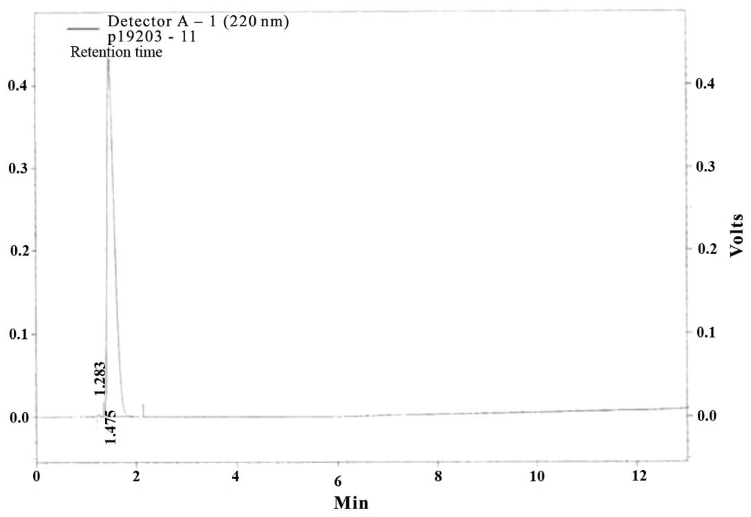
Preparation of peptide
The peptide PPY, found in Porphyra yezoensis,
was synthesized by the Peptron (Daejeon, Korea). Purification of
PPY was performed using a Shimadzu Prominence HPLC apparatus and
controlled using the software package Class-VP, 6.14 (Kyoto,
Japan). A C18 column (Shiesido Capcell Pak) in 0.1% TFA/water and a
gradient of 10–70% acetonitrile in 0.1% TFA, with a flow rate of 1
nm/min and UV detection at 220 nm was used.
Cell culture
MCF-7 human breast cancer cells were obtained from
the Korean Cell Line Bank (KCLB) and grown in RPMI-1640 medium
supplemented with 10% fetal bovine serum, 100 μg/ml penicillin and
100 ng/ml streptomycin at 37°C in a humidified atmosphere with 5%
CO2.
Cell proliferation assay
Cell proliferation was estimated using a CellTiter
96 aqueous non-radioactive cell proliferation assay (Promega,
Madison, WI, USA), which is based on the cleavage of
3-(4,5-dimethylthiazol-2-yl)-5-(3-carboxymethoxy-phenyl)-2H-tetrazolium
(MTS) into a formazan product that is soluble in cell culture
medium. Cells were seeded onto 96-well plates at 2×10⁴
cells per well in 100 μl medium and allowed to attach for 24 h. The
cell monolayer was washed with phosphate-buffered saline (PBS) to
remove unattached cells. The attached cells were maintained in
serum-free medium (SFM) for 12 h and then washed with PBS. Cells
were then incubated with fresh SFM containing various
concentrations (0–500 ng/ml) of peptide for 24 h. Subsequently, the
cells were incubated with 10 μg/ml MTS solution for 30 min, and the
absorbance of each well was measured at 490 nm using a SpectraMAX
340-pc multi-plate reader (Molecular Devices, Sunnyvale, CA,
USA).
DAPI staining assay
Cell were washed with PBS and fixed with 3.7%
paraformaldehyde in PBS for 10 min at room temperature. Fixed cells
were washed with PBS and stained with 2.5 μg/ml
4,6-diamidio-2-phenylindole (DAPI) solution for 10 min at room
temperature. The cells were washed twice with PBS and analyzed
using a fluorescent microscope.
Western blot analysis
Proteins (50 μg/ml) from cell lysate were separated
using 7.5–15% SDS-PAGE and transferred to a polyvinylidene fluoride
(PVDF) membrane (Millipore, Billerica, MA, USA). The membranes were
blocked with 1% bovine serum albumin (BSA) in TBS-T (10 mM
Tris-HCl, 150 mM NaCl, pH 7.5, 0.1% Tween-20) and then incubated
overnight with the indicated primary antibodies (diluted 1:1,000)
in TBS-T containing 1% BSA with gentle shaking at 4°C. The
secondary antibody was peroxidase-conjugated goat anti-mouse or
anti-rabbit (diluted 1:10,000). Signals were detected using an ECL
western blotting kit (Amersham, Piscataway, NJ, USA).
Apoptosis assay
The Annexin V and Dead Cell Assay was performed
utilizing the Muse™ Cell Analyzer from Millipore following the
manufacturer’s instructions. Briefly, after the indicated
treatments, the cells were incubated with Annexin V and Dead Cell
Reagent (7-aminoactinomycin D; 7-AAD), and the dead/late apoptotic,
early apoptotic and live cells were counted.
Results
Synthesized peptide from Porphyra
yezoensis
Purified peptides were analysis as KKAAE, and the
molecular mass was determined to be 546 Da (Fig. 1).
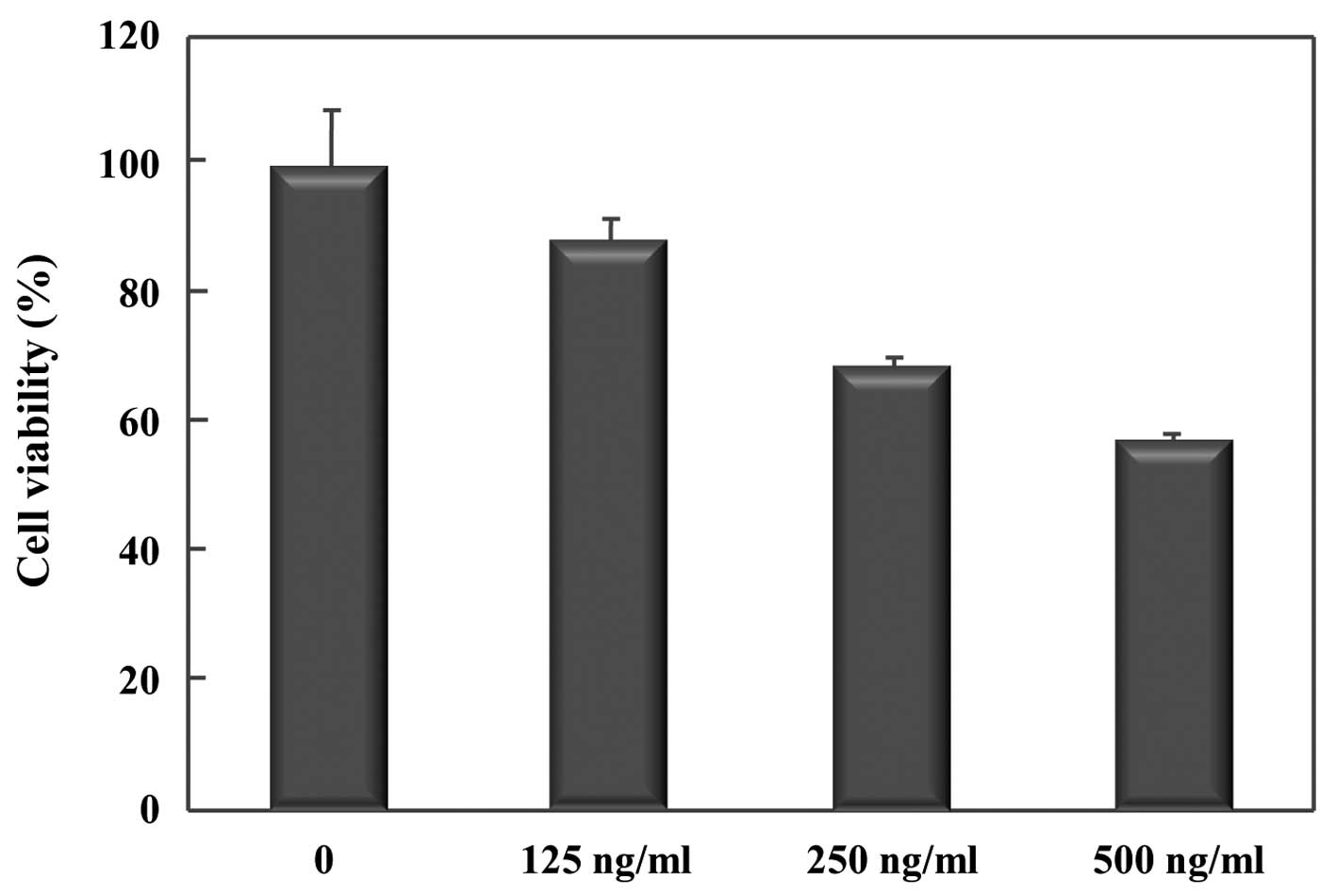
Inhibitory effect of PPY on proliferation
of MCF-7 cells
PPY inhibited proliferation of MCF-7 breast cancer
cells, as determined by the MTS assay. This assay revealed that PPY
induced growth inhibition occurred in a dose-dependent manner
(Fig. 2), and treatment with the
highest concentration of peptide (500 ng/ml) for 24 h resulted in
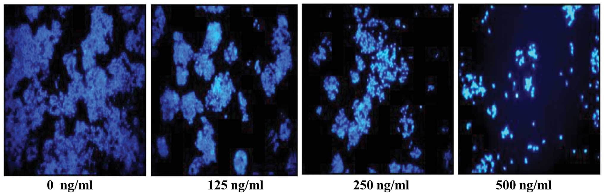
60% inhibition of cell growth. In addition to growth inhibition,
PPY treatment of MCF-7 cells decreased the relative cell numbers,
which was also concentration-dependent manner. This decrease is
attributable to the induction of apoptotic cell death by PPY, as
determined by a DAPI assay (Fig.
3). These conclusions were further supported through cell
morphology observations, which indicated that cells treated with
PPY revealed to decrease in number compared with untreated cells.
DAPI staining showed that PPY inhibited the proliferation of MCF-7
cells in a time-dependent manner. The DAPI assay also confirmed
that PPY treatment induced cell death. Taken together, these
results demonstrate that PPY induces both growth inhibition and
apoptosis in MCF-7 cells.
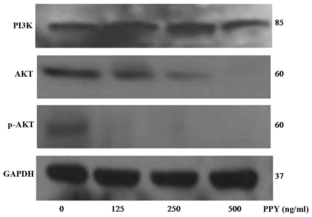
PPY induced phosphorylation of the
PI3K-Akt pathway
The phosphatidylinositol 3-kinases (PI3K)/Akt
pathway is mainly associated with cell growth and is a critically
important regulator of cell differentiation and proliferation. This
prompted us to examine the potential involvement of this pathway in
PPY induced inhibition of MCF-7 cell proliferation. This study
examined whether PPY influenced the activation of p85, a subunit of
PI3K. MCF-7 cells treated with PPY had a decreased level of Akt
phosphorylation/activation, compared with untreated cells.
Moreover, PPY treatment decreased the activation of p85 (Fig. 4). These results suggest that the
inhibition of the PI3K/Akt pathway is at least part of the
mechanism by which PPY inhibits MCF-7 cell proliferation.
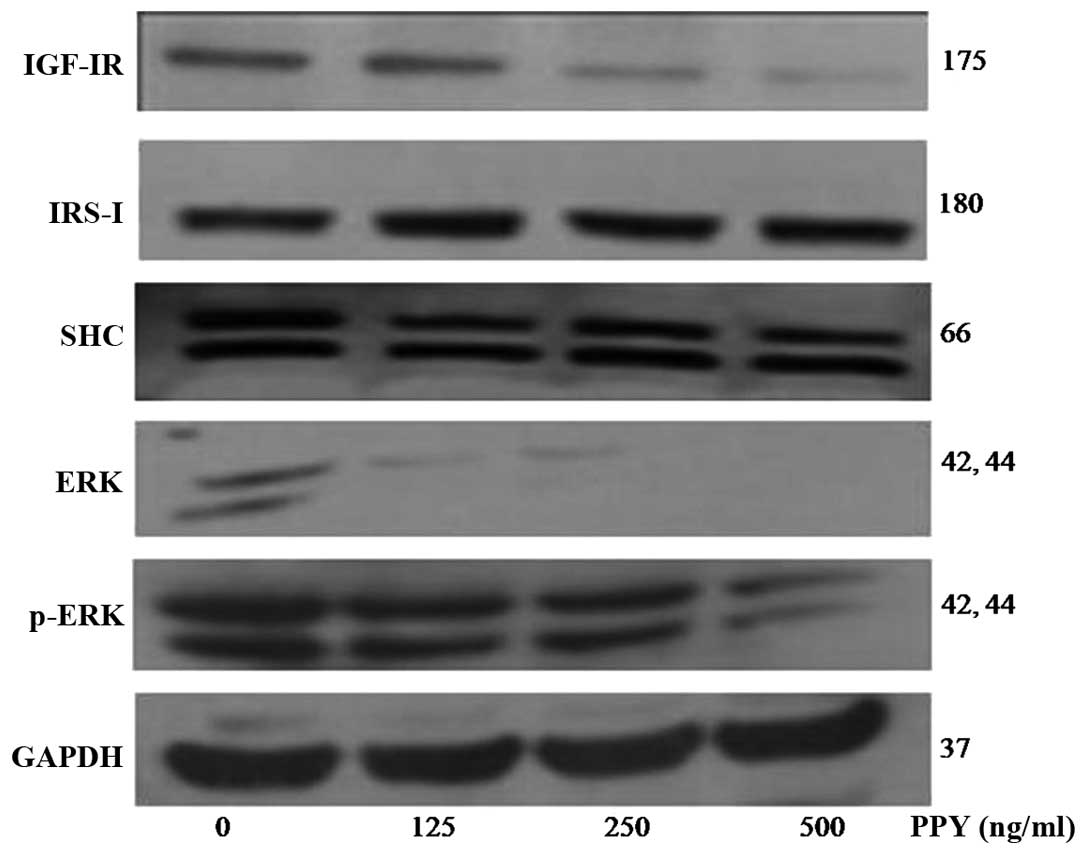
PPY affects the expression of IGF-IR
binding proteins in MCF-7 cells
To further investigate the mechanism of PPY induced
growth inhibition, additional components of the IGF signaling
pathway were examined. The signaling activity of IGF-IR is a
crucial regulator of apoptosis and cell proliferation and IGF-IR
activation results in auto-phosphorylation (8). Therefore, the effect of PPY on IGF-IR
expression was examined. MCF-7 cells were treated with different
concentrations of PPY for 24 h; the protein expression of
apoptosis-associated proteins is shown in Fig. 4. Expression levels of IGF-IR and
IRS-I were decreased by PPY treatment in a dose-dependent manner.
Total ERK protein expression decreased in cells treated with 125
ng/ml PPY, and phosphorylation of ERK was inhibited by PPY.
Consistent with these findings, treatment with PPY resulted in
activation of the intrinsic apoptosis pathway, which can be induced
by decreased ERK and phospho-ERK in MCF-7 cells (Fig. 5). Induction of apoptosis is a
predominant mechanism by which chemotherapeutic agents exert
cytotoxicity (15). The data
presented here provide evidence for the apoptotic effect of PPY in
MCF-7 cells.
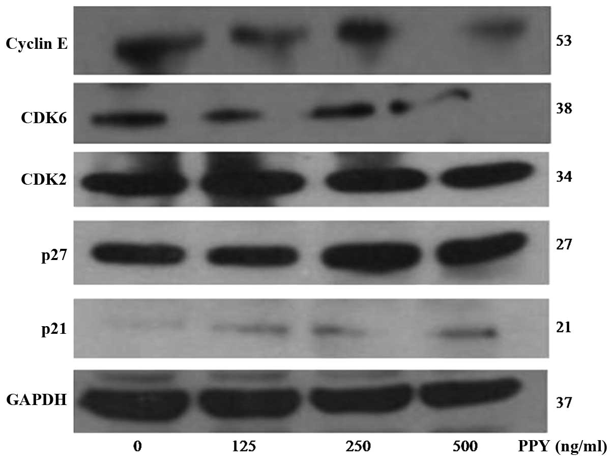
PPY affects the expression of cell
cycle-related proteins in MCF-7 cells
Cell cycle progression is a highly ordered and
tightly regulated process that involves multiple checkpoints that
monitor extracellular growth signals, cell size and DNA integrity.
Deregulation of the cell cycle has been recognized as a hallmark of
cancer progression in most malignant tumors. Furthermore, cell
cycle deregulation has been shown to induce an aberrant form of
mitosis called mitotic catastrophe and it may also be involved in
triggering apoptosis (3). The cdk
inhibitors p21 and p27 suppress the activity of the
pro-proliferative cyclin E/Cdk complex. Cyclin E is highly
expressed during the G1 to S phase transition, and Cdks, including
Cdk2, Cdk4, Cdk6, are critical regulators during phase transitions.
Accordingly, we examined whether PPY treatment affected critical
cell cycle regulators. Incubation of MCF-7 cells with PPY resulted
in a dose-dependent decrease in the expression levels of the
pro-proliferative proteins cyclin E and cdk6, while levels of Cdk2
were largely unchanged. In contrast, expression of p21 and p27
increased in response to PPY treatment dose-dependently (Fig. 6). Given that reduced expression of
p27 has been observed in several human cancers, and p27 is
associated with the induction of apoptosis in cancer cells
(16), our findings suggest that
PPY may modulate the sub-G1 arrest via upregulation of p21 and p27
and downregulation of Cdk6 and cyclin E.
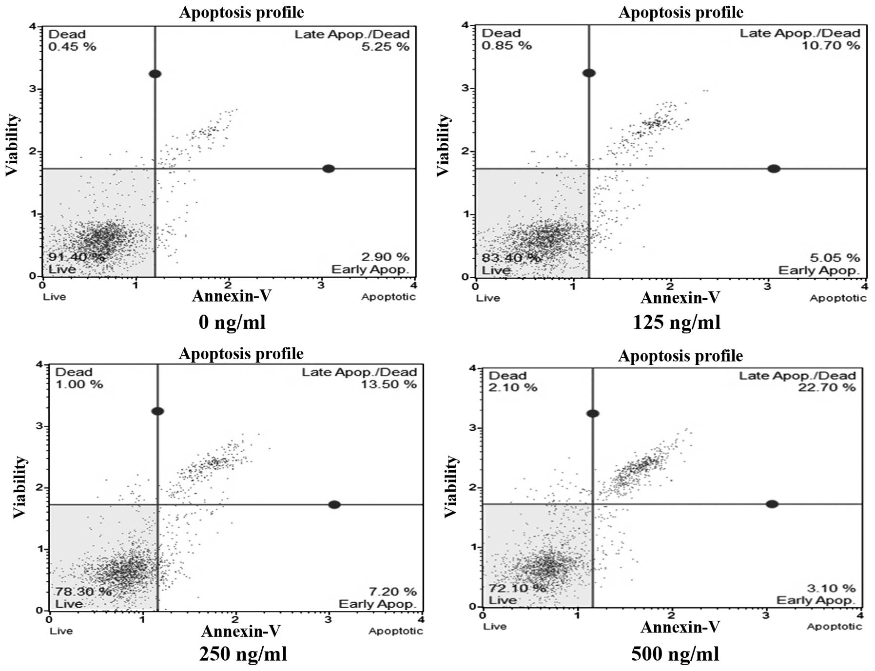
PPY induces apoptosis in MCF-7 cells
During early apoptosis, the externalization of the
phospholipid phosphatidylserine occurs at the cell membrane and can
be detected by Annexin V; this reagent can therefore be used to
detect cells undergoing apoptosis (17). Using an Annexin V/7-AAD apoptosis
assay, PPY treatment was found to induce apoptosis of MCF-7 cells
in a dose-dependent manner. A control cell population was comprised
of 8.15% apoptotic cells, 0.45% necrotic cells and 91.40% living
cells (Fig. 7). Treatment with
125, 250 and 500 ng/ml PPY for 24 h resulted in 15.75, 20.70 and
25.80% apoptotic cells, respectively. This indicates that even at
the highest concentration, cell death via apoptosis still occurs,
which is desirable from a chemotherapeutic standpoint.
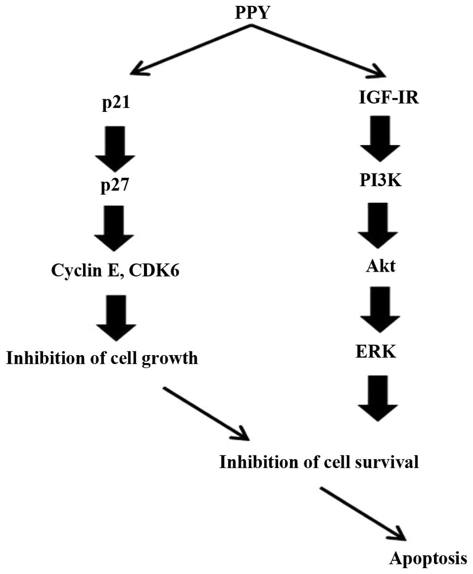
In conclusion, this study investigated the effective
PPY on the inhibition of MCF-7 cell proliferation, as well as the
possible mechanisms of growth inhibition. This study showed PPY
apoptotic cells and identified regulation of the IGF-IR signaling
pathway in MCF-7 cells (Fig.
8).
Discussion
Various seaweed types have high levels of nutrients
and other potentially beneficial components that may be useful for
the treatment of various diseases. In particular, the antitumor and
antibacterial activities of seaweeds have been widely studied
(2).
The aim of present study was to determine whether
PPY could inhibit the growth of MCF-7 breast cancer cells, as well
as the underlying mechanism. Several mechanisms were identified.
First, treatment of MCF-7 cells with PPY induced the cells to
undergo apoptosis, which was confirmed by examining the nuclear
morphology (using a DAPI staining assay). The cells treated with
PPY appeared to decrease in number compared with untreated cells.
This observation was confirmed using an apoptosis assay that
quantitatively demonstrated that PPY induces apoptosis in MCF-7
cells dose-dependently.
In addition to apoptosis, cell population size is
also influenced by regulation of proliferation. Stimulation of cell
proliferation and division is mediated by multiple signaling
pathways induced by tyrosine kinases, including MAPK and PI3K
(18). Tyrosine kinase receptors
transduce extracellular growth signals to the nucleus using signal
transduction pathways, including the Ras/Raf/MAPK and PI3K/Akt
pathways (22). PI3K is activated
by the growth factor EGF (19),
and following its activation, Akt is recruited to the cytoplasmic
surface of the cell membrane (20,21).
This study demonstrates that, in addition to inducing apoptosis,
PPY also inhibits the PI3K/Akt pathway in MCF-7 cells. The PI3K/Akt
pathway has been identified as key player in cell survival
(23,24). Akt also functions in normal growth,
as evidenced by Akt-knockout mice, which show retarded growth
(25,26).
The variety of ways in which apoptosis can be
induced suggests that wild-type IGF-IR and its ligands may have
widespread anti-apoptotic effects via many death signals (27). IGF-IR levels have been found
elevated in breast cancer compared with non-malignant tumors or
normal epithelium (28). In breast
cancer cell lines, IGF-IR is often co-expressed with autocrine
IGF-like mitogens that promote cell proliferation (29). The present study indicates that PPY
induces apoptosis in MCF-7 cells by downregulating the expression
of IGF-IR and IRS-I, which initiates the extrinsic apoptosis
pathway and the active forms of SHC were detected. Signaling
through IGF-IR stimulates proliferation and promotes angiogenesis
and metastasis, and there is now abundant evidence indicating that
signaling through the IGF-IR pathway is important for survival of
breast cancers as well as cancer cell lines, such as MCF-7 cells
(30). Our results show that PPY
treatment was effective for growth inhibition and induction of
apoptosis in MCF-7 cells.
A model regarding gossypol-induced cell cycle arrest
of breast cancer cells has been proposed, which involved the p53,
p21, cyclin D1 and Rb cell cycle proteins (31). In response to DNA damage, the cell
cycle checkpoints and cell death signals are activated to stop cell
growth and to halt the genetically modified cells from multiplying.
Damaged cells stop DNA replication at the G1 phase, presumably
activating the repair system before the next cell cycle begins
(16). Cell cycle progression is a
highly ordered and tightly regulated process that involves the
sequential activation and inhibition of cyclin-Cdk complexes. There
is accumulating evidence that manipulation of the cell cycle may
prevent or induce an apoptotic response depending on the cellular
context (32). The accumulation of
sub-G1 phase cells induced by PPY treatment led us to examine the
expression levels of cell cycle regulators. The observed
upregulation of Cdk inhibitors and downregulation of cyclin E and
Cdk6 reveal a mechanistic explanation for the growth inhibitory
effects of this peptide.
In conclusion, our studies investigated the effects
of the PPY peptide on the growth of MCF-7 cells, and we have shown
here that PPY inhibits MCF-7 cell growth by inducing apoptosis,
inhibiting proliferative signaling by antagonizing the IGF-I, and
inducing cell cycle arrest by altering expression of key cell cycle
modulators.
Acknowledgements
This research was supported by Basic Science
Research Program through the National Research Foundation of Korea
(NRF) funded by the Ministry of Education (grant no.
2012R1A6A1028677).
References
|
1
|
Go H, Hwang HJ and Nam TJ: Polysaccharides
from Capsosiphon fulvescens stimulate the growth of IEC-6
cells by activating the MAPK signaling pathway. Mar Biotechnol.
13:433–440. 2011.
|
|
2
|
Yoshizawa Y, Ametani A, Tsunehiro, Nomura
K, Itoh M, Fukui F and Kaminogawa S: Macrophage stimulation
activity of the polysaccharide fraction from a marine alga
(Porphyra yezoensis): structure-function relationships and
improved solubility. Biosci Biotechnol Biochem. 59:1933–1937. 1995.
View Article : Google Scholar : PubMed/NCBI
|
|
3
|
He L, Huang A, Shen S, Niu J and Wang G:
Comparative analysis of microRNAs between sporophyte and
gametophyte of Porphyra yezoensis. Comp Funct Genomics.
912843:2012
|
|
4
|
Shen S, Zhang G, Li Y, Wang L, Xu P and Yi
L: Comparison of RNA expression profiles on generation of
Porphyra yezoensis(Rhodophyta), based on suppression
subtractive hybridization (SSH). BMC Res Notes.
4:4282011.PubMed/NCBI
|
|
5
|
Butler AA, Blakesley VA, Poulaki V, Tsokos
M, Wood TL and LeRoith D: Stimulation of tumor growth by
recombinant human insulin-like growth factor-I (IGF-I) is dependent
on the dose and the level of IGF-I receptor expression. Cancer Res.
58:3021–3027. 1998.
|
|
6
|
Rubini M, Hongo A, D’Ambrosio C and
Baserga R: The IGF-I receptor in mitogenesis and transformation of
mouse embryo cells: role of receptor number. Exp Cell Res.
230:284–292. 1997. View Article : Google Scholar : PubMed/NCBI
|
|
7
|
Reiss K, Valentinis B, Tu X, Xu SQ and
Baserga R: Molecular markers of IGF-I-mediated mitogenesis. Exp
Cell Res. 242:361–372. 1998. View Article : Google Scholar : PubMed/NCBI
|
|
8
|
Butler AA, Yakar S, Gewolb IH, Karas M,
Okubo Y and LeRoith D: Insulin-like growth factor-I receptor signal
transduction: at the interface between physiology and cell biology.
Comp Biochem Physiol B Biochem Mol Biol. 121:19–26. 1998.
View Article : Google Scholar : PubMed/NCBI
|
|
9
|
Go H, Hwang HJ and Nam TJ: A glycoprotein
from Laminaria japonica induces apoptosis in HT-29 colon
cancer cells. Toxicol In Vitro. 24:1546–1553. 1998.
|
|
10
|
Khan N, Adhami VM and Mukhtar H: Apoptosis
by dietary agents for prevention and treatment of cancer. Biochem
Pharmacol. 76:1333–1339. 2008. View Article : Google Scholar : PubMed/NCBI
|
|
11
|
Burz C, Berindan-Neagoe I, Balacescu O and
Irimie A: Apoptosis in cancer: key molecular signaling pathways and
therapy targets. Acta Oncol. 48:811–821. 2009. View Article : Google Scholar : PubMed/NCBI
|
|
12
|
Johnson DG and Walker CL: Cyclins and cell
cycle checkpoints. Annu Rev Pharmacol Toxicol. 39:295–312. 1999.
View Article : Google Scholar : PubMed/NCBI
|
|
13
|
Weinstein IB: Disorders in cell circuitry
during multistage carcinogenesis: the role of homeostasis.
Carcinogenesis. 21:857–864. 2000. View Article : Google Scholar : PubMed/NCBI
|
|
14
|
Chan-Hui PY and Weaver R: Human
mitogen-activated protein kinase kinase kinase mediates the
stress-induced activation of mitogen-activated protein kinase
cascades. Biochem J. 336:599–609. 1998.PubMed/NCBI
|
|
15
|
Wang HJ, Tashiro S, Onodera S and Ikejima
T: Inhibition of insulin-like growth factor 1 receptor signaling
enhanced silibinin-induced activation of death receptor and
mitochondrial apoptotic pathways in human breast cancer MCF-7
cells. J Pharmacol Sci. 107:260–269. 2008. View Article : Google Scholar
|
|
16
|
Naumann U, Weit S, Rieger L, Meyermann R
and Weller M: p27 modulates cell cycle progression and
chemosensitivity in human malignant glioma. Biochem Biophys Res
Commun. 261:890–896. 1999. View Article : Google Scholar : PubMed/NCBI
|
|
17
|
Vangestel C, Peeters M, Oltenfreiter R,
D’Asseler Y, Staelens S, Van Steenkiste M, Philippé J, Kusters D,
Reutelingsperger C, Van Damme N and Van de Wiele C: In vitro and in
vivo evaluation of [99mTc]-labeled tricarbonyl His-Annexin A5 as an
imaging agent for the detection of phosphatidylserine-expressing
cells. Nucl Med Biol. 37:965–975. 2010.
|
|
18
|
Khandwala HM, McCutcheon IE, Flyvbjerg A
and Friend KE: The effects of insulin-like growth factors on
tumorigenesis and neoplastic growth. End Rev. 21:215–244. 2000.
View Article : Google Scholar
|
|
19
|
White MF and Kahn CR: The insulin
signaling system. J Biol Chem. 269:1–4. 1994.
|
|
20
|
Dews M, Prisco M, Peruzzi F, Romano G,
Morrione A and Baserga R: Domains of the insulin-like growth factor
I receptor required for the activation of extracellular
signal-regulated kinase. Endocrinology. 141:1289–1300.
2000.PubMed/NCBI
|
|
21
|
White MF: The IRS-signaling system: a
network of docking proteins that mediate insulin and cytokine
action. Recent Prog Horm Res. 53:119–138. 1998.
|
|
22
|
Scaltriti M and Baselga J: The epidermal
growth factor receptor pathway: a model for targeted therapy. Clin
Cancer Res. 12:5268–5272. 2006. View Article : Google Scholar
|
|
23
|
Kandel ES and Hay N: The regulation and
activities of the multifunctional serine/threonine kinase Akt/PKB.
Exp Cell Res. 253:210–229. 1999. View Article : Google Scholar : PubMed/NCBI
|
|
24
|
Parrizas M, Saltiel AR and LeRoith D:
Insulin-like growth factor I inhibits apoptosis using
phosphatidylinositol 3′-kinase and mitogen-activated protein kinase
pathways. J Biol Chem. 272:154–161. 1997.
|
|
25
|
McCurdy CE and Cartee GD: Akt2 is
essential for the full effect of calorie restriction on
insulin-stimulated glucose uptake in skeletal muscle. Diabetes.
54:1349–1356. 2005. View Article : Google Scholar : PubMed/NCBI
|
|
26
|
Hwang HJ, Kwon MJ and Nam TJ:
Chemoprotective effect of insulin-like growth factor I against
acetaminophen-induced cell death in Chang liver cells via ERK1/2
activation. Toxicology. 230:76–82. 2007. View Article : Google Scholar : PubMed/NCBI
|
|
27
|
Cui Q, Yu JH, Wu JN, Tashiro S, Onodera S,
Minami M and Ikejima T: P53-mediated cell cycle arrest and
apoptosis through a caspase-3-independent, but caspase-9-dependent
pathway in oridonin-treated MCF-7 human breast cancer cells. Acta
Pharmacol Sin. 28:1057–1066. 2007. View Article : Google Scholar : PubMed/NCBI
|
|
28
|
Quinn KA, Treston AM, Unsworth EJ, Miller
MJ, Vos M, Grimley C, Battey J, Mulshine JL and Cuttitta F:
Insulin-like growth factor expression in human cancer cell lines. J
Biol Chem. 271:11477–11483. 1996. View Article : Google Scholar : PubMed/NCBI
|
|
29
|
Ibrahim YH and Yee D: Insulin-like growth
factor-I and breast cancer therapy. Clin Cancer Res. 11:944s–950s.
2005.PubMed/NCBI
|
|
30
|
Shen G, Xu C, Chen C, Hebbar V and Kong
AN: P53-independent G1 cell cycle arrest of human colon carcinoma
cells HT-29 by sulforaphane is associated with induction of
p21cip1and inhibition of expression of cyclin D1. Cancer
Chemother Pharmacol. 57:317–327. 2006. View Article : Google Scholar : PubMed/NCBI
|
|
31
|
Hu ZY, Sun J, Zhu XF, Yang D and Zeng YX:
ApoG2 induces cell cycle arrest of nasopharyngeal carcinoma cells
by suppressing the c-Myc signaling pathway. J Transl Med. 7:742009.
View Article : Google Scholar : PubMed/NCBI
|
|
32
|
Pucci B, Kasten M and Giodano A: Cell
cycle and apoptosis. Neoplasia. 2:291–299. 2000. View Article : Google Scholar
|