Introduction
Oral mucositis is a common adverse event in
chemotherapy and radiotherapy against human head and neck cancers
(1,2), and results from the damage of the
mucosal lining of the gastrointestinal tract, especially the oral
and oropharyngeal mucosa (3).
Previously, mucositis was considered to arise as a consequence of
epithelial injury (4–6), i.e., it was thought that chemotherapy
and radiotherapy non-specifically kill the rapidly proliferating
cells of the basal cell layer, thereby abolishing the ability of
the layer to renew itself. In the case of radiotherapy-induced
mucositis, the cell death was attributed to DNA strand breaks in
the oral basal epithelial cells, while in chemotherapy-induced
mucositis it was attributed to direct basal cell damage (non-DNA
injury) caused by the drugs permeating the cells from the
submucosal blood supply (3).
Although the clinical symptoms of oral mucositis,
such as ulceration of the mucosal epithelium, pain, infection, and
swallowing dysfunction, are almost all the results of epithelial
injury (7), accumulating evidence
indicate that the clinical manifestations of this condition are
attributable to a series of interactive biological events that
involve all of the cells and tissues of the mucous membrane
(8–10). For example, morphological
observations suggest that damage in the submucosal endothelium and
connective tissue occur first, followed by injury of the epithelial
cells (9). Moreover, it has been
reported that endothelial damage (endothelial toxicity) might be
the initiating event in the radiotherapy-induced mucositis
(10), indicating that several
chemotherapeutic agents, including 5-FU and cisplatin, also
similarly exert their endothelial toxicity (11,12).
Therefore, chemotherapy- and radiotherapy-induced oral mucositis is
initiated by direct damage to basal epithelial cells and cells in
the underlying tissues.
Chemotherapy induces non-DNA damage in the cells,
e.g., basal epithelial cells, through a variety of mechanisms, some
of which are mediated by the generation of reactive oxygen species
(ROS) (13). Although a moderate
increase in ROS can promote cell proliferation and differentiation
(14,15), excessive amounts of ROS can cause
oxidative damage to lipids, proteins and DNA (16), thereby leading to cell death or
abnormal cell growth (17).
Maintenance of the ROS level in cells is thus crucial for normal
growth and survival. To achieve such maintenance, the cells control
ROS levels by balancing ROS generation with their elimination by
ROS-scavenging systems such as intracellular redox-balancing genes
[heme oxygenase-1 (HO-1)], phase II detoxifying genes
[NAD(P) H:quinone oxidoreductase-1 (NQO-1)], and genes
encoding transporters (multidrug resistant proteins) (18). Many of these genes contain an
enhancer sequence known as the antioxidant response element (ARE)
(19–21), and are enhanced by the
transcription factor NF-E2-related factor 2 (Nrf2). Based on the
functions of these ARE-containing genes, it seems likely that
activation of Nrf2 target genes would stimulate the detoxication of
xenobiotics, such as chemopreventive drugs, and protect cells from
ROS-driven apoptosis (22).
A vitamin E constituent may be one such candidate
agent derived from natural sources with great potential for
preventing the cell death induced by anticancer drugs. Vitamin E is
a general term representing a family of compounds that is further
divided into two subgroups: tocopherols and tocotrienols (23). Although tocopherols and
tocotrienols exist in α, β, γ and δ form, the two differ
structurally in that tocopherols contain a saturated phytyl chain,
whereas tocotrienols possess an unsaturated side chain. Thus far,
tocopherols have been studied extensively, while very little is
known about tocotrienols. Previous studies including ours have
shown that tocotrienols are more potent antioxidant agents than
tocophenols (24), and that
γ-tocotrienol enhances the chemosensitivity of human oral cancer
cells to docetaxel (25).
Importantly, γ-tocotrienol exerts significant anti-proliferative
effects in malignant cells, but not in normal cells (26). Therefore, it is likely that the
oral mucositis caused by chemotherapeutic agents could be prevented
by a low dose of γ-tocotrienol through the detoxification of ROS in
basal epithelial cells.
In the present study, we demonstrate that
simultaneous treatment of human oral epithelial (RT7) cells with
5-FU and γ-tocotrienol suppressed the 5-FU-induced generation of
ROS, leading to the amelioration of cell growth. We also found that
the inhibition of ROS generation in RT7 cells was due to the
stabilization of 5-FU-mediated activation of Nrf2, which resulted
in the enhanced production of the ROS-scavenging enzymes HO-1 and
NQO-1.
Materials and methods
Cells and media
RT7, an immortalized human oral keratinocyte cell
line, was established by transfection of human telomerase reverse
transcriptase (hTERT) and E7, as previously described (27), and the metastatic human oral cancer
cell line (B88) was previously established in our laboratory
(28). RT7 and B88 cells,
respectively, were cultured in keratinocyte serum-free medium (SFM)
(Gibco-BRL, Gaithersburg, MD, USA) that included 25 μg/ml bovine
pituitary extract, 0.05 ng/ml epidermal growth factor, 100 U/ml
penicillin and 100 μg/ml streptomycin, and in DMEM supplemented
with 10% fetal bovine serum (both from Gibco-BRL), 100 U/ml
penicillin and 100 μg/ml streptomycin in the presence of 5%
CO2 in an incubator at 37°C.
In vitro cell growth assay
Cells (3×10³ cells/well) were grown on
96-well plates (Falcon; Becton-Dickinson Labware, Lincoln Park, NJ,
USA) in SFM and DMEM in the presence or absence of 5-FU (1, 2, 5,
10 μg/ml) (Taiho Pharmaceutical Co., Ltd., Tokyo, Japan) and
γ-tocotrienol (10 nM) (with a purity exceeding 98.7%; Eisai Food
& Chemical Co., Tokyo, Japan) alone, or both for 3 days. In
addition, RT7 cells were also treated with 5-FU and a ROS
scavenger, N-acetyl cysteine (NAC) (Sigma-Aldrich, St. Louis, MO,
USA), to inhibit ROS generation. Thereafter, 10 μl of 5 mg/ml
3-(4,5-dimethyl-thiazol-2-yl)-2,5-diphenyltetrazolium bromide (MTT)
were added to each well and the cells were incubated for 4 h. The
blue dye taken up by the cells was dissolved in dimethyl sulfoxide
(100 μg/ml), and the absorbance was measured with a Titertek
Multiscan spectrophotometer (Flow Laboratories, Irvine, UK) at 570
nm. All assays were run in triplicate.
Measurement of ROS
2′,7′-Dichlorofluorescein diacetate
(H2DCF-DA; Molecular Probes, Eugene, OR, USA) was used
to analyze the intracellular ROS level. RT7 cells were incubated
with 5-FU, NAC, or γ-tocotrienol alone, or with a combination of
5-FU and NAC for 48 h. Adherent cells were washed with PBS and
stained with 2 μM DCFH-DA diluted in PBS at 37°C in the dark for 30
min (29). The DCFH-DA dye
oxidized by ROS can be excited by a 488-nm laser. RT7 cells were
washed twice with PBS before flow cytometric analysis using a
FACSCalibur flow cytometer and data were analyzed by CellQuest
software (both from Becton-Dickinson, East Rutherford, NJ,
USA).
Nuclear and cytosolic extract
preparations
RT7 cells were seeded on 100-mm plastic petri dishes
(Falcon; Becton-Dickinson Labware). Twenty-four hours after
seeding, the cells were treated with either 5-FU, γ-tocotrienol, or
both for 24 h, and then the nuclear extracts were obtained
according to a previously described method (30). The cells were washed twice with
ice-cold PBS before being resuspended in 400 μl of ice-cold lysis
buffer consisting of 10 mM
N-2-hydroxyethylpiperazine-N′-2-ethanesulfonic acid (HEPES) (pH
7.9), 10 mM KCl, 0.1 mM ethylenediaminetetraacetate (EDTA), 0.1 mM
ethylene glycol-bis (2-aminoethylether)-N,N,N′,N′-tetraacetic acid
(EGTA), 0.5 mM dithiothreitol (DTT), 0.5 mg/ml benzamidine and 2
mg/ml aprotinin for 15 min. Nonidet P-40 was added to a final
concentration of 0.3%, and the lysates were vortexed before being
pelleted in a microfuge. The supernatants of this centrifugation
were designated cytosolic extracts. Each nuclear pellet was
resuspended in 50 μl of extraction buffer consisting of 10 mM HEPES
(pH 7.9), 400 mM NaCl, 10 mM KCl, 0.1 mM EDTA, 0.1 mM EGTA, 1 mM
DTT, 0.5 mM phenyl-methylsulfonyl fluoride and 2 mg/ml aprotinin
and then placed on ice for 30 min. The nuclear extracts were
pelleted, and the supernatants were designated nuclear extracts.
The protein concentrations contained in samples were determined
using a Bio-Rad protein assay kit (Bio-Rad, Hercules, CA, USA).
Western blot analysis of Nrf2, Kelch-like
ECH-associated protein 1 (Keap1) and β-actin proteins
Cytosolic extracts (20 μg) were subjected to
electrophoresis on 10% SDS-polyacrylamide gels, then transferred
onto nylon membranes. The membranes were blocked with 3% bovine
serum albumin and incubated with each of the following antibodies
(all from Santa Cruz Biotechnology, Inc., Santa Cruz, CA, USA):
anti-Nrf2, anti-Keap1 and anti-β-actin. After intervening rinses
with PBS, the antibodies were detected using a chemiluminescence
western blot analysis kit (Amersham Pharmacia Biotech, Tokyo,
Japan) according to the manufacturer’s instructions.
Immunofluorescence staining for Nrf2
protein
Cells grown on coverglasses were washed with PBS
three times, fixed in acetone at 4°C for 10 min, and incubated for
1 h at 37°C with mouse polyclonal antibody to Nrf2 (Abcam,
Cambridge, MA, USA) at a dilution of 1:200. After three rinses with
PBS, the coverglasses were incubated for 1 h with
fluorescein-conjugated goat anti-mouse IgG (1:200 dilution; Abcam).
The coverglasses were then mounted with PermaFluor™ Aqueous
Mounting Medium (Lab Vision Corporation, Fremont, CA, USA). To
establish a negative control, the primary antibody was omitted.
RNA isolation and quantitative real-time
PCR
Total cellular RNA was isolated after the RT7 cells
were treated with TRIzol reagent plus 5-FU, or γ-tocotrienol or
both (Invitrogen Life Technologies, Carlsbad, CA, USA). The cDNA
was synthesized from 5 μg of total RNA using an Advantage cDNA PCR
kit (Clontech Laboratories, Inc., Palo Alto, CA, USA). For the
quantitative real-time PCR, equal aliquots (1 μl) of cDNA were
amplified according to the manufacturer’s TaqMan universal (50 μl)
PCR master mix protocol using an ABI PRISM 7300 RT-PCR system
(Applied Biosystems Japan, Ltd., Tokyo, Japan). The primer set and
TaqMan probe mixture used for the PCR were purchased from Applied
Biosystems Japan, Ltd. (HO-1: Hs01110250_m1; NQO-1: Hs02512143_s1).
The data were normalized using RT-PCR β-actin primers
(Hs99999903_m1; Applied Biosystems Japan, Ltd.)
Statistical analysis
The statistical analysis was performed using the
Mann-Whitney U test; P<0.05 was considered to indicate
statistical significance.
Results
Optimal concentration of 5-FU used in
this study
The concentrations of chemotherapeutic drugs,
including 5-FU, used in the clinical setting would be determined
based on the balance between the cytotoxicity to cancer cells and
non-cytotoxicity to normal cells. Therefore, in this study, we used
an immortalized normal human oral keratinocyte (RT7) and a human
oral cancer cell (B88) line to determine the optimal dose of 5-FU
in our system. The growth inhibitory response of both cell lines to
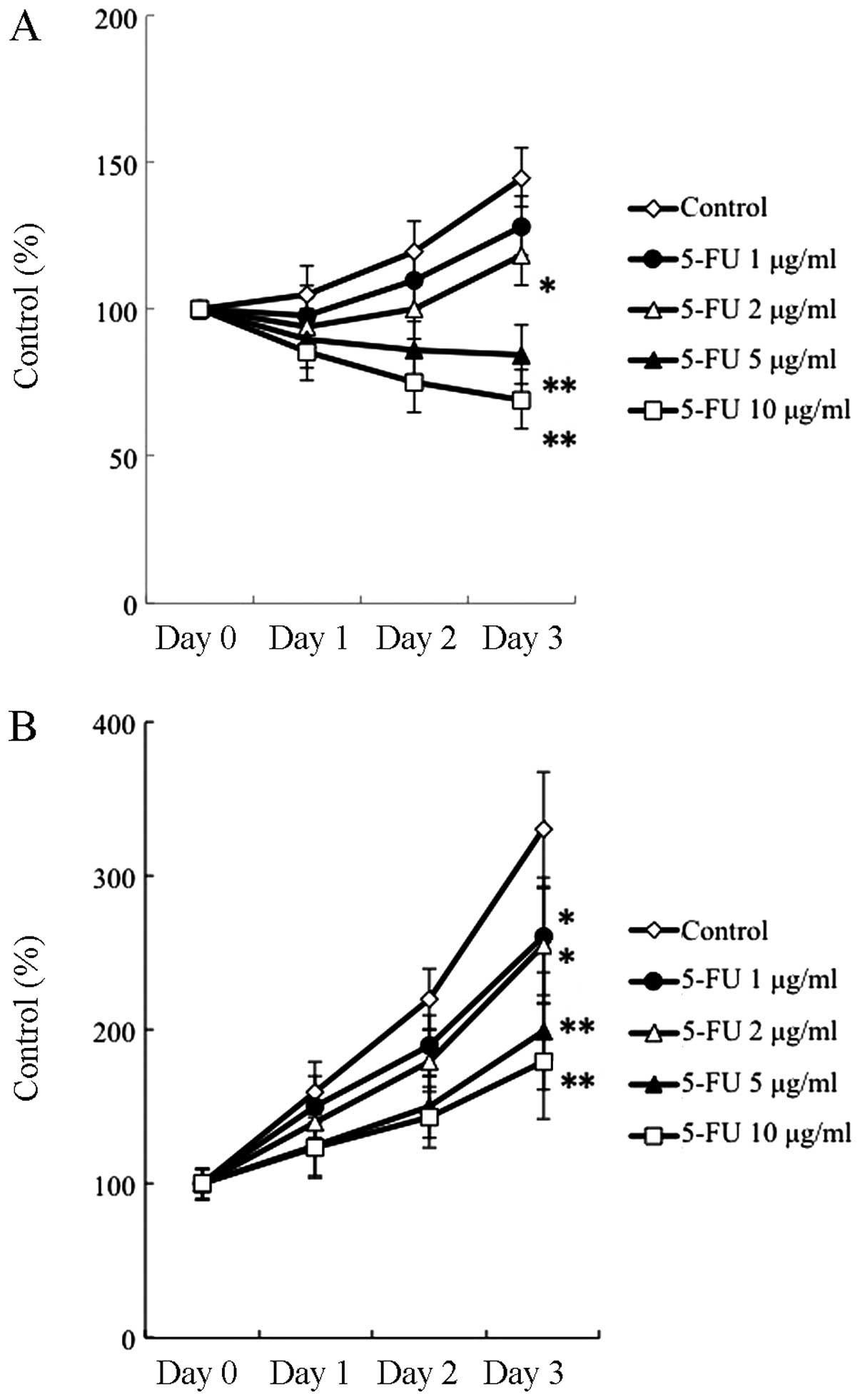
5-FU was investigated by MTT assay for 3 days. As shown in Fig. 1, 5-FU at the concentrations of 5
and 10 μg/ml had a significant cytotoxic effect on B88 cells,
inducing an apparent growth inhibitory response as compared to that
in untreated cells, whereas the same concentrations of 5-FU did not
exert cytotoxic or cytostatic effects on RT7 cells. Based on this
finding, we chose a 5-FU concentration of 10 μg/ml for the use in
this study.
Growth-suppressive effect of 5-FU on RT7
cells via ROS generation
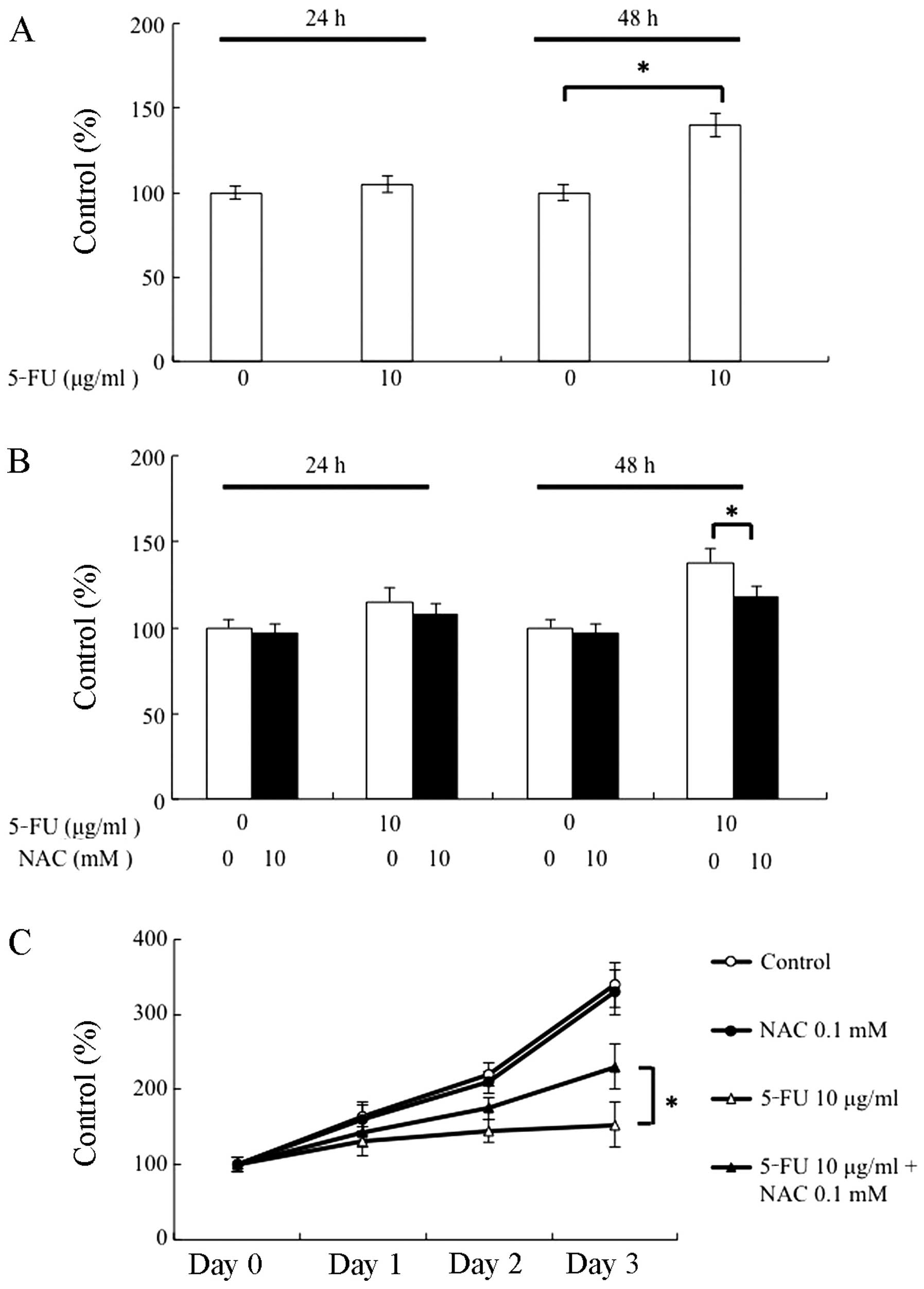
To determine whether or not the 5-FU treatment would
stimulate ROS generation in RT7 cells, we measured the levels of
ROS using the ROS detection dye DCFH-DA. No increase in ROS was
detected at 24 h after treatment with 10 μg/ml of 5-FU (Fig. 2A). Further incubation with 10 μg/ml
of 5-FU for 48 h resulted in a significant increase in ROS
production in RT7 cells. To examine whether NAC, a ROS scavenger,
actually abolishes ROS, we measured the ROS level using the method
described above. When RT7 cells were treated with both 5-FU and NAC
(0.1 mM) for 24 h, the generation of ROS was not affected. However,
ROS production was significantly suppressed at 48 h after treatment
with NAC (Fig. 2B). Since ROS
affects the suppression of cell growth, we examined the effect of
NAC on the growth of RT7 cells. As shown in Fig. 2C, although NAC alone did not affect
the growth of RT7 cells, the cell growth that was suppressed by
5-FU was significantly restored through the elimination of ROS in
RT7 cells. Therefore, these results indicate that growth
suppression by 5-FU may be at least partly due to the generation of
ROS from RT7 cells.
Amelioration of 5-FU-induced growth
suppression by γ-tocotrienol via ROS inhibition
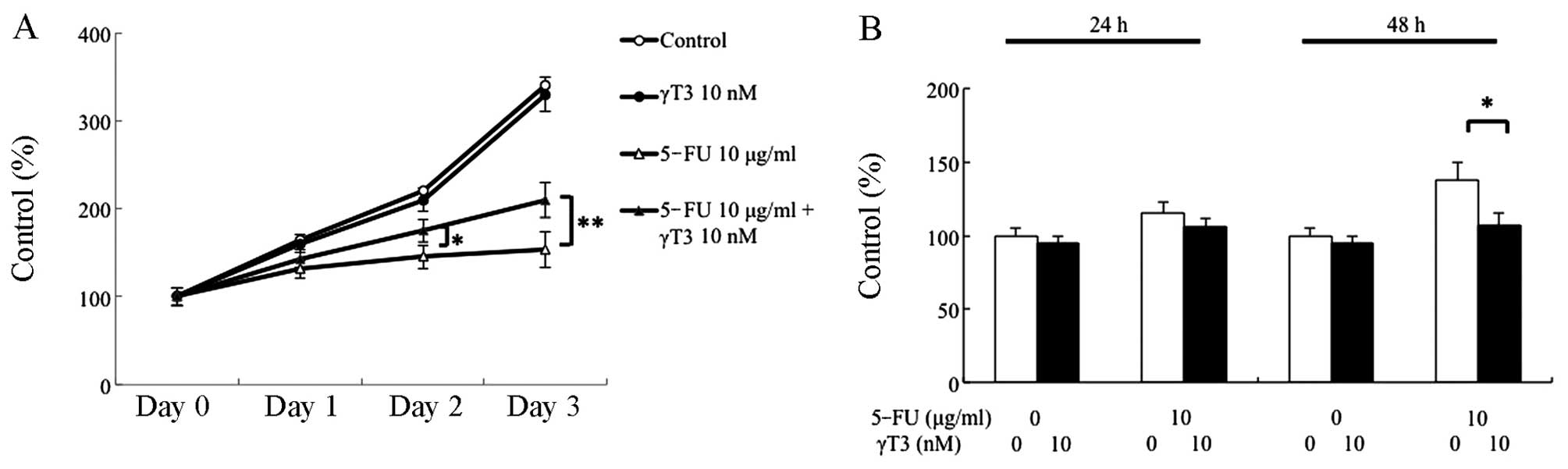
The growth response of RT7 cells to γ-tocotrienol
(10 nM) was investigated by MTT assay for 3 days. As can be seen in
Fig. 3A, γ-tocotrienol alone did
not affect RT7 cell growth when compared to the growth of the
control cells. Whether or not γ-tocotrienol can restore the
suppressive effect of 5-FU on RT7 cell growth was also examined.
The dose of γ-tocotrienol (10 nM) that did not affect cell growth
when used alone resulted in an enhanced recovery of cell growth
when used in combination with 5-FU. In addition, although 5-FU
alone stimulated the generation of ROS in RT7 cells, combined
treatment with 5-FU and γ-tocotrienol significantly inhibited the
production of ROS at 48 h (Fig.
3B).
Effect of 5-FU and γ-tocotrienol on the
expression of Nrf2 and Keap1 proteins
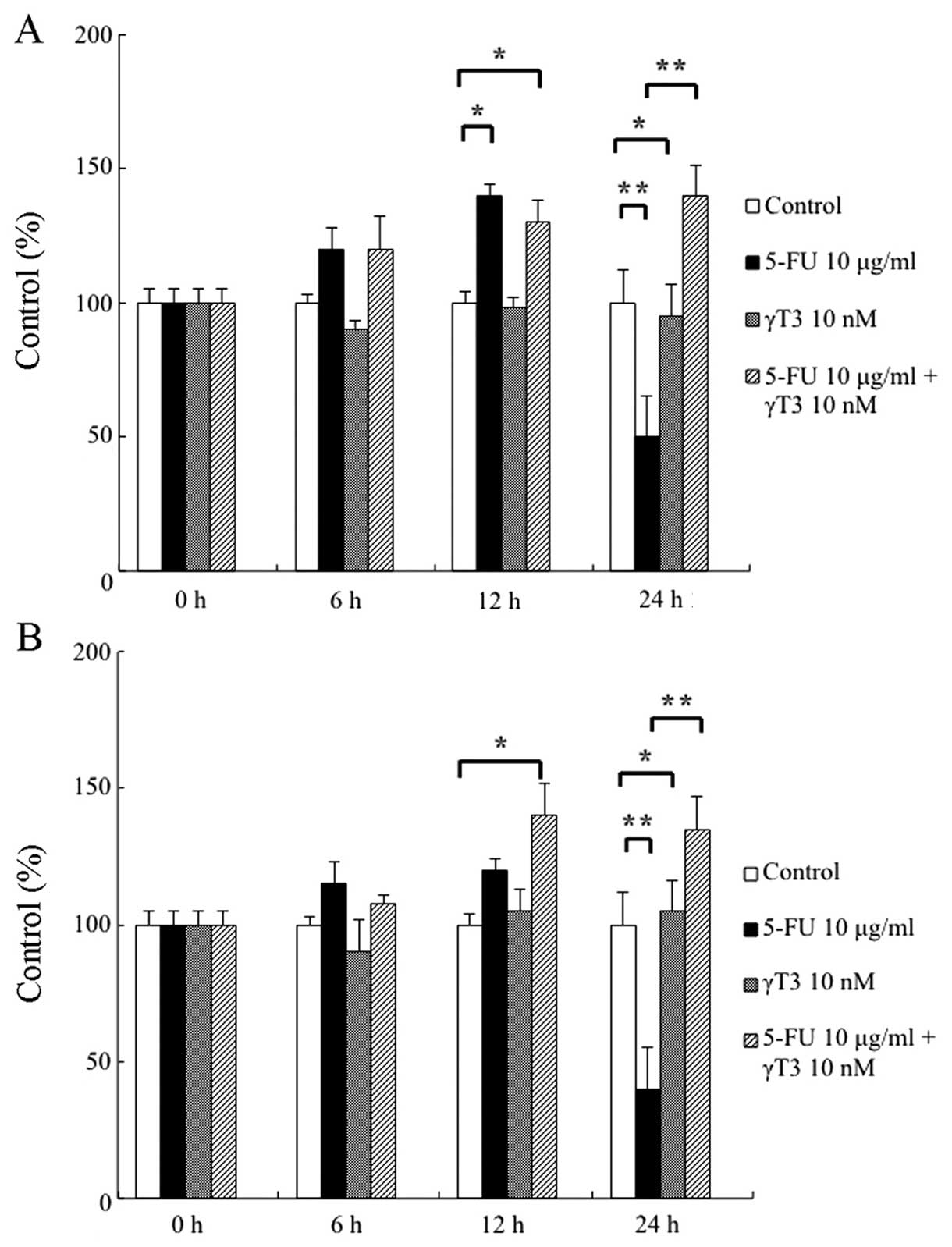
We examined the expression of the proteins Nrf2, a
master transcriptional factor of various cytoprotective genes
against oxidative stress, and Keap1, a negative regulator of Nrf2,
by treatment with 5-FU, γ-tocotrienol, or both. As shown in
Fig. 4A, 5-FU induced the nuclear
accumulation of Nrf2 in RT7 cells for up to 12 h; however, no
apparent effect was observed in γ-tocotrienol-treated cells. Of
note, the simultaneous treatment of cells with 5-FU and
γ-tocotrienol led to sustained nuclear accumulation of Nrf2 for up
to 24 h. As shown in Fig. 4B, the
expression of Keap1 was inversely correlated with that of Nrf2:
5-FU inhibited Keap1 expression at 6 and 12 h after treatment,
followed by reappearance at 24 h. Although γ-tocotrienol had no
effect on the expression of Keap1 in RT7 cells, combined treatment
of cells with 5-FU and γ-tocotrienol suppressed the expression of
Keap1 for up to 24 h after treatment. Therefore, the sustained
activation of 5-FU-induced Nrf2 expression may have been due to the
continuous degradation of Keap1 through the combined treatment with
5-FU and γ-tocotrienol in RT7 cells.
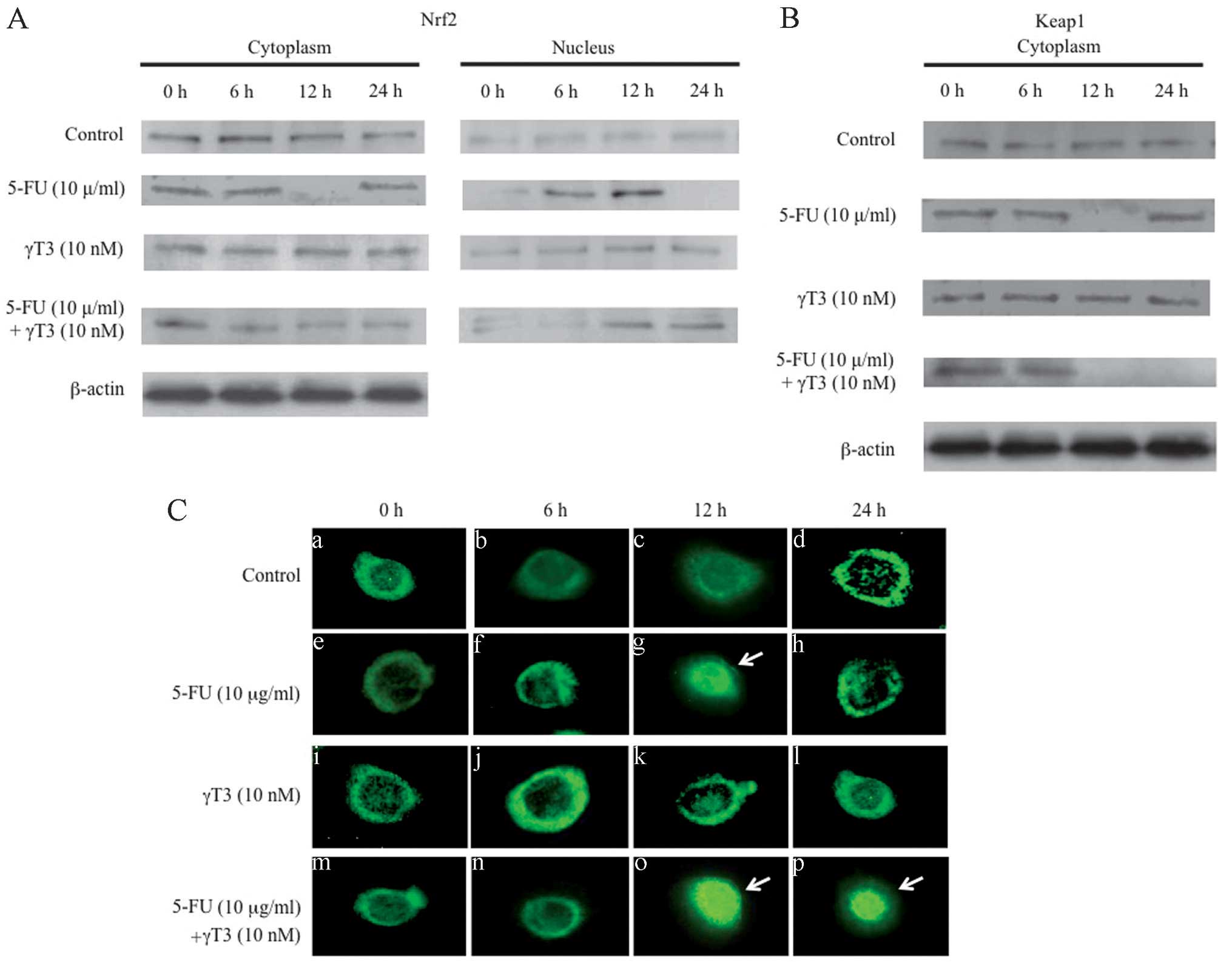 | Figure 4Expression of the NF-E2-related factor
2 (Nrf2) protein and Kelch-like ECH-associated protein 1 (Keap1) in
RT7 cells treated with 5-FU (10 μg/ml), γ-tocotrienol (γT3, 10 nM),
or both. (A) Western blot analysis of Nrf2 protein present in the
cytoplasm and nucleus in the cells treated with 5-FU,
γ-tocotrienol, or both for 24 h. Cytosolic and nuclear extracts
were subjected to western blot analysis to detect the expression
levels of Nrf2 protein. Although 5-FU stimulated the nuclear
localization of Nrf2 for up to 12 h after treatment, γ-tocotrienol
had no effect on the cytoplasmic localization of Nrf2. On the other
hand, simultaneous treatment of cells with both agents
significantly stimulated the nuclear localization of Nrf2 for up to
24 h. As a loading control for the protein samples, the expression
of β-actin is also shown. Results are representative of three
independent experiments. (B) Western blot analysis for the
detection of Keap1 in the cytoplasm. Cells were treated with 5-FU,
γ-tocotrienol, or both for 24 h. Cytosolic fractions were analyzed
for the detection of Keap1. Although 5-FU degraded the cytosolic
Keap1 at 12 h after treatment, γ-tocotrienol had no effect on
cytoplasmic localization of the Keap1. On the other hand,
simultaneous treatment of cells with both agents significantly
inhibited the cytoplasmic localization of Keap1 for up to 24 h. As
a loading control for the protein samples, the expression of
β-actin is also shown. Results are representative of three
independent experiments. (C) Indirect immunofluorescence microscopy
of Nrf2 protein in RT7 cells treated with or without (control, a–d)
5-FU (e–h), γ-tocotrienol (i–l), or both (m–p) for 24 h. The
expression of activated Nrf2 was specifically observed in the
nuclei of 5-FU-treated cells at 12 h (arrow, g). However, activated
Nrf2 protein was continuously detected in the nuclei of the cells
treated with both agents for 24 h (arrows, o and p). |
Expression of HO-1 and NQO-1 mRNA by
treatment with 5-FU and γ-tocotrienol
To examine the effect of 5-FU and γ-tocotrienol on
the expression of the detoxifying enzymes HO-1 and NQO-1 in RT7
cells, we used quantitative RT-PCR analysis. As shown in Fig. 5A and B, although 5-FU treatment at
12 h was associated with a significant increase in the expression
of these enzymes, the expression of both enzymes was remarkably
suppressed when 5-FU treatment was continued for up to 24 h. When
γ-tocotrienol was used, no significant changes were observed in the
mRNA expression levels of these enzymes. Importantly, combined
treatment of cells with 5-FU and γ-tocotrienol increased the
expression levels of HO-1 and NQO-1 mRNA for up to 24 h. These
results indicate that expression of the detoxifying enzymes HO-1
and NQO-1 is likely regulated by a master regulatory transcription
factor, Nrf2 (31).
Discussion
Oral mucositis is a common adverse event caused by
antineoplastic radiation (radiotherapy) and drug therapies
(chemotherapy) for patients with head and neck cancer, and is
associated with severe adverse symptomatic health and economic
outcomes (32,33). Thus far, although understanding of
the pathobiology of this unfavorable condition has increased
rapidly over the past decade, there remains no effective way to
prevent or treat mucositis (3).
Therefore, the objective of the present study was to investigate
whether or not γ-tocotrienol, a component of vitamin E, can improve
the cell survival of human oral keratinocytes against 5-FU-induced
cell toxicity. The results showed that γ-tocotrienol suppressed the
5-FU-induced generation of ROS, leading to the potentiation of RT7
cell growth, and sustained the upregulated expression of Nrf2 by
5-FU, as well as the expression of the detoxifying enzymes HO-1 and
NQO-1.
A key challenge to the development of drugs for
ameliorating chemotherapy- and radiotherapy-induced oral mucositis
is to ensure that they target normal tissue effectively, but do not
diminish the tumoricidal effect of the antineoplastic drugs.
Therefore, we investigated the optimal concentrations of 5-FU and
γ-tocotrienol using human oral cancer cells and normal human oral
keratinocytes, and determined that concentrations of 10 μg/ml and
10 nM, respectively, were most effective. Although the
concentrations of drugs used in this study showed no significant
cytotoxic effects on the growth of normal keratinocytes, the growth
of cancer cells was clearly suppressed. At present, several
possible mechanism-based treatments for oral mucositis are
undergoing trial (3). Some of
these approaches target ROS, which are ubiquitous in the tissues of
mucosal injury, for cytoprotective intervention in the oral mucosa
(34). Thus, we analyzed the
effects of a free-radical scavenger, γ-tocotrienol, for its ability
to reduce ROS levels. Our results showed that γ-tocotrienol could
reduce 5-FU-induced ROS generation, leading to the sustained
survival of oral keratinocytes.
Chemotherapy induces non-DNA injury in the
epithelial cells through a variety of mechanisms, some of which are
mediated by the generation of ROS (3). ROS also activate several
injury-producing pathways, such as the nuclear factor-κB (NF-κB)
and p53 pathways, in epithelial cells (35). In the present study, we
demonstrated that γ-tocotrienol could suppress 5-FU-induced ROS
generation, which in turn suggested that γ-tocotrienol induced the
inhibition of NF-κB in RT7 cells. Since we have previously shown
that γ-tocotrienol suppressed the activation of NF-κB induced by a
chemotherapeutic agent (docetaxel) in oral cancer cells (25), mechanism involved in the cell
survival of RT7 by γ-tocotrienol might involve regulation of the
balance between NF-κB-related pro-apoptotic and anti-apoptotic
genes (3).
Cell membrane-bound molecules released during
chemotherapy (lipid peroxidation) also result in the upregulation
of genes, including those encoding c-Jun and c-Jun amino-terminal
kinase (JNK) (35,36). Therefore, these molecules
upregulate other transcription factors, such as Nrf2 (37). In addition, it is well known that
many chemotherapeutic agents are able to stabilize the expression
of Nrf2 by inhibiting Nrf2 degradation, and thereby enhance the
protein level of Nrf2 and activate the Nrf2-dependent antioxidant
response (38,39). In this study, we have shown that
although 5-FU treatment of RT7 cells actually resulted in the
activation of Nrf2 for up to 12 h, the combined treatment with 5-FU
and γ-tocotrienol led to the sustained activation of Nrf2 for up to
24 h. In addition, the expression of Keap1, an E3 ubiquitin ligase
that promotes proteasome-dependent degradation of Nrf2 (40), was inhibited by the 24 h
combination treatment, indicating that γ-tocotrienol could activate
Nrf2 by suppressing the expression of Keap1 in RT7 cells.
Consistent with the above results, a previously reported
quantitative RT-PCR analysis clearly demonstrated that expression
of the antioxidant enzymes HO-1 and NQO-1 was Nrf2-dependent
(41), suggesting that the
improved survival of RT7 cells was attributable to the sustained
reduction of ROS by γ-tocotrienol.
In conclusion, the results of the present study
indicate that γ-tocotrienol potentiates and sustains the
5-FU-induced expression of Nrf2 in human keratinocytes, leading to
the continuous degradation of 5-FU-generated ROS via the augmented
production of ROS-scavenging enzymes, followed by the improved cell
growth of keratinocytes. Based on our present results,
well-designed animal and clinical studies will be needed for the
potential translation of our preclinical findings in patients with
oral cancer receiving chemotherapy and radiotherapy.
Acknowledgements
This study was supported by a Grant-in-Aid from the
Ministry of Education, Culture, Sports, Science, and Technology of
Japan.
References
|
1
|
Keefe DM, Schubert MM, Elting LS, et al:
Updated clinical practice guidelines for the prevention and
treatment of mucositis. Cancer. 109:820–831. 2007. View Article : Google Scholar : PubMed/NCBI
|
|
2
|
Le QT, Kim HE, Schneider CJ, et al:
Palifermin reduces severe mucositis in definitive chemoradiotherapy
of locally advanced head and neck cancer: a randomized,
placebo-controlled study. J Clin Oncol. 29:2808–2814. 2011.
View Article : Google Scholar : PubMed/NCBI
|
|
3
|
Sonis ST: The pathobiology of mucositis.
Nat Rev Cancer. 4:277–284. 2004. View
Article : Google Scholar : PubMed/NCBI
|
|
4
|
Bloomer WD and Hellman S: Normal tissue
responses to radiation therapy. N Engl J Med. 293:80–83. 1975.
View Article : Google Scholar : PubMed/NCBI
|
|
5
|
Lockhart PB and Sonis ST: Relationship of
oral complications to peripheral blood leukocyte and platelet
counts in patients receiving cancer chemotherapy. Oral Surg Oral
Med Oral Pathol. 48:21–28. 1971. View Article : Google Scholar
|
|
6
|
Sonis ST, Lindquist L, Van Vugt A, et al:
Prevention of chemotherapy-induced ulcerative mucositis by
transforming growth factor beta 3. Cancer Res. 54:1135–1138.
1994.PubMed/NCBI
|
|
7
|
Henke M, Alfonsi M, Foa P, et al:
Palifermin decreases severe oral mucositis of patients undergoing
postoperative radiochemotherapy for head and neck cancer: a
randomized, placebo-controlled trial. J Clin Oncol. 29:2815–2820.
2011. View Article : Google Scholar : PubMed/NCBI
|
|
8
|
Sonis ST: Mucositis as a biological
process: a new hypothesis for the development of
chemotherapy-induced stomatotoxicity. Oral Oncol. 34:39–43. 1998.
View Article : Google Scholar : PubMed/NCBI
|
|
9
|
Sonis ST, Peterson RL, Edwards LJ, et al:
Defining mechanisms of action of interleukin-11 on the progression
of radiation-induced oral mucositis in hamsters. Oral Oncol.
36:373–381. 2000. View Article : Google Scholar : PubMed/NCBI
|
|
10
|
Paris F, Fuks Z, Kang A, et al:
Endothelial apoptosis as the primary lesion initiating intestinal
radiation damage in mice. Science. 293:293–297. 2001. View Article : Google Scholar : PubMed/NCBI
|
|
11
|
Kinhult S, Albertsson M, Eskilsson J and
Cwikiel M: Effects of probucol on endothelial damage by
5-fluorouracil. Acta Oncol. 42:304–308. 2003. View Article : Google Scholar : PubMed/NCBI
|
|
12
|
Kuenen BC, Levi M, Meijers JC, et al:
Potential role of platelets in endothelial damage observed during
treatment with cisplatin, gemcitabine, and the angiogenesis
inhibitor SU5416. J Clin Oncol. 21:2192–2198. 2003. View Article : Google Scholar : PubMed/NCBI
|
|
13
|
Nogueira V and Hay N: Molecular pathways:
reactive oxygen species homeostasis in cancer cells and
implications for cancer therapy. Clin Cancer Res. 19:4309–4314.
2013. View Article : Google Scholar : PubMed/NCBI
|
|
14
|
Boonstra J and Post JA: Molecular events
associated with reactive oxygen species and cell cycle progression
in mammalian cells. Gene. 337:1–13. 2004. View Article : Google Scholar : PubMed/NCBI
|
|
15
|
Schafer FQ and Buettner GR: Redox
environment of the cell as viewed through the redox state of the
glutathione disulfide/glutathione couple. Free Radic Biol Med.
30:1191–1212. 2001. View Article : Google Scholar : PubMed/NCBI
|
|
16
|
Perry G, Raina AK, Nunomura A, Wataya T,
Sayre LM and Smith MA: How important is oxidative damage? Lessons
from Alzheimer’s disease. Free Radic Biol Med. 28:831–834. 2000.
View Article : Google Scholar : PubMed/NCBI
|
|
17
|
Toyokuni S, Okamoto K, Yodoi J and Hiai H:
Persistent oxidative stress in cancer. FEBS Lett. 358:1–3. 1995.
View Article : Google Scholar : PubMed/NCBI
|
|
18
|
Trachootham D, Alexandre J and Huang P:
Targeting cancer cells by ROS-mediated mechanisms: a radical
therapeutic approach? Nat Rev Drug Discov. 8:579–591. 2009.
View Article : Google Scholar : PubMed/NCBI
|
|
19
|
Kensler TW, Wakabayashi N and Biswal S:
Cell survival responses to environmental stresses via Keap1–2-ARE
pathway. Annu Rev Pharmacol Toxicol. 47:89–116. 2007. View Article : Google Scholar
|
|
20
|
Yates MS, Tauchi M, Katsuoka F, et al:
Pharmacodynamic characterization of chemopreventive triterpenoids
as exceptionally potent inducers of Nrf2-regulated genes. Mol
Cancer Ther. 6:154–162. 2007. View Article : Google Scholar : PubMed/NCBI
|
|
21
|
MacLeod AK, McMahon M, Plummer SM, et al:
Characterization of the cancer chemopreventive NRF2-dependent gene
battery in human keratinocytes: demonstration that the KEAP1-NRF2
pathway, and not the BACH1-NRF2 pathway, controls cytoprotection
against electrophiles as well as redox-cycling compounds.
Carcinogenesis. 30:1571–1580. 2009. View Article : Google Scholar : PubMed/NCBI
|
|
22
|
Hui KF, Lam BH, Ho DN, Tsao SW and Ching
AK: Borte-zomib and SAHA synergistically induce ROS-driven
caspase-dependent apoptosis of nasopharyngeal carcinoma and block
replication of Epstein-Barr virus. Mol Cancer Res. 12:747–758.
2013.
|
|
23
|
Wada S: Cancer preventive effects of
vitamin E. Curr Pharm Biotechnol. 13:156–164. 2012. View Article : Google Scholar
|
|
24
|
Kamal-Eldin A and Appelqvist LA: The
chemistry and anti-oxidant properties of tocopherols and
tocotrienols. Lipids. 31:671–701. 1996. View Article : Google Scholar : PubMed/NCBI
|
|
25
|
Kani K, Momota Y, Harada M, et al:
γ-tocotrienol enhances the chemosensitivity of human oral cancer
cells to docetaxel through the downregulation of the expression of
NF-κB-regulated anti-apoptotic gene products. Int J Oncol.
42:75–82. 2013.
|
|
26
|
Wada S, Satomi Y, Murakoshi M, Noguchi N,
Yoshikawa T and Nishino H: Tumor suppressive effects of tocotrienol
in vivo and in vitro. Cancer Lett. 229:181–191. 2005. View Article : Google Scholar : PubMed/NCBI
|
|
27
|
Fujimoto R, Kamata N, Yokoyama K, et al:
Establishment of immortalized human oral keratinocytes by gene
transfer of a telomerase component. J Jpn Oral Muco Membr. 8:1–8.
2002.(In Japanese). View Article : Google Scholar
|
|
28
|
Tamatani T, Azuma M, Aota K, Yamashita T,
Bando T and Sato M: Enhanced IkappaB kinase activity is responsible
for the augmented activity of NF-kappaB in human head and neck
carcinoma cells. Cancer Lett. 171:165–172. 2001. View Article : Google Scholar : PubMed/NCBI
|
|
29
|
McDonald JT, Kim K, Norris AJ, et al:
Ionizing radiation activates the Nrf2 antioxidant response. Cancer
Res. 70:8886–8895. 2010. View Article : Google Scholar : PubMed/NCBI
|
|
30
|
Imbert V, Rupec RA, Livolsi A, et al:
Tyrosine phosphorylation of I kappa B-alpha activates NF-kappa B
without proteolytic degradation of I kappa B-alpha. Cell.
86:787–798. 1996. View Article : Google Scholar : PubMed/NCBI
|
|
31
|
DeNicola GM, Karreth FA, Humpton TJ, et
al: Oncogene-induced Nrf2 transcription promotes ROS detoxification
and tumorigenesis. Nature. 475:106–109. 2011. View Article : Google Scholar : PubMed/NCBI
|
|
32
|
Peterman A, Cella D, Glandon G, et al:
Mucositis in head and neck cancer: economic and quality-of-life
outcomes. J Natl Cancer Inst Monogr. 29:45–51. 2001. View Article : Google Scholar : PubMed/NCBI
|
|
33
|
Elting LS, Cooksley CD, Chambers MS and
Garden AS: Risk, outcomes, and costs of radiation-induced oral
mucositis among patients with head-and-neck malignancies. Int J
Radiat Oncol Biol Phys. 68:1110–1120. 2007. View Article : Google Scholar : PubMed/NCBI
|
|
34
|
Koukourakis MI: Amifostine in clinical
oncology: current use and future applications. Anticancer Drugs.
13:181–209. 2002. View Article : Google Scholar : PubMed/NCBI
|
|
35
|
Criswell T, Leskov K, Miyamoto S, Luo G
and Boothman DA: Transcription factors activated in mammalian cells
after clinically relevant doses of ionizing radiation. Oncogene.
22:5813–5827. 2003. View Article : Google Scholar : PubMed/NCBI
|
|
36
|
Davis RJ: Signal transduction by the JNK
group of MAP kinases. Cell. 103:239–252. 2000. View Article : Google Scholar : PubMed/NCBI
|
|
37
|
Braun S, Hanselmann C, Gassmann MG, et al:
Nrf2 transcription factor, a novel target of keratinocyte growth
factor action which regulates gene expression and inflammation in
the healing skin wound. Mol Cell Biol. 22:5492–5505. 2002.
View Article : Google Scholar : PubMed/NCBI
|
|
38
|
Surh YJ: Cancer chemoprevention with
dietary phytochemicals. Nat Rev Cancer. 3:768–780. 2003. View Article : Google Scholar : PubMed/NCBI
|
|
39
|
Zhang DD: Mechanistic studies of the
Nrf2-Keap1 signaling pathway. Drug Metab Rev. 38:769–789. 2006.
View Article : Google Scholar : PubMed/NCBI
|
|
40
|
Furukawa M and Xiong Y: BTB protein Keap1
targets antioxidant transcription factor Nrf2 for ubiquitination by
the Cullin 3-Roc1 ligase. Mol Cell Biol. 25:162–171. 2005.
View Article : Google Scholar :
|
|
41
|
Jiang T, Chen N, Zhao F, et al: High
levels of Nrf2 determine chemoresistance in type II endometrial
cancer. Cancer Res. 70:5486–5496. 2010. View Article : Google Scholar : PubMed/NCBI
|