Introduction
Cutaneous squamous cell carcinoma (SCC) is a
metastatic cancer that originates from the squamous cells located
in the suprabasal epidermis. Keratoacanthoma (KA) is characterized
by a nodular exo-endophytic lesion with a keratin-filled crater; at
a well-developed stage, the large, proliferating, pale pink cells
have a glassy appearance (1,2). KA
was originally believed to be a benign neoplasm that showed
complete resolution within a few months. However, Hodak et
al asserted that KA should be classified as a subtype of SCC
with a low-grade malignancy (3).
Weedon et al regarded KA as a type of benign squamous
proliferation that can show malignant transformation into SCC
(4). Kossard et al proposed
follicular SCC and infudibular SCC, a new variant of SCC, and these
variants may refine the classification of KA (5). Misago et al considered these
two variants of SCC to be similar and to represent the same
neoplastic disease; also, that SCC with follicular differentiation
was clinicopathologically distinct from KA (6). These studies by others indicate that
during the histopathological diagnosis of a cutaneous tumor, the
differential diagnosis of SCC with crateriform architecture and KA
is often difficult, and a reliable marker to differentiate these
pathological lesions has not been found.
The insulin-like growth factor 2 (IGF2) mRNA-binding
protein (IMP) family consists of IMP1, IMP2, and IMP3. IMP3 is also
known as L523S and K-homology (KH) domain-containing protein
overexpressed in cancer (KOC) (7–9).
IMP3 binds to and regulates IGF-2 transcripts, and is involved in
the posttranscriptional regulation of cell proliferation during
embryogenesis (8). The expression
of IMP3 in normal tissues such as placenta, ovary, testis, internal
root sheath of hair follicles, pituitary gland, and lymph node
germinal centers has been demonstrated (7,8,10–13).
Liao et al described how IMP3 was a translational activator
of IGF-2 leader-3 mRNA and promoted cell proliferation by inducing
the translation of IGF-2 mRNA in human K562 leukemia cells
(14). IMP3 over-expression has
been demonstrated in various tumors, such as squamous cell
carcinoma, melanoma and lung cancer (15–23).
In cutaneous cancer, it was claimed that IMP3 was a
diagnostic clue to cutaneous melanocytic neoplasms because of its
expression in malignant melanomas, but not in benign melanocytic
nevi, even when dysplastic features were present (17,19).
Recently, Sheen et al confirmed that IMP3 expression was a
poor prognostic factor in melanomas, especially acral lentiginous
melanoma (ALM), and promoted migration and invasion of melanoma
cells (18). Moreover, IMP3 was
helpful in distinguishing benign intranodal nevi from metastatic
melanoma in sentinel lymph node biopsy specimens (20). Soddu et al suggested IMP3
may be suitable for a differential diagnosis between KA and SCC
based on IMP3 immunohistochemical findings (24). However, understanding the role of
IMP3 in cutaneous SCC and KA using cell and molecular biological
approaches has not been well studied. In this study, we confirmed
that IMP3 expression promoted cell proliferation, migration and
invasion in SCC cell lines using siRNA. In addition, Ki67 labeling
indexes (LI) and IMP3 staining patterns in SCC and KA tissues were
also examined.
Materials and methods
Cell culture
Human SCC cell lines (HSC-1, HSC-5) (25,26)
were obtained from the Japanese Collection of Research Bioresources
(Osaka, Japan), and the immortalized human keratinocyte cell line,
HaCaT, was purchased from CLS Cell Lines Service GmbH (Eppelheim,
Germany). HSC-1 and HSC-5 cells were cultured in RPMI-1640 (Gibco,
Grand island, NY, USA) medium supplemented with 10%
heat-inactivated fetal bovine serum (FBS; Nichirei Biosciences
Inc., Tokyo Japan), and HaCaT cells were cultivated in DMEM medium
supplemented with 10% FBS, at 37°C under a humidified 5%
CO2 atmosphere.
Reverse transcription-quantitative
polymerase chain reaction (RT-qPCR)
A total of 2.5×10⁵ cells were seeded in
60-mm dishes and cultured for 48 h. Total RNA was extracted by Fast
Pure RNA kit, and 1 μg of total RNA was used for reverse
transcription using a High Capacity cDNA Reverse Transcription Kit
following the manufacturer's protocol (Applied Biosystems, Foster
City, CA, USA). We performed RT-qPCR for IMP3 and 18S rRNA (as an
internal standard) using a StepOnePlus Real-Time PCR system (Life
Technologies, Carlsbad, CA, USA) with specific primers (18S: Hs
03928990_g1, IMP3: Hs 00559907_g1, Applied Biosystems) and a TaqMan
probe (Applied Biosystems). Cycling conditions were as follows: 20
sec at 95°C, and then 40 cycles of 1 sec at 95°C and 20 sec at
60°C. RT-qPCR results are expressed as the ratio of target mRNA to
18S rRNA. Gene expression levels were measured in triplicate. MVP
human skin total RNA (Agilent Technologies, Santa Clara, CA, USA)
was used for comparing the IMP3 mRNA expression level of normal
skin and cell lines.
Western blotting
To extract total protein for determining IMP3
protein expression levels, cells were lysed in urea/thiourea buffer
containing 7 M urea, 2 M thiourea, 3%
3-[3-(cholamidopropyl)-dimethylammonio]-1-propanesulphonate
(CHAPS), and 1% Triton X-100. Cell lysates were centrifuged at 3000
rpm for 10 min at 4°C and the supernatants were centrifuged at
15,000 x g for 30 min at 4°C. The resulting supernatants were
collected as a cell extract, and the protein concentration was
measured using the Bradford method. An equal amount of protein for
each cell extract was loaded and separated by 10% sodium dodecyl
sulfate-polyacrylamide gel electrophoresis (SDS-PAGE) and then
electrophorectically blotted onto a poly-vinylidene difluoride
(PVDF) membrane (Immobilon-P, EMD Millipore, Billerica, MA, USA).
Blots were blocked for 1 h with 5% skim milk in Tris-buffered
saline (TBS) containing 0.2 M Tris-HCl, 150 mM NaCl, and 0.01%
Tween-20, and then incubated with anti-human IMP3 monoclonal mouse
antibody (1:1000; Dako, M3626, clone: 69.1, Glostrup, Denmark) or
anti-β-actin monoclonal mouse antibody (1:10,000; Sigma-Aldrich,
clone: AC-74, St. Louis, MO, USA), followed by overnight incubation
at 4°C.
After 30 min washing in TBS with 0.01% Triton X-100,
the blots were incubated with a 1:10,000 dilution of horse-radish
peroxidase-conjugated secondary antibody (A106PU, American Qualex
Antibodies, San Clemente, CA, USA) for 1 h at room temperature
(RT). Immunoreactive products were visualized using a SuperSignal
West Dura Extended Duration Substrate (Thermo Fisher Scientific,
Waltham, MA, USA) for IMP3, and a Super Signal West Pico
Chemiluminescence substrate (Thermo Fisher Scientific) for β-actin.
Experiments were performed in triplicate.
Knockdown of IMP3 expression in HSC-1,
HSC-5 and HaCaT cells
Short-interfering RNA (siRNA) was used to determine
the influence of IMP3 in cellular kinetics such as cell
proliferation, migration, and invasion. HSC-1, HSC-5 and HaCaT
cells were transfected by Lipofectamine® RNAiMAX Reagent
(Invitrogen/ThermoFisher Scientific, Carlsbad, CA, USA) with 5 nM
of silencer pre-designed siRNA for the knockdown of IMP3 expression
(siIMP3; #4392420, Ambion/ThermoFisher Scientific), or 5 nM of
silencer negative control siRNA for the negative control (siCtrl;
#4390844, Ambion) according to the manufacturer's protocol. Total
RNA and total protein were extracted, respectively, after 48 and 72
h of siRNA treatment. Experiments were performed in triplicate.
Cell proliferation assay
Cells were seeded in 96-well plates at a density of
5,000 cells per well, followed by culturing at 37°C in a humidified
5% CO2 atmosphere after siRNA treatments: siRNA for IMP3
(siIMP3) and siRNA for negative control (siCtrl), as described
above. After 24, 48, 72 and 96 h growth, cells were incubated with
WST-8 cell counting reagent (Dojindo, Kumamoto, Japan) for 2 h at
37°C. The optical density of the culture solution in each well were
determined at 450 nm using an ELISA plate reader (model 680, BioRad
Laboratories, Hercules, CA, USA). Experiments were performed in
triplicate.
Cell migration and invasion assays
In vitro migration and invasion assays were
carried out using BioCoat control inserts and BioCoat
Matrigel-coated inserts with BioCoat chambers (BD Bioscience,
Franklin Lakes, NJ, USA), respectively. After siRNA treatment,
cells were harvested and suspended in serum-free RPMI-1640 for
HSC-1 and HSC-5 cells, and DMEM for HaCaT cells. The cells were
applied to the surface of control or Matrigel-coated inserts at a
density of 1×10⁵ cells per insert, and culture medium
with 10% FBS was added to the lower chamber to serve as
chemoattractant. After 24 h incubation for HSC-1 and HSC-5 cells,
and 36 h of incubation for HaCaT cells at 37°C in a humidified 5%
CO2 atmosphere, migrating and invading cells were
stained with Diff-Quick stain™ (Sysmex Corp., Kobe, Japan). Stained
cells on the outer surface in each of five fields per inserts were
counted using bright field microscopy (Olympus, Tokyo, Japan) and a
20× objective. Experiments were performed in triplicate.
Formalin-fixed paraffin-embedded (FFPE)
tissue samples
A total of eight cases of KA and 15 cases of SCC,
which were classified on a conventional or actinic keratosis basis
between 2009 to 2015 and procured from the archives at Nippon
Medical School from 2009 to 2015; six were male and two were female
for KA, and 15 were male and five were female for SCC. The mean age
was 61.75 (38–75) years for KA and 78.4 (53–92) years for SCC. A
total of eight KA were excised from face (4/8), trunk (3/8) and
extremity (1/8). A total of 15 SCC were excised from face (10/15),
head (3/15) and extremity (2/15). The mean of tumor size of KA and
SCC were, respectively, 14.14 mm (ranged from 9 to 21 mm) and 18.43
mm (ranged from 6 to 49 mm). All 23 patients were stage I or II
classified in the UICC-TNM staging system 7 without recurrent and
metastatic lesions. KA tissue samples showed a characteristic
architectural pattern (an exo-endophytic lesion with a central
keratotic horn), with the involvement of unclear epithelial lips on
both sides by the lesions themselves. A lobule of tumor consisted
of large pale, pink cells with a glassy appearance and without
nuclear atypia. SCC tissue samples showed elevated lesions with
hyperkeratosis and acanthosis. The neoplastic lobules consisted of
squamoid cells, which showed nuclear atypia in the dermis. This
study was carried out in accordance with the principles embodied in
the Declaration of Helsinki, 2013, and the Japanese Society of
Pathology Ethics Committee. Informed consent for the use of skin
tissues was obtained from all the patients.
Immunostaining and scoring
Tissue sections from a total of eight cases of KA
and 15 cases of SCC were used for immunostaining. After
deparaffinization, sections were pretreated in an autoclave at
121°C for 15 min in EDTA (pH 8.0) for IMP-3 staining, and 10 mM
citrate buffer (pH 6.0) for Ki-67 staining. Endogenous peroxidase
was blocked using 0.3% hydrogen peroxide in methanol for 30 min.
The sections were then incubated with anti-human IMP3 monoclonal
mouse antibody (1:200, Dako, clone: 69.1, Tokyo, Japan) and
anti-Ki-67 antibody (1:100, Dako, clone: MIB-1) in
phosphate-buffered saline containing 1% bovine serum albumin at 4°C
overnight. The sections were further incubated with Simple Stain
MAX-PO (M;NichireiBiosciencesInc.)for30minandperoxidaseactivity was
visualized by diaminobenzidine. The sections were then
counterstained with Mayer's hematoxylin. Semi-quantitative
measurements were used for comparing IMP3 expression levels and the
Ki-67 LI of KA and SCC tissue sections as described below. A Ki-67
LI was determined by selecting five equivalent fields in KA and SCC
specimens as a percentage of Ki-67-positive cells. In addition, the
Ki-67 LI of the tumor-free margin (TFM) adjacent to SCC and KA
tissues was determined. To determine the localization and
expression level of IMP3-positive cells in KA and SCC sections, the
intensity and percentage of IMP3-positive cells were determined
using the H-score method (27).
H-score formula = 3 x percentage of strongly staining cytoplasm +2
x percentage of moderately staining cytoplasm + percentage of
weakly staining cytoplasm. The Ki-67 LI and H-score in each basal
cell layer and suprabasal cell layer were determined. The
suprabasal cell layer was prescribed by the cells located 1 mm from
squamous metaplasia in SCC specimens. Ki-67 LI and H-scores were
evaluated by two investigators (A.K. and M.K.) in a blinded
manner.
Statistical analysis
All data are shown using a two-sided 95% confidence
interval. Statistical comparisons between and among the groups were
made using two-way ANOVA, Sidak's multiple comparison test, or a
Mann-Whitney U-test. A value of P<0.05 was considered
significant. All statistical analyses were performed using GraphPad
Prism version 5 (GraphPad Software, La Jolla, CA, USA).
Results
IMP3 expression in SCC cell lines and
HaCaT cells
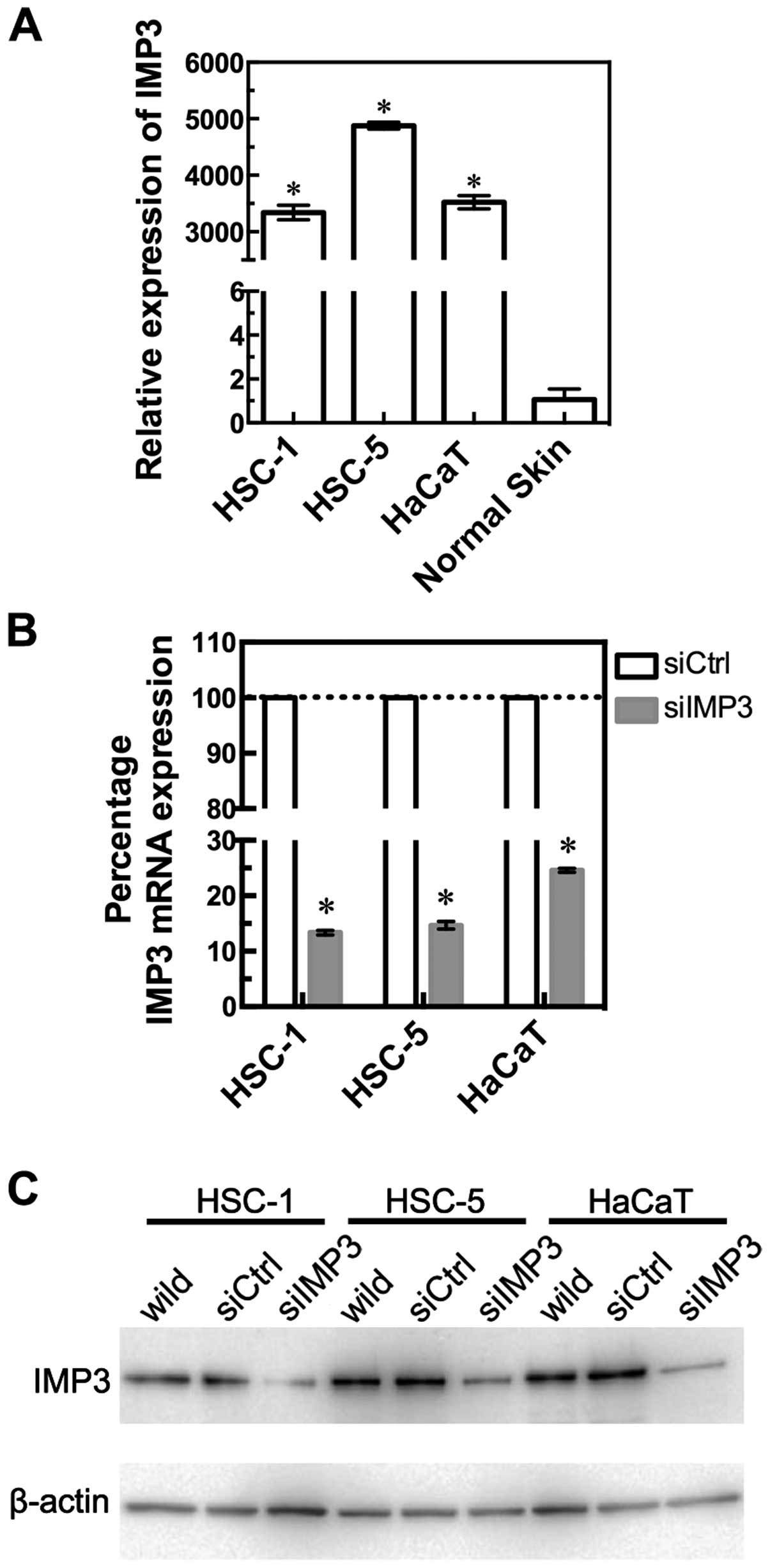
We confirmed IMP3 expression levels in two SCC cell
lines, HSC-1 and HSC-5, and HaCaT cells by RT-qPCR (Fig. 1A). HSC-1, HSC-5, and HaCaT cells
showed significantly higher levels of IMP3 mRNA, compared with
normal skin (HSC-1: P<0.0001, HSC-5: P<0.0001, HaCaT:
P<0.0001, Mann-Whitney U-test, Fig.
1A). To examine the effects of IMP3 inhibition, we transfected
IMP3 siRNA (siIMP3) into HSC-1, HSC-5, and HaCaT cells. RT-qPCR
revealed that IMP3 mRNA expression was significantly decreased in
siIMP3 cells of these three cell lines (Fig. 1B). Thus, siIMP3-transfected cells
were used in the following experiments to examine the inhibitory
effects of IMP3.
IMP3 protein expression in
siIMP3-transfected SCC and HaCaT cells
Western blot analysis revealed that the expression
level of the 70 kDa IMP3 protein was also downregulated in
siIMP3-transfected HSC-1, HSC-5, and HaCaT cells compared to
control siRNA (siCtrl)-transfected cells (Fig. 1C).
IMP3 siRNA downregulated SCC and HaCaT
cell proliferation
To study the effects of reduced IMP3 expression on
cell proliferation, siIMP3 and siCtrl-transfected cell lines were
compared using WST-8 cell counting reagent. The cell proliferation
of siIMP3-transfected HSC-1 cells was significantly decreased at 72
and 96 h (P<0.0001 for both, Sidak's multiple comparison test,
two-way ANOVA; Fig. 2A HSC-1)
compared with siCtrl-transfected cells. The cell proliferation of
siIMP3-transfected HSC-5 and HaCaT cells also tended to be
significantly decreased at 96 h (HSC-5: P<0.0001; HaCaT:
P=0.0008, Sidak's multiple comparison test; Fig. 2A).
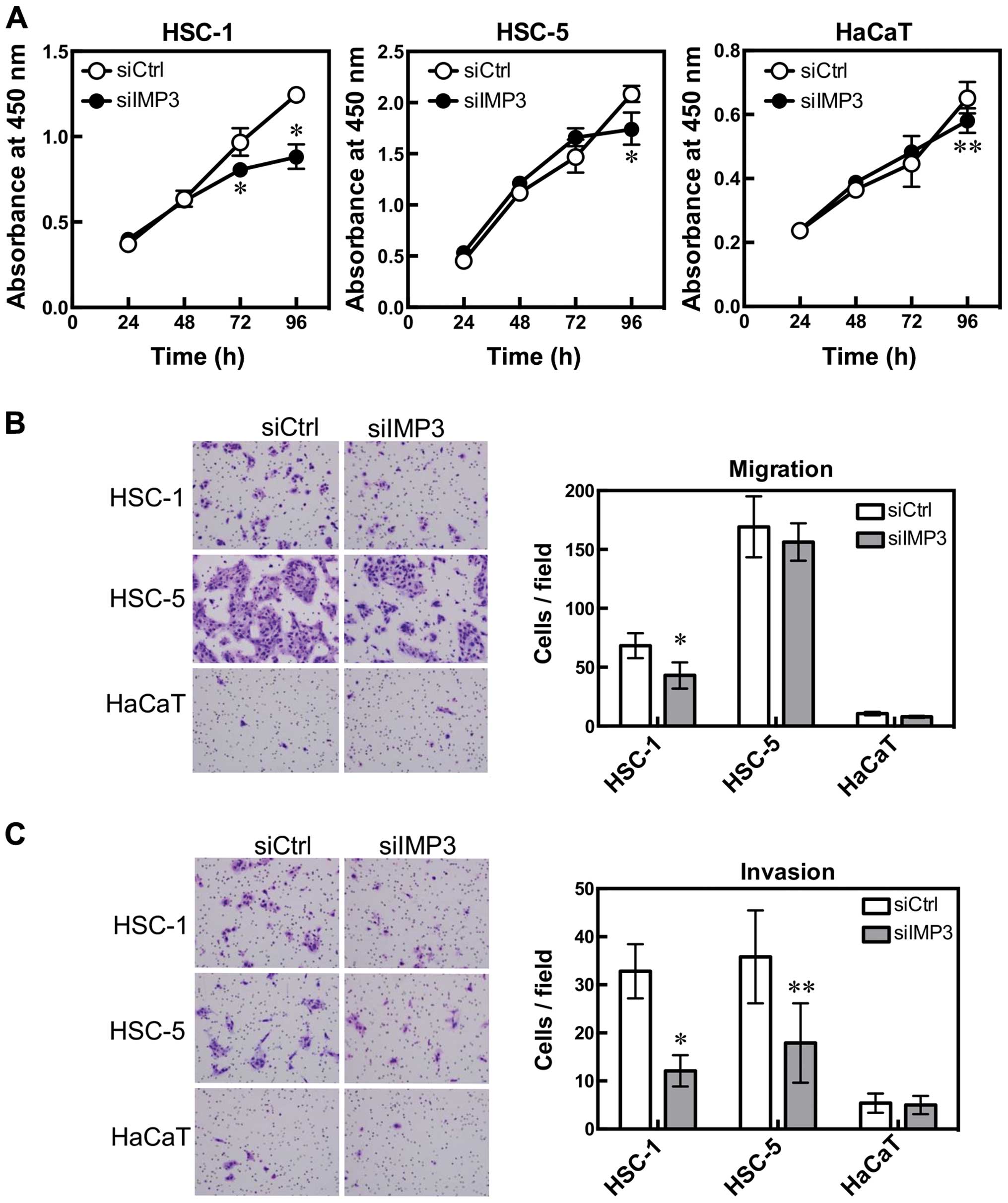 | Figure 2IMP3 is associated with cell
proliferation, migration and invasion. HSC-1, HSC-5 and HaCaT cells
were transfected with IMP3 siRNA and cell proliferation, migration
and invasion were measured. Cell proliferation was determined by
WST-8 cell counting reagent. HSC-1: *P<0.0001 for 72
and 96 h, Sidak's multiple comparison test, and two-way ANOVA; and
HSC-5: *P<0.0001 for 96 h; HaCaT:
**P=0.0008 for 96 h, Sidak's multiple comparison test,
compared with si-control (siCtrl)-transfected cells (A). Cell
migration was measured using BioCoat chambers. HSC-1:
*P=0.0010, Mann-Whitney U-test, compared with
siCtrl-transfected cells. HSC-5 and HaCaT cells were not affected
(B). Cell invasion was determined using Matrigel-coated inserts in
Boyden chambers. HSC-1: *P<0.0001; HSC-5:
**P=0.0063, Mann-Whitney U-test compared with
siCtrl-transfected cells (C). Results are expressed as mean ±95%
confidence interval. (B and C) Diff-Quick-stained cells.
Magnification, x200. |
IMP3 siRNA downregulated SCC cell
migration and invasion
To clarify the effect of IMP3 on cell migration and
invasion, we used a modified Boyden chamber. The number of cells
that migrated from the inside to the outside of the chamber was
significantly decreased in siIMP3-transfected HSC-1 cells (HSC-1:
P=0.0010, Mann-Whitney U-test, Fig.
2B). The invasion assay using Matrigel-coated Boyden chambers
showed that the number of invading cells decreased significantly
for siIMP3-transfected HSC-1 and HSC-5 cells (HSC-1: P<0.0001,
HSC-5: P=0.0063, Mann-Whitney U-test, Fig. 2C). HaCaT cells were not affected by
IMP3 knockdown in either the cell migration or invasion assay.
Immunohistochemical analysis of IMP3 and
Ki-67
We performed immunohistochemical staining of IMP3
and Ki-67 in SCC and KA tissues (Figs.
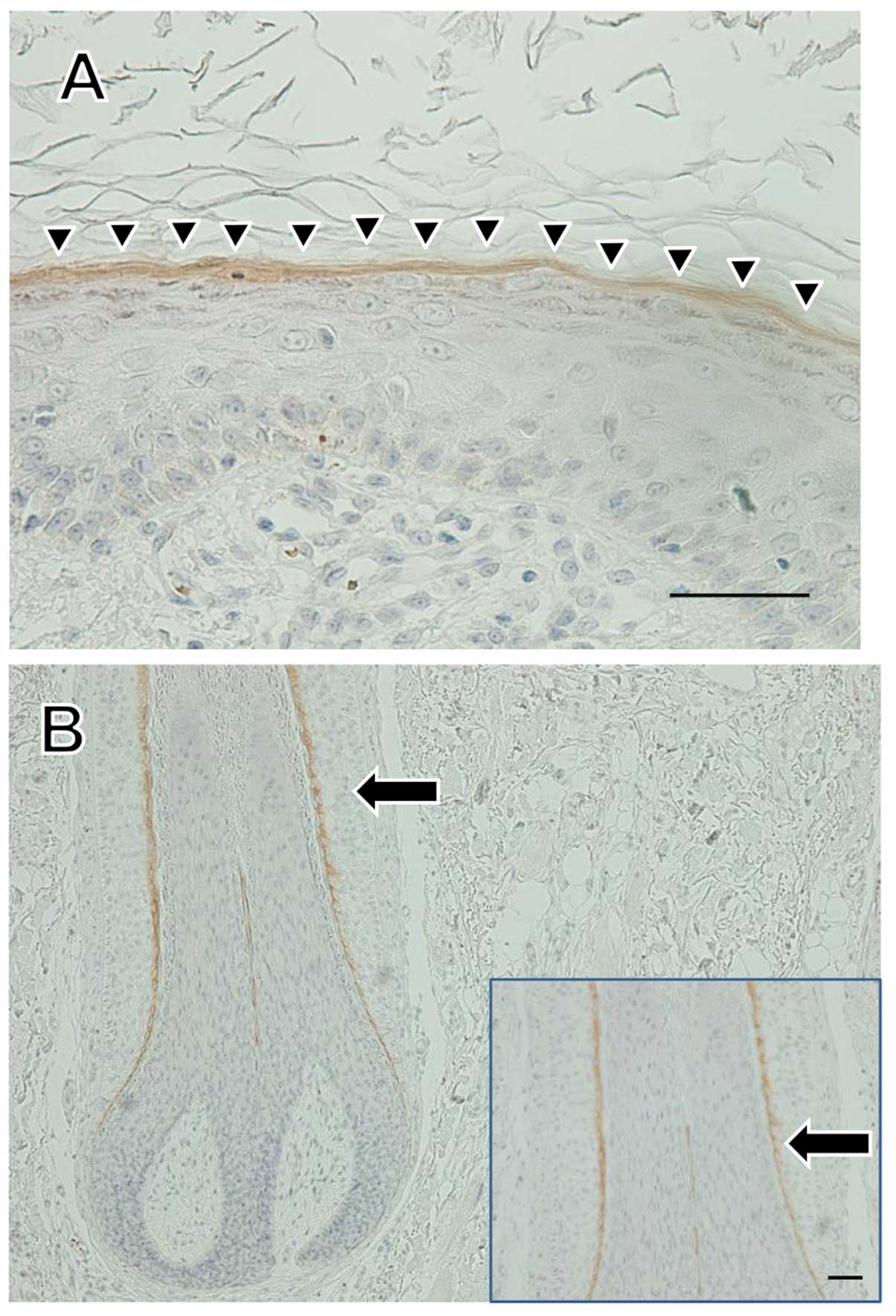
3, 5 and 6). IMP3 expression was not seen in normal
skin except in the internal root sheath and granular layer
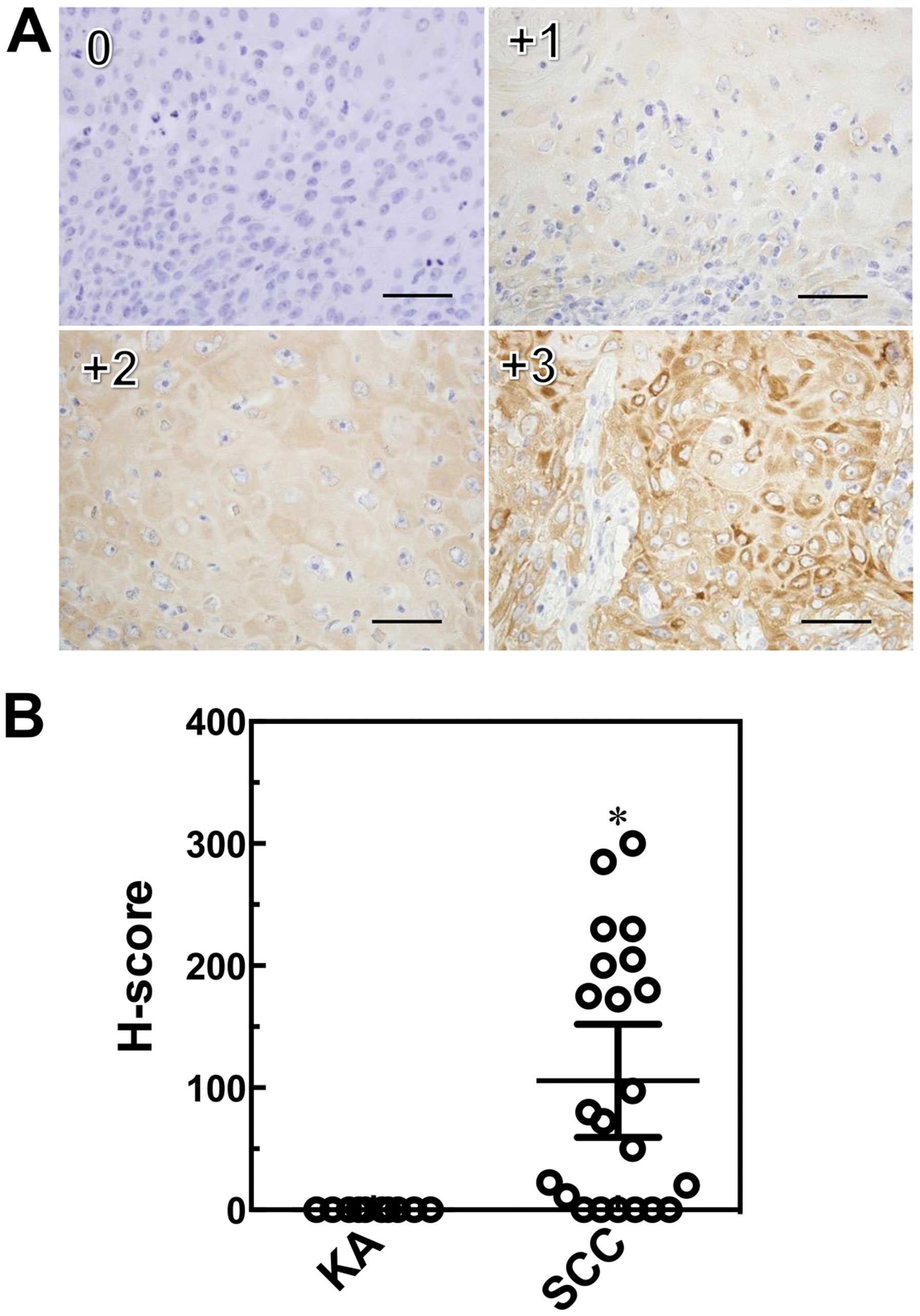
(Fig. 4). A total of ten cases of
SCC (66.7%) were positive for IMP3 and, in contrast, all KA cases
were negative for IMP3. IMP-3 expression was assessed as an H-score
(Fig. 5A). The H-score for SCC
tissues was significantly higher than that for KA tissues
(P=0.0003, Mann-Whitney U-test: Fig.
5B).
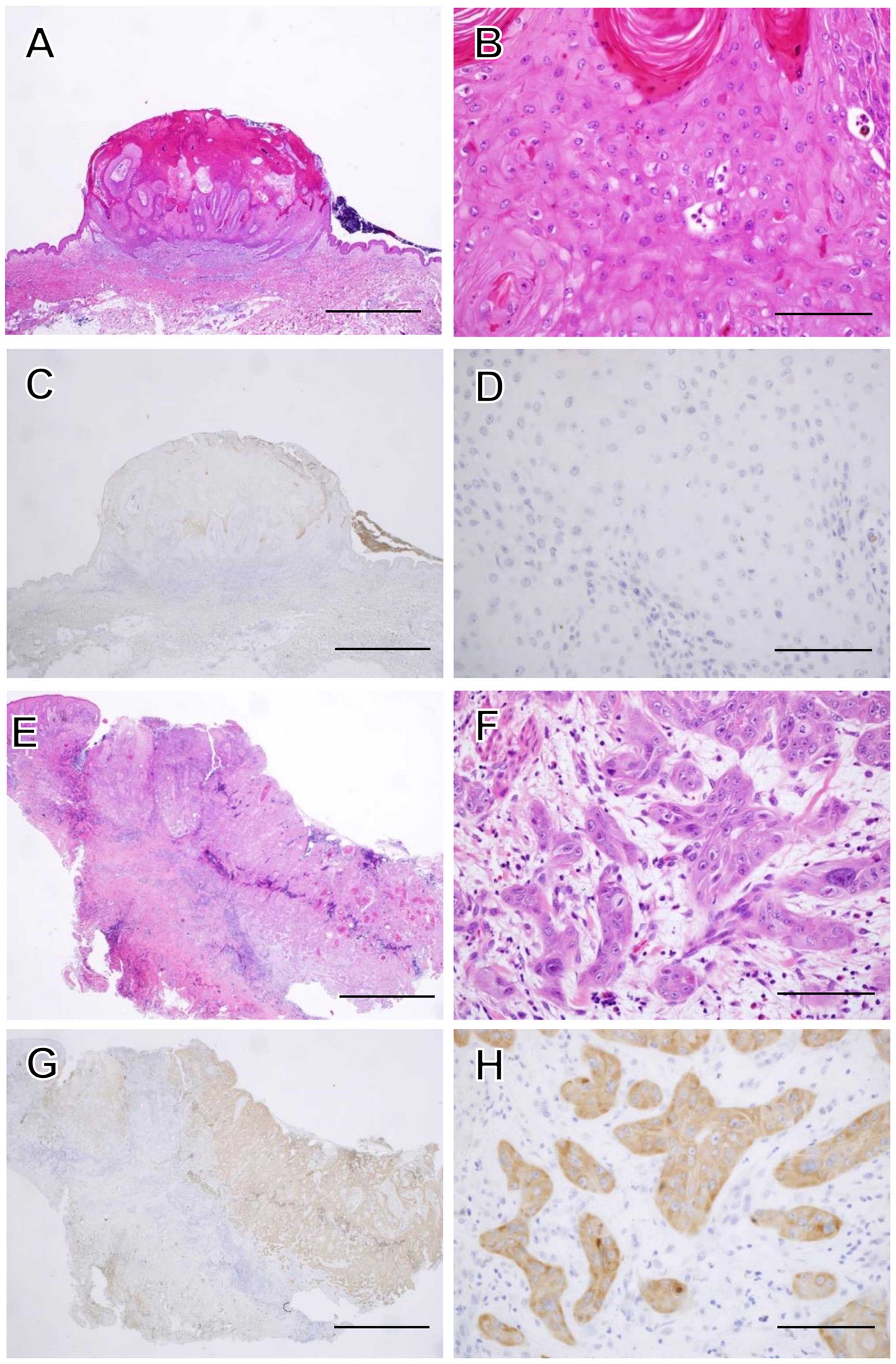 | Figure 3Histopathological features of KA and
SCC. A magnified histological section showing a characteristic
architectural pattern (an exo-endophytic lesion with a central
keratotic horn) with the involvement of unclear epithelial lips on
both sides by the lesions themselves (A). A close-up view of the KA
of a lobule made up of large, pale pink cells with a glassy
appearance without nuclear atypia (B). IMP3 was not expressed in
the KA at all (C and D). A magnified histological section showed an
elevated lesion with hyperkeratosis and acanthosis (E). In a
close-up view of a SCC section, the neoplastic lobules consisted of
squamoid cells, which showed nuclear atypia in the dermis (F). IMP3
was expressed diffusely in the neoplastic lobules of SCC (G and H).
Hematoxylin and eosin (H&E) staining was used (A, B, E and F).
(C, D, G and H) Sections were stained with anti-IMP3 antibody and
peroxidase activity visualized by diaminobenzidine; sections were
counterstained with Mayer's hematoxylin. The scale bar is 2 mm (A,
C, E and G), and 100 μm (B, D, F and H). |
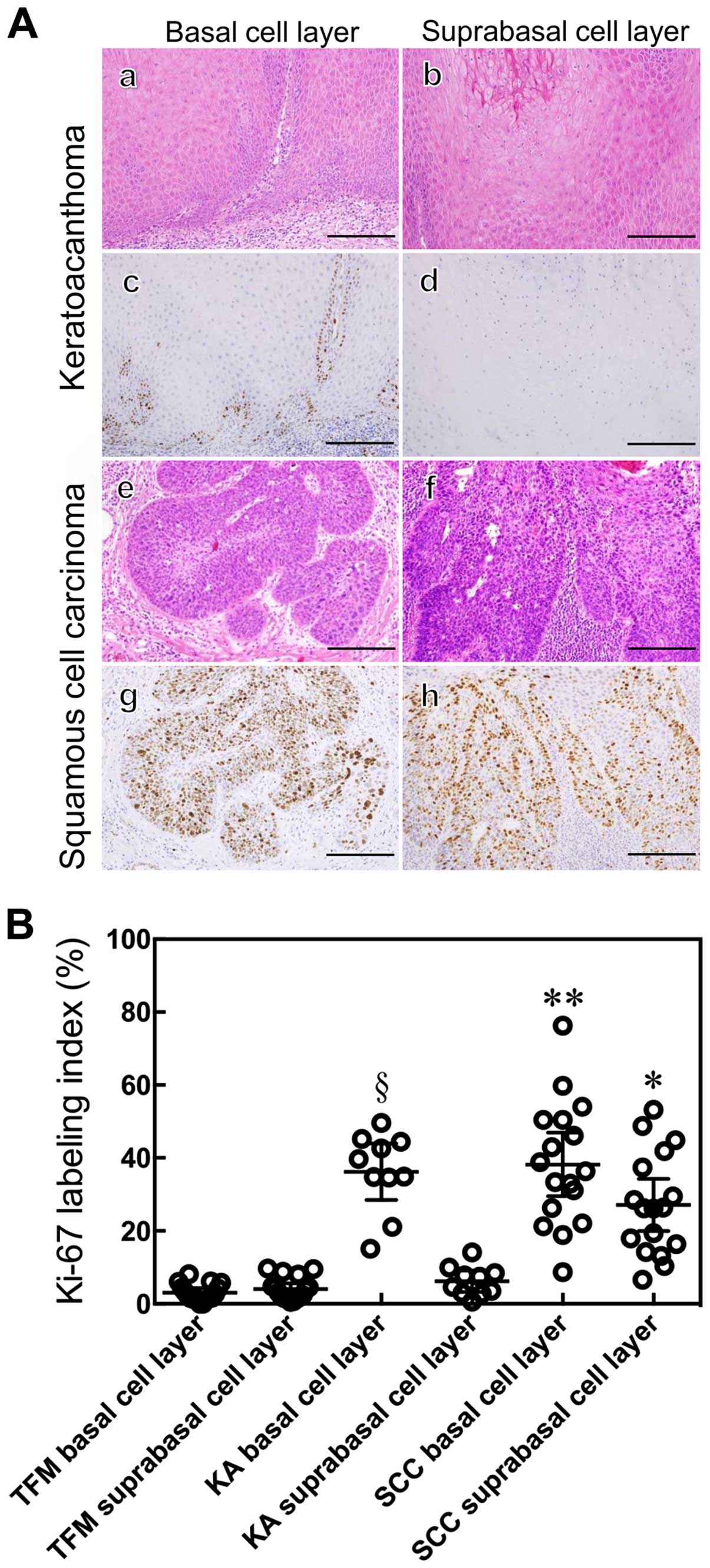
The Ki-67 LI of the suprabasal cell layer of SCC
tissues was higher than that of KA and TFM tissues (P<0.0001,
Mann-Whitney U-test: Fig. 6B). The
Ki-67 LI for the basal cell layer of SCC was higher than that for
the suprabasal cell layers of SCC (P=0.0445, Mann-Whitney U-test;
Fig. 6B) and for the basal cell
layers of KA (P=0.8428, Mann-Whitney U-test; Fig. 6B). The Ki-67 LI for the basal cell
layer of KA was higher than that for the suprabasal cell layer of
KA and for the basal cell layer of TFM (P<0.0001, Mann-Whitney
U-test; Fig. 6B).
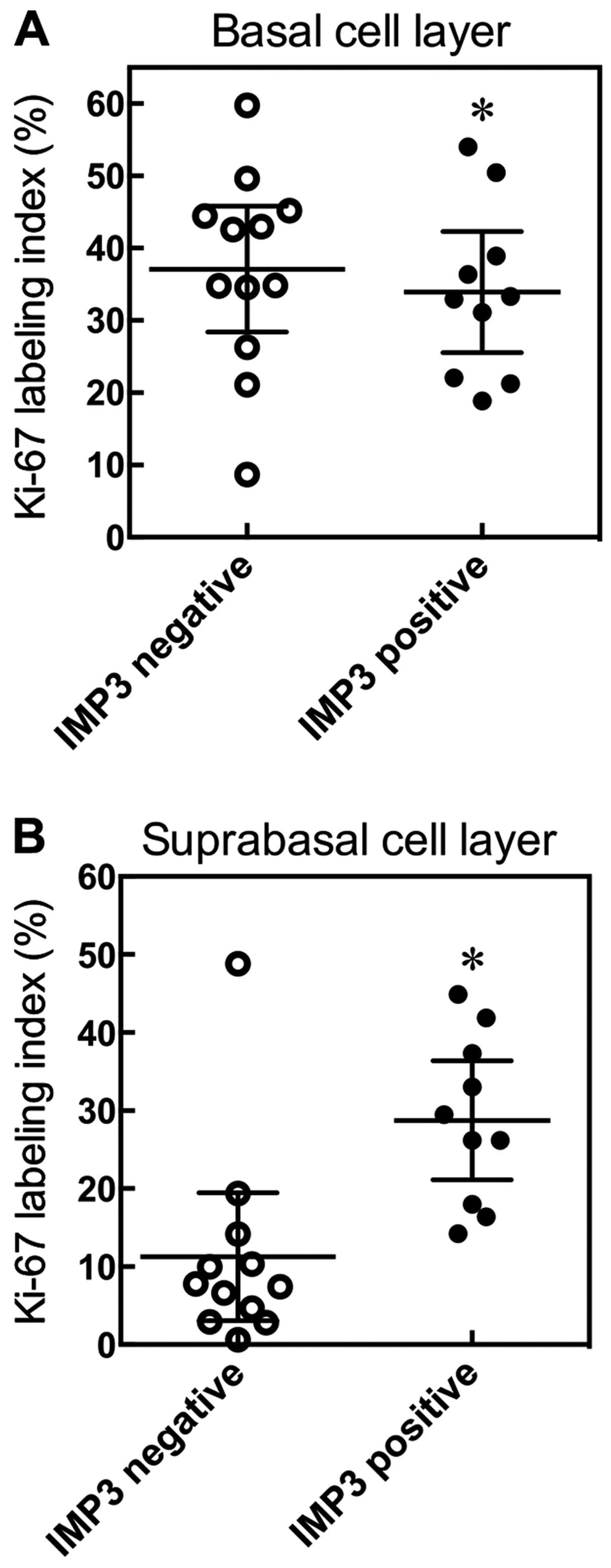
To examine the relationship between Ki-67 and IMP3,
we divided SCC cases into two groups, an IMP3-positive group and an
IMP3-negative group. We examined, in detail, the Ki-67 LI of basal
and suprabasal cell layers in SCC. We found there was no
significant difference in the Ki-67 LI of the basal cell layer
between the IMP3-positive and -negative tumor groups (P=0.4465,
Mann-Whitney U-test; Fig. 7A).
However, the IMP3-positive group tended to show a high Ki-67 LI for
the suprabasal cell layer (P=0.0011, Mann-Whitney U-test; Fig. 7B).
Discussion
IMP3 is thought of as a bona fide oncofetal protein
that is overexpressed and is involved in cell proliferation,
migration, and invasion in several kinds of tumors (18,23,28,29).
In regard to skin squamous neoplasms, the role of IMP3 in cutaneous
SCC, using various cell biological and molecular biological
approaches, has not been well studied. Moreover, we show that
expression of IMP3 mRNA was significantly overexpressed in SCC cell
lines compared with TFM and KA. We found that IMP3 was expressed
not only in the internal root sheath, but also in the granular
layer of normal skin. To our knowledge, these results are the first
to report on the role of IMP3 in SCC.
Knockdown of IMP3 using siRNA reduced IMP3 mRNA and
protein expression levels in the human SCC cell lines, HSC-1 and
HSC-5, and in the immortalized human keratinocyte cell line, HaCaT.
Moreover, knockdown of IMP3 reduced the proliferation of HSC-1
cells, and also significantly inhibited invasion by HSC-1 and HSC-5
cells. However, the knockdown of IMP3 in HaCaT cells did not
influence cell invasion, in spite of the high level of IMP3 mRNA
found in normal cells. These results raise the possibility that the
kinetics of downstream signaling by IMP3 target genes differ in
each cell line. With this in mind, we plan to undertake a detailed
analysis of the cell signaling and target gene expression involved
in IMP3 cell invasion in our next study. These data also suggest
that IMP3 may promote cell invasion by cutaneous SCC cells, and may
partially induce their cell proliferation. In this study, IMP3
expression did not correlate with clinicopathological variables
such as patient age, gender, maximum diameter of tumors,
ulceration, and tumor thickness in SCC.
Since all SCC cases were stage I or II without
recurrent and metastatic lesions, our next study may focus on the
late-stage cases, recurrent cases and metastasis cases. With regard
to the relationship between IMP3 expression and the biological
behavior of SCC from other organs, there are several reports
suggesting that IMP3 expression might be associated with the lymph
nodal and tumor stage of tumor invasion, as well as with overall
survival in oral SCC (30,31), also shown to be an independent
prognostic factor in cervical SCC (32). Moreover, the high expression of
IMP3 has been associated with lymph nodal metastasis and poor
patient outcome in tongue SCC (33). Taken together, IMP3 may play an
important role in the biological behavior of cutaneous SCC, and may
be used as new therapeutic target for SCC.
KA was originally considered to be a benign
neoplasm. However, a few cases of metastasis purported to originate
from KAs have been reported, and many reports describe KA as a
variant of SCC with low-grade malignancy (3,34).
Moreover, Sánchez Yus et al reported the first formal study
on the ‘transformation’ of a subset of KAs into SCCs (35), and Weedon et al regarded KA
as a benign lesion that could undergo malignant transformation into
SCC (4). Weedon et al
described how KA differed markedly from SCC in morphology,
biological behavior, and outcome when perineural and/or intravenous
invasion were present, and believed that KA was not a variant of
SCC (36). Ni et al
demonstrated that mRNA levels of mitogen-activated protein kinase 1
(MAPK1) and caspase-14 (CASP14) were upregulated in SCC, while
those of BAG (Bcl-2-associated athanogene) family molecular
chaperone regulator 1 (BAG1) and matrix metalloproteinase-14
(MMP14) were downregulated in SCC, and suggested that SCC and KA
were molecularly distinct entities (37). Recently, Soddu et al showed
that IMP3 immunohistochemistry was useful in distinguishing KA from
cutaneous SCC (24).
We demonstrated that IMP3 plays a role in cell
proliferation and invasion in cutaneous SCC. In the
histopathological diagnosis of a cutaneous tumor, distinguishing
between SCC with a crateriform architecture and KA is often
difficult; we suggest IMP3 may be a suitable marker. In our
immunohistochemical study, IMP3 was observed in the internal root
sheath and also in the granular layer of normal skin. Pryor et
al also reported the expression of IMP3 in the internal root
sheath (11). However, to our
knowledge, this is the first report of the expression of IMP3 in
the granular layer. Previous reports showed that IMP3 regulate
CD44s expression in pancreatic cancer and hepatocellular carcinoma
(23,38). Liu et al found that the
expression of syndecan, a cell surface proteoglycan like CD44,
switched from the granular cell layer to the basal and lower
spinous cell layers in irradiated mouse skin. They suggested that
CD44 and syndecan play an important role in the proliferative
process (39). Hyaluronan (HA)
binds to CD44, and HA/CD44-mediated activation of RhoGTPase
signaling leads to the regulation of keratinocyte activities and
various epidermal functions, such as, proliferation, migration and
differentiation (40). In our
study, the tumor thickness of IMP3-positive SCC tended to be
thicker than that of IMP3- negative SCC (data not shown). Thus, the
relationship between IMP3 and CD44-related molecules may be our
next focus.
Ki-67 is a cell cycle-regulating protein that is
expressed during all phases of the cell cycle except G0. The
cellular expression of Ki-67 provides a measure of the growth
fraction of a tumor (41). We
demonstrated that the Ki-67 LI of SCC was significantly higher than
that of KA, as reported previously by others (42,43).
However, a comparison between basal cell and suprabasal cell layers
in Ki-67 LI analysis had not yet been made prior to this study. We
demonstrate that the Ki-67 LI in the suprabasal cell layer of SCC
was significantly higher than that in the suprabasal cell layer of
KA, which indicates that the proliferative potential of suprabasal
cells of SCC is higher than those of KA. We also found that
IMP3-positive cells in the suprabasal cell layer tended to have a
high Ki-67 LI. These are novel findings. IMP3 regulates the
expression of cyclines D1, D3 and G1 (44). Lin et al demonstrated that
the Ki-67 LI was significantly correlated with IMP3 expression in
colon cancer (45). Similar to
these findings, IMP3 may play an important role in the cell cycle
of cutaneous SCC.
Other groups have described the mechanism of cell
proliferation and invasion of IMP3 expression in cells. Recent
studies have revealed that IMP3 promoted the progression of
melanoma by regulating high mobility group AT-hook 2 (HMGA2)
(18), the cell migration of renal
cell carcinoma by activation of the NF-κB pathway (28), cell invasion and migration after
epithelial to mesenchymal transition (EMT) (29), and matrix adhesion, cell motility
and invasion of pancreatic carcinoma by enhancing CD44 and the
transcription of kinesin KIF11 expression (23).
In conclusion, IMP3 may play an important role in
the biological behavior of cutaneous SCC, and may be used as new
therapeutic target for SCC. Moreover, the usefulness of IMP3 as a
marker for distinguishing between SCC and KA warrants further
evaluation.
Acknowledgements
This study was supported, in part, by grants-in-aid
for the Clinical Rebiopsy Bank Project for Comprehensive Cancer
Therapy Development to Z.N. from the Ministry of Education,
Culture, Sport, Science and Technology, Japan (MEXT), 2013–2017
(S13110022) and a grant-in-aid for scientific research (C, no.
25461716 to S.A.) from JSPS KAKENHI.
References
|
1
|
Schwartz RA: The keratoacanthoma: A
review. J Surg Oncol. 12:305–317. 1979. View Article : Google Scholar : PubMed/NCBI
|
|
2
|
Misago N, Inoue T, Koba S and Narisawa Y:
Keratoacanthoma and other types of squamous cell carcinoma with
crateriform architecture: Classification and identification. J
Dermatol. 40:443–452. 2013. View Article : Google Scholar : PubMed/NCBI
|
|
3
|
Hodak E, Jones RE and Ackerman AB:
Solitary keratoacanthoma is a squamous-cell carcinoma: Three
examples with metastases. Am J Dermatopathol. 15:332–342;
discussion 343–352. 1993. View Article : Google Scholar : PubMed/NCBI
|
|
4
|
Weedon DD, Malo J, Brooks D and Williamson
R: Squamous cell carcinoma arising in keratoacanthoma: A neglected
phenomenon in the elderly. Am J Dermatopathol. 32:423–426. 2010.
View Article : Google Scholar : PubMed/NCBI
|
|
5
|
Kossard S, Tan KB and Choy C:
Keratoacanthoma and infundibulocystic squamous cell carcinoma. Am J
Dermatopathol. 30:127–134. 2008. View Article : Google Scholar : PubMed/NCBI
|
|
6
|
Misago N, Inoue T, Toda S and Narisawa Y:
Infundibular (follicular) and infundibulocystic squamous cell
carcinoma: A clinicopathological and immunohistochemical study. Am
J Dermatopathol. 33:687–694. 2011. View Article : Google Scholar : PubMed/NCBI
|
|
7
|
Müeller-Pillasch F, Lacher U, Wallrapp C,
Micha A, Zimmerhackl F, Hameister H, Varga G, Friess H, Büchler M,
Beger HG, et al: Cloning of a gene highly overexpressed in cancer
coding for a novel KH-domain containing protein. Oncogene.
14:2729–2733. 1997. View Article : Google Scholar : PubMed/NCBI
|
|
8
|
Nielsen J, Christiansen J, Lykke-Andersen
J, Johnsen AH, Wewer UM and Nielsen FC: A family of insulin-like
growth factor II mRNA-binding proteins represses translation in
late development. Mol Cell Biol. 19:1262–1270. 1999. View Article : Google Scholar : PubMed/NCBI
|
|
9
|
Simon R, Bourne PA, Yang Q, Spaulding BO,
di Sant'Agnese PA, Wang HL and Xu H: Extrapulmonary small cell
carcinomas express K homology domain containing protein
overexpressed in cancer, but carcinoid tumors do not. Hum Pathol.
38:1178–1183. 2007. View Article : Google Scholar : PubMed/NCBI
|
|
10
|
Hammer NA, Hansen T, Byskov AG, Rajpert-De
Meyts E, Grøndahl ML, Bredkjaer HE, Wewer UM, Christiansen J and
Nielsen FC: Expression of IGF-II mRNA-binding proteins (IMPs) in
gonads and testicular cancer. Reproduction. 130:203–212. 2005.
View Article : Google Scholar : PubMed/NCBI
|
|
11
|
Pryor JG, Simon RA, Bourne PA, Spaulding
BO, Scott GA and Xu H: Merkel cell carcinoma expresses K homology
domain-containing protein overexpressed in cancer similar to other
high-grade neuroendocrine carcinomas. Hum Pathol. 40:238–243. 2009.
View Article : Google Scholar
|
|
12
|
Righi A, Zhang S, Jin L, Scheithauer BW,
Kovacs K, Kovacs G, Goth MI, Korbonits M and Lloyd RV: Analysis of
IMP3 expression in normal and neoplastic human pituitary tissues.
Endocr Pathol. 21:25–31. 2010. View Article : Google Scholar
|
|
13
|
King RL, Pasha T, Roullet MR, Zhang PJ and
Bagg A: IMP-3 is differentially expressed in normal and neoplastic
lymphoid tissue. Hum Pathol. 40:1699–1705. 2009. View Article : Google Scholar : PubMed/NCBI
|
|
14
|
Liao B, Hu Y, Herrick DJ and Brewer G: The
RNA-binding protein IMP-3 is a translational activator of
insulin-like growth factor II leader-3 mRNA during proliferation of
human K562 leukemia cells. J Biol Chem. 280:18517–18524. 2005.
View Article : Google Scholar : PubMed/NCBI
|
|
15
|
Clauditz TS, Wang CJ, Gontarewicz A,
Blessmann M, Tennstedt P, Borgmann K, Tribius S, Sauter G, Dalchow
C, Knecht R, et al: Expression of insulin-like growth factor II
mRNA-binding protein 3 in squamous cell carcinomas of the head and
neck. J Oral Pathol Med. 42:125–132. 2013. View Article : Google Scholar
|
|
16
|
Wang T, Fan L, Watanabe Y, McNeill PD,
Moulton GG, Bangur C, Fanger GR, Okada M, Inoue Y, Persing DH, et
al: L523S, an RNA-binding protein as a potential therapeutic target
for lung cancer. Br J Cancer. 88:887–894. 2003. View Article : Google Scholar : PubMed/NCBI
|
|
17
|
Pryor JG, Bourne PA, Yang Q, Spaulding BO,
Scott GA and Xu H: IMP-3 is a novel progression marker in malignant
melanoma. Mod Pathol. 21:431–437. 2008. View Article : Google Scholar : PubMed/NCBI
|
|
18
|
Sheen YS, Liao YH, Lin MH, Chu CY, Ho BY,
Hsieh MC, Chen PC, Cha ST, Jeng YM, Chang CC, et al: IMP-3 promotes
migration and invasion of melanoma cells by modulating the
expression of HMGA2 and predicts poor prognosis in melanoma. J
Invest Dermatol. 135:1065–1073. 2015. View Article : Google Scholar
|
|
19
|
Yu L, Xu H, Wasco MJ, Bourne PA and Ma L:
IMP-3 expression in melanocytic lesions. J Cutan Pathol.
37:316–322. 2010. View Article : Google Scholar
|
|
20
|
Mentrikoski MJ, Ma L, Pryor JG, McMahon
LA, Yang Q, Spaulding BO, Scott GA, Wang HL and Xu H: Diagnostic
utility of IMP3 in segregating metastatic melanoma from benign nevi
in lymph nodes. Mod Pathol. 22:1582–1587. 2009. View Article : Google Scholar : PubMed/NCBI
|
|
21
|
Takata A, Takiguchi S, Okada K, Takahashi
T, Kurokawa Y, Yamasaki M, Miyata H, Nakajima K, Mori M and Doki Y:
Expression of insulin-like growth factor-II mRNA-binding protein-3
as a marker for predicting clinical outcome in patients with
esophageal squamous cell carcinoma. Oncol Lett. 8:2027–2031.
2014.PubMed/NCBI
|
|
22
|
Zhang J, Ou Y, Ma Y, Zheng L, Zhang X, Xia
R, Kong F, Shen Y, Wang S and Lin L: Clinical implications of
insulin-like growth factor II mRNA-binding protein 3 expression in
non-small cell lung carcinoma. Oncol Lett. 9:1927–1933.
2015.PubMed/NCBI
|
|
23
|
Pasiliao CC, Chang CW, Sutherland BW,
Valdez SM, Schaeffer D, Yapp DT and Ng SS: The involvement of
insulin-like growth factor 2 binding protein 3 (IMP3) in pancreatic
cancer cell migration, invasion, and adhesion. BMC Cancer.
15:2662015. View Article : Google Scholar : PubMed/NCBI
|
|
24
|
Soddu S, Di Felice E, Cabras S,
Castellanos ME, Atzori L, Faa G and Pilloni L: IMP-3 expression in
keratoacanthomas and squamous cell carcinomas of the skin: an
immunohistochemical study. Eur J Histochem. 57:e62013. View Article : Google Scholar : PubMed/NCBI
|
|
25
|
Kondo S, Hozumi Y and Aso K: Autocrine
secretion of an EGF-like substance by a cell line (HSC-1) derived
from a human skin squamous cell carcinoma. J Dermatol Sci.
2:161–165. 1991. View Article : Google Scholar : PubMed/NCBI
|
|
26
|
Hozumi Y, Kondo S, Shimoura T and Aso K:
Human squamous cell carcinoma from skin: Establishment and
characterization of a new cell line (HSC-5). J Dermatol.
17:143–148. 1990. View Article : Google Scholar : PubMed/NCBI
|
|
27
|
Abdou AG, Maraee AH, El-Sayed EM and
Elnaidany NF: Immunohistochemical expression of ezrin in cutaneous
basal and squamous cell carcinomas. Ann Diagn Pathol. 15:394–401.
2011.PubMed/NCBI
|
|
28
|
Pei X, Li M, Zhan J, Yu Y, Wei X, Guan L,
Aydin H, Elson P, Zhou M, He H, et al: Enhanced IMP3 expression
activates NF-κB pathway and promotes renal cell carcinoma
progression. PLoS One. 10:e01243382015. View Article : Google Scholar
|
|
29
|
Su P, Hu J, Zhang H, Li W, Jia M, Zhang X,
Wu X, Cheng H, Xiang L and Zhou G: IMP3 expression is associated
with epithelial-mesenchymal transition in breast cancer. Int J Clin
Exp Pathol. 7:3008–3017. 2014.PubMed/NCBI
|
|
30
|
Lin CY, Chen ST, Jeng YM, Yeh CC, Chou HY,
Deng YT, Chang CC and Kuo MY: Insulin-like growth factor II
mRNA-binding protein 3 expression promotes tumor formation and
invasion and predicts poor prognosis in oral squamous cell
carcinoma. J Oral Pathol Med. 40:699–705. 2011. View Article : Google Scholar : PubMed/NCBI
|
|
31
|
Kim KY, Li S, Cha JD, Zhang X and Cha IH:
Significance of molecular markers in survival prediction of oral
squamous cell carcinoma. Head Neck. 34:929–936. 2012. View Article : Google Scholar
|
|
32
|
Wei Q, Yan J, Fu B, Liu J, Zhong L, Yang Q
and Zhao T: IMP3 expression is associated with poor survival in
cervical squamous cell carcinoma. Hum Pathol. 45:2218–2224. 2014.
View Article : Google Scholar : PubMed/NCBI
|
|
33
|
Li HG, Han JJ, Huang ZQ, Wang L, Chen WL
and Shen XM: IMP3 is a novel biomarker to predict metastasis and
prognosis of tongue squamous cell carcinoma. J Craniofac Surg.
22:2022–2025. 2011. View Article : Google Scholar : PubMed/NCBI
|
|
34
|
Schwartz RA: Keratoacanthoma: A
clinico-pathologic enigma. Dermatol Surg. 30:326–333.
2004.PubMed/NCBI
|
|
35
|
Sánchez Yus E, Simón P, Requena L, Ambrojo
P and de Eusebio E: Solitary keratoacanthoma: A self-healing
proliferation that frequently becomes malignant. Am J
Dermatopathol. 22:305–310. 2000. View Article : Google Scholar : PubMed/NCBI
|
|
36
|
Weedon D, Malo J, Brooks D and Williamson
R: Keratoacanthoma: Is it really a variant of squamous cell
carcinoma? ANZ J Surg. 80:129–130. 2010. View Article : Google Scholar : PubMed/NCBI
|
|
37
|
Ni C, Su A, Ra S, Li X, Cochran A and
Binder S: Squamous cell carcinoma arising from keratoacanthoma: An
evaluation by RT-PCR. Am J Dermatopathol. Jun 2–2015.Epub ahead of
print. View Article : Google Scholar : PubMed/NCBI
|
|
38
|
Hu S, Wu X, Zhou B, Xu Z, Qin J, Lu H, Lv
L, Gao Y, Deng L, Yin J, et al: IMP3 combined with CD44s, a novel
predictor for prognosis of patients with hepatocellular carcinoma.
J Cancer Res Clin Oncol. 140:883–893. 2014. View Article : Google Scholar : PubMed/NCBI
|
|
39
|
Liu K, Kasper M, Bierhaus A, Langer S,
Peterson I, Müller M and Trott KR: Differential expression of CD44s
and CD44v10 proteins and syndecan in normal and irradiated mouse
epidermis. Histochem Cell Biol. 107:159–167. 1997. View Article : Google Scholar : PubMed/NCBI
|
|
40
|
Bourguignon LY: Matrix
hyaluronan-activated CD44 signaling promotes keratinocyte
activities and improves abnormal epidermal functions. Am J Pathol.
184:1912–1919. 2014. View Article : Google Scholar : PubMed/NCBI
|
|
41
|
Brown DC and Gatter KC: Ki67 protein: The
immaculate deception? Histopathology. 40:2–11. 2002. View Article : Google Scholar : PubMed/NCBI
|
|
42
|
Ribeiro D, Narikawa S and Marques ME:
Expression of apoptotic and cell proliferation regulatory proteins
in keratoacanthomas and squamous cell carcinomas of the skin.
Pathol Res Pract. 204:97–104. 2008. View Article : Google Scholar
|
|
43
|
Scola N, Segert HM, Stücker M, Altmeyer P,
Gambichler T and Kreuter A: Ki-67 may be useful in differentiating
between keratoacanthoma and cutaneous squamous cell carcinoma. Clin
Exp Dermatol. 39:216–218. 2014. View Article : Google Scholar : PubMed/NCBI
|
|
44
|
Rivera Vargas T, Boudoukha S, Simon A,
Souidi M, Cuvellier S, Pinna G and Polesskaya A:
Post-transcriptional regulation of cyclins D1, D3 and G1 and
proliferation of human cancer cells depend on IMP-3 nuclear
localization. Oncogene. 33:2866–2875. 2014. View Article : Google Scholar
|
|
45
|
Lin L, Zhang J, Wang Y, Ju W, Ma Y, Li L
and Chen L: Insulin-like growth factor-II mRNA-binding protein 3
predicts a poor prognosis for colorectal adenocarcinoma. Oncol
Lett. 6:740–744. 2013.PubMed/NCBI
|