Introduction
Hepatobiliary and pancreatic cancer (HPC) is a major
threat to human health, with an increasing incidence over the last
few years. Surgery, chemotherapy and radiotherapy are currently the
mainstay of treatment for HPC. Although surgery is the first
treatment of choice, early-stage cancer is diagnosed in a limited
number of patients (1,2). Furthermore, the hepatobiliary and
pancreatic systems exhibit a complex anatomical structure and serve
important physiological functions. Consequently, HPC symptoms lack
specificity and have typically reached an advanced stage at
diagnosis. Radical resection of HPC is not typically performed, and
the majority of HPCs are associated with a poor prognosis (3,4).
Chemotherapy and radiotherapy are the primary treatments for
advanced-stage disease; however, these treatments may result in
major health issues and are associated with severe
treatment-related toxicity. Furthermore, the overall health status
of HPC patients is generally poor, which may prevent them from
being eligible for these standard therapies (2,5).
In addition to the common factors affecting the
prognosis of cancer patients (disease stage, tumor size, lymph node
metastasis and radical surgery), individual differences in the
immune factors among patients also affect prognosis. As all cancer
patients exhibit varying degrees of low immunity and a significant
proportion are in an immunosuppressed state, recovering anticancer
immunity is a possible approach to cancer treatment. Autologous
immune cell therapy has been the fourth most commonly used
treatment method for cancer and it is widely used in clinical
practice. Immunotherapy stimulates the body's own immune system to
fight cancer cells. It has been demonstrated that antigen-specific
T-cell dysfunction occurs in several cancer patients (6–9). As a
result, tumor cells may escape immune surveillance (10). Restoring proper immune system function
may be a viable option for improving cancer treatment and
immunotherapy may be a promising and safe approach to cancer
eradication (11).
The aim of the present study was to investigate the
cellular immune response to autoimmune cell therapy in patients
with HPC in terms of delayed-type hypersensitivity (DTH),
improvement in the quality of life (QOL), safety and prolonged
survival.
Patients and methods
Patients
All the patients were treated at the Department of
Oncology, Tianjin Union Medicine Center (Tianjin, China). HPC
patients who received immunotherapy, including an autologous
dendritic cell (DC) vaccine and cytokine-induced killer (CIK) cell
therapy comprised the immunotherapy group (I group). All the
patients provided informed consent prior to DC-CIK treatment and
were required to meet the following inclusion criteria: i) A
cytological or histological diagnosis of HPC; ii) hospitalization
between January 4, 2012 and December 31, 2012; iii) adequate kidney
and liver function and normal coagulation; iv) a Karnofsky
performance status score of >60; and v) expected survival of
>3 months.
The control group included patients who satisfied
the abovementioned inclusion criteria, but did not receive
immunotherapy [non-immunotherapy (NI) group]. The follow-up for all
the patients was completed on November 14, 2013. The median
survival time (MST) was compared between the two groups and the
benefit of the DC vaccine and CIK cell-based immunotherapy regimens
was evaluated in terms of DTH and improvement in QOL.
Study design
This retrospective study was an open-label,
parallel-group, single-institution, non-randomized study and
complied with the class III medical techniques policy of the
Ministry of Health of China, as described in the guidelines for
treatment with autologous immune cells (T cells, natural killer
cells) (12,13). The study protocol was approved by the
Ethics Committee of the Tianjin Union Medicine Center. Prior to
treatment, all the patients were asked to provide written informed
consent.
The DC vaccine and CIK cell therapy schedule was as
follows: The day on which peripheral blood mononuclear cells
(PBMCs) were collected was defined as day 0. On day 8,
1×10⁷ DCs were infused intravenously once/week for 6
weeks, and on day 11, 1×10⁹ CIK cells were infused
intravenously once/day for 4 days (Table
I).
 | Table I.Regimen of dendritic cells (DCs) +
cytokine-induced killer (CIK) cell therapy. |
Table I.
Regimen of dendritic cells (DCs) +
cytokine-induced killer (CIK) cell therapy.
| Day | Therapy | Functions |
|---|
| 0 | Collection of
leukocyte fraction |
|
| 8 | Reinfusion of DC1
i.v. | Vaccination 1 |
| 11 | Reinfusion of CIK1
i.v. | Against CC |
| 12 | Reinfusion of
CIK2+CIK3 i.v. | Against CC |
| 13 | Reinfusion of CIK4
i.v. | Against CC |
| 14 | Reinfusion of CIK4+
CIK5 i.v. | Against CC |
| 15 | Reinfusion of DC2
i.v. | Vaccination 2 |
| 22 | Reinfusion of DC3
i.v. | Vaccination 3 |
| 29 | Reinfusion of DC4
i.v. | Vaccination 4 |
| 36 | Reinfusion of DC5
i.v. | Vaccination 5 |
| 43 | Reinfusion of DC6
i.d. | Vaccination 6 |
Preparation of cells
Using the Fresenius Kabi system (Fresenius Kabi AG,
Bad Homburg, Germany), PBMCs were collected from each patient under
electrocardiographic monitoring. The cells were cultured overnight,
then adherent cells (including monocytes) and suspension cells
(including lymphocytes) were collected separately (14–16).
Preparation of the DC vaccine
The DC vaccine was prepared according to the
following protocol: Isolated PBMCs were resuspended in a serum-free
culture medium (Hyclone, Logan, UT, USA). The PBMCs were then
seeded into a 175-cm3 flask to a concentration of
2–4×10⁶/ml, divided among three bottles and then placed
in a 37°C, 5% CO2 incubator (Thermo Fisher Scientific,
Waltham, MA, USA). The cell concentration and the number of bottles
used may be adjusted according to the number of cells. After 2 h of
incubation, the 175-cm3 flasks were gently agitated; the
suspension cells were discarded (or collected and used for
culturing CIK cells), then washed 1–2 times with serum-free medium
and, following addition of the DC medium, placed again in the 37°C,
5% CO2 incubator. Autologous serum,
granulocyte-macrophage colony-stimulating factor, interleukin
(IL)-4 and tumor necrosis factors (all from PeproTech EC, Ltd.,
London, UK) were added on the 3rd day. The tumor antigen
(Peprotech, Inc., Rocky Hill, NJ, USA) was added on the 5th day and
the ripening factor was added following addition of the tumor
antigen. Flat blood tests were performed and the flasks were again
placed in the incubator for 2 h. After the flasks were washed twice
with serum-free 1640 medium, the DC culture solution was added and
the flasks were placed back in the incubator. DC cells were
collected between the 7th and 8th days and then washed three times
with saline. The quality of the preparation was determined at the
same time.
Preparation of CIK cells
The CIK cells were prepared according to the
following protocol: The culture bottles were pre-coated with a 5
µg/ml final concentration of CIK reagents before preparing the CIK
cells, then placed in a 4°C environment overnight and washed with
phosphate-buffered saline (PeproTech EC, Ltd.) 2–3 times on day 0.
The CIK cells were resuspended in the initial CIK cell medium at a
concentration of 2–3×10⁶/ml and spread into a culture
bag according to the number of cells. Autologous plasma was then
added and cultured in a 37°C, 5% CO2 incubator
(PeproTech EC, Ltd.). IL-2, interferon 2γ (both from PeproTech EC,
Ltd.) and CD3 monoclonal antibody (100 µg/ul; cat. no 20150605;
Beckman Coulter, Inc., Brea, CA, USA) were added using 5-ml
disposable syringes with needles. To ensure that all factors were
added in the culture bag, the aspiration was repeated five times,
after which the culture bags were placed in the 37°C, 5%
CO2 incubator. Samples were prepared and counted every
2–3 days using 2-ml disposable syringes with needles. Appropriate
additional CIK expansion bottle medium was added according to the
results of the count in order to maintain a CIK cell concentration
of 1–3×10⁶/ml and the total number of cells was recorded
at the same time.
Cell quality control
The immune phenotype markers of HLA2DR, CD86 and
CD83 were used for DCs; CD3, CD8 and CD56 were used for CIK cells.
The bacteria, endotoxin and fungus levels of the cultured samples
satisfied the release criteria for infusion (Table II).
| Table II.Immune phenotype markers. |
Table II.
Immune phenotype markers.
| Cell types | Early separation | Reagents | Cell surface
phenotype |
|---|
| DCs | Adherent cells
(monocytes) | GM-CSF, IL-4, TNF,
Aga | CD83+,
CD86+, HLA2DR+ |
| CIK cells | Suspension cells
(lymphocytes) | IFN2, CD3McAb,
IL-2 | CD3+,
CD8+, CD56+ |
DTH testing, QOL and safety
To determine DTH, 4 µg of tumor lysate was injected
intradermally 1 week after the last infusion and the results were
read 48 h later. The diameter of the induration corresponded to the
result, i.e., >10 mm was considered as strongly positive, 5–10
mm as positive, 2–5 mm as weakly positive and <2 mm as negative.
Fever, insomnia, anorexia, arthralgia and skin rash were evaluated
as adverse effects during therapy. Certain patients developed ≥2
events. The general status in terms of physical strength, appetite,
sleep and body weight were evaluated as QOL indices. QOL scores
were defined as follows: 0, none of the 4 indices; 1, any one of
the 4 indices; 2, any 2 of the 4 indices; 3, any 3 of the 4
indices; and 4, all indices. The degrees of 4, 3, 2 and 1 were
considered as an improvement in the general status (Table III) (15,16).
| Table III.DTH, QOL and safety (data loss,
n=5). |
Table III.
DTH, QOL and safety (data loss,
n=5).
| Results | Degree | No. (%) |
|---|
| DTH | >10 mm | 12 (16.5) |
|
| 5–10 mm | 12 (16.5) |
|
| 2–5 mm | 18 (25.0) |
|
| <2 mm | 30 (42.0) |
| QOL | 4 | 9
(13.0) |
|
| 3 | 13 (18.0) |
|
| 2 | 13 (18.0) |
|
| 1 | 12 (16.0) |
|
| 0 | 25 (35.0) |
| Fever | >39°C | 9
(13.0) |
|
| 38–39°C | 11 (15.0) |
|
| <38°C | 10 (14.0) |
|
| Cold symptoms | 5 (7.0) |
|
| None | 37 (51.0) |
| Skin rash | Local
injection | 8
(11.0) |
|
| Systemic | 1 (1.0) |
|
| None | 63 (88.0) |
| Arthralgia | – | 22 (31.0) |
| Anorexia | – | 12 (17.0) |
| Insomnia | – | 18 (25.0) |
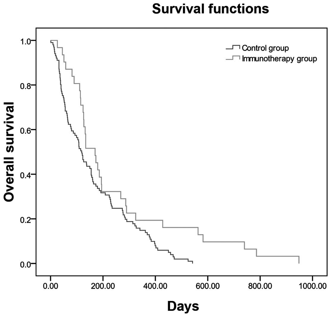
Overall survival
Overall survival was measured from the time of
enrollment to the date of death and compared between groups using
the Kaplan-Meier method (Table IV,
Fig. 1).
| Table IV.Comparison of median survival time
(MST) between the immunotherapy (I) and the non-immunotherapy (NI)
control groups. |
Table IV.
Comparison of median survival time
(MST) between the immunotherapy (I) and the non-immunotherapy (NI)
control groups.
| Groups | No. | MST, days | ΔMST, days | χ2 | P-value |
|---|
| I | 31 | 134 | 126 | 37.121 | 0.014 |
| NI | 101 | 115 |
|
|
|
Data collection and statistical
analysis
Clinical data were collected from the Inpatients
Electronic Medical Records of Tianjin Union Medicine Center,
re-analyzed and documented for use with the EpiData software,
version 3.02 (Epidata Association, Odense, Denmark). Statistical
analyses were performed using the SAS statistical software package
(SAS Institute Inc., Cary, NC, USA). The MST curves were calculated
using the Kaplan-Meier method. The associations between variables
were compared using the Pearson's Chi-square test. P<0.05 was
considered to indicate a statistically significant difference.
Results
Patient characteristics
A total of 407 patients (270 men and 137 women) were
enrolled in this study, with a median age of 66.4±11.5 years
(range, 37–93 years). Among these, 157 patients had hepatocellular
cancer, 46 had gallbladder cancer, 54 had cholangiocarcinoma and
150 had pancreatic cancer. Of the 407 patients, 124 underwent
primary tumor resection, 33 received radiotherapy, 44 received
chemotherapy and 36 received Chinese medicine treatment. All these
treatments were administered within the 3 months preceding the
initiation of immunotherapy (Table
V). The characteristics of the patients in the two groups were
well-balanced with respect to the evaluated factors, including age,
gender, diagnostic methods and tumor type. The treatment strategies
also did not differ significantly between the two groups.
| Table V.Patient characteristics. |
Table V.
Patient characteristics.
|
|
| Groups, no.
(%) |
|---|
|
|
|
|
|---|
|
Characteristics | Total (n=407) | I 77 (19.0) | NI 330 (81.0) |
|---|
| Age, years |
|
|
|
|
Range | 37–93 | 37–86 | 40–93 |
| Mean ±
SD | 66.4±11.5 | 61.6±12.2 | 67.5±11.1 |
| Gender |
|
|
|
|
Male | 270 | 52 (19.0) | 218 (81.0) |
|
Female | 137 | 25 (18.0) | 112 (82.0) |
| Tumor
location |
|
|
|
|
Hepatocellular Ca | 157 | 36 (23.0) | 121 (77.0) |
|
Gallbladder Ca | 46 | 6
(12.0) | 40 (88.0) |
|
CholangioCa | 54 | 6
(11.0) | 48 (89.0) |
|
Pancreatic Ca | 150 | 29 (19.0) | 121 (81.0) |
| Treatment baseline
immunotherapy |
|
|
|
|
Surgery | 124 | 10 (8.0) | 114 (92.0) |
|
Radiotherapy | 33 | 15 (45.0) | 18 (55.0) |
|
Chemotherapy | 44 | 21 (48.0) | 23 (52.0) |
| Chinese
herbal treatment | 36 | 23 (64.0) | 13 (36.0) |
|
DC-CIK | 77 | 77 (100.0) | 0
(0.0) |
DTH, QOL and adverse effects
A DTH skin test used to assess the immune response
to cell therapy for all patients and the results were recorded for
72 of the 77 patients in the I group. Twelve patients (16.5%) had a
strongly positive response; 12 (16.5%) had a weakly positive
response; 18 (25%) had a positive result; and 30 (42%) patients
failed to show an immune response. Thus, 42 (58%) patients in total
exhibited a positive effect. QOL was recorded for 72 of the 77
patients in the I group. Of these, 9 (13%) patients exhibited
improvements in all QOL events; 13 (18%) exhibited improvements in
3 events; another 13 (18%) exhibited improvements in 2 events; 12
(16%) exhibited improvement in 1 event; and 25 (35%) exhibited no
improvements of the QOL factors. Adverse effects were assessed in
72 of the 77 patients in the I group. Of these, 37 (51%) did not
have fever or cold symptoms and 5 (7%) developed cold symptoms. A
total of 30 patients developed fever: Low fever occurred in 10
(14%), moderate fever in 11 (15%) and high fever in 9 patients
(13%). A total of 12 patients (17%) developed anorexia; 18 (25%)
developed insomnia; 22 (31%) developed arthralgia; and 8 (11%)
developed skin rash (located at the intradermal injection site in 7
cases and on the upper as well as the lower body in 1 case;
Table III).
Comparison of the MST between the I
and NI groups
The MST differed between the I and NI groups (134
vs. 115 days, respectively; Table
IV) and it was significantly longer (19 days) in the I group
compared with that in the NI group (P=0.014). The overall survival
curves for the I and NI groups are shown in Fig. 1.
Discussion
Biological treatment will likely become a major part
of standard cancer therapy and autologous cell immunotherapy is one
of the most important tumor biological treatments. DCs were first
identified in 1973 by Steinman and Cohn, revealing the enormous
potential of these cells in immunotherapy (17). Steinman was awarded the 2011 Nobel
Prize in medicine and physiology, focusing attention once again on
DCs. DCs may prime a primary as well as a secondary immune response
against cancer and function as antigen-presenting cells (18). DCs are currently considered the most
powerful antigen-presenting cells in the body; they effectively
present tumor antigens to T lymphocytes and function as a bridge
between the tumor cells and T cells (7,19). CIK
cells exhibit strong antitumor activity and non-major
histocompatibility complex-restricted tumor-killing characteristics
(10,20). A number of studies suggest that CIK
cells co-cultured with DCs exhibit stronger tumoricidal effects and
have already produced better results in different tumor types
(21,22). CIK cells have cytotoxic properties and
are able to kill tumor cells directly as well as indirectly,
through stimulation of the host immune system. CIK cells thus have
the potential to eradicate residual cancer cells, thereby
preventing recurrence following tumor resection. These properties
have led to CIK cells being included in immunotherapy strategies
against cancer (1,23–25).
Immunotherapy is commonly used together with surgery, chemotherapy,
radiotherapy, traditional Chinese medicine and other cancer
therapies (26–29). Immune cell therapy may alleviate the
adverse effects of chemotherapy and surgery to promote the recovery
of the patient's immunity. Several cancer patients with
advanced-stage disease are unable to tolerate surgery and
radiotherapy, whereas multicycle chemotherapy may also cause severe
bone marrow suppression, drug resistance and other adverse effects.
Thus, these patients are not suitable for further chemotherapy and
are only able to receive supportive treatment. Therefore, there is
an urgent need to improve the QOL of these patients and enable them
to continue to receive effective therapy (30,31).
Adoptive cell immunotherapy provides a novel approach to solving
this problem. In our study, the MST was significantly different
between the I and NI groups. Combining immunotherapy with surgery,
radiotherapy and chemotherapy may exert a synergistic effect on
survival compared with the each method alone. Immunotherapy is a
new promising approach to cancer therapy. DC-CIK adoptive
immunotherapy bears great potential and its antitumor efficacy has
repeatedly been demonstrated in clinical practice. With the rapid
advances in molecular biology, immunology and other biological
disciplines, the therapeutic potential of DC-CIK is expected to be
further developed.
In conclusion, our findings demonstrated that
autoimmune cell therapy is a viable treatment option for HPC
patients. The QOL and overall survival of these patients were
improved, without severe adverse effects (19,23,32). The
mortality rate was lower in the I group compared with that in the
NI group and the overall survival was prolonged, proving that
immunotherapy is an effective method for cancer treatment (10,33).
Acknowledgements
The present study was partially supported by the
Tianjin Municipal Science and Technology Commission (grant no.
12ZCDZSY17100). The authors would like to thank Shanghai Claison
Biotechnology Co. Ltd, Shanghai, China, for providing the method of
preparation of the immunocytes.
Glossary
Abbreviations
Abbreviations:
|
CIK
|
cytokine-induced killer
|
|
DC
|
dendritic cell
|
|
DTH
|
delayed-type hypersensitivity
|
|
HPC
|
hepatobiliary and pancreatic
cancer
|
|
I
|
immunotherapy
|
|
NI
|
non-immunotherapy
|
|
IL-4
|
interleukin-4
|
|
MST
|
median survival time
|
|
PBMC
|
peripheral blood mononuclear cell
|
|
QOL
|
quality of life
|
References
|
1
|
Tan G, Zhang X, Feng H, Luo H and Wang Z:
The therapeutic effect of cytokine-induced killer cells on
pancreatic cancer enhanced by dendritic cells pulsed with K-ras
mutant peptide. Clin Dev Immunol. 2011:6493592011. View Article : Google Scholar : PubMed/NCBI
|
|
2
|
Wong HH and Lemoine NR: Biological
approaches to therapy of pancreatic cancer. Pancreatology.
8:431–461. 2008. View Article : Google Scholar : PubMed/NCBI
|
|
3
|
Ghanaati H, Alavian SM, Jafarian A, et al:
Imaging and imaging-guided interventions in the diagnosis and
management of hepatocellular carcinoma (HCC) - review of evidence.
Iran J Radiol. 9:167–177. 2012. View Article : Google Scholar : PubMed/NCBI
|
|
4
|
Dhanasekaran R, Limaye A and Cabrera R:
Hepatocellular carcinoma: Current trends in worldwide epidemiology,
risk factors, diagnosis and therapeutics. Hepat Med. 4:19–37.
2012.PubMed/NCBI
|
|
5
|
Pardee AD and Butterfield LH:
Immunotherapy of hepatocellular carcinoma: Unique challenges and
clinical opportunities. Oncoimmunology. 1:48–55. 2012. View Article : Google Scholar : PubMed/NCBI
|
|
6
|
Wang HY and Wang RF: Enhancing cancer
immunotherapy by intracellular delivery of cell-penetrating
peptides and stimulation of pattern-recognition receptor signaling.
Adv Immunol. 114:151–176. 2012. View Article : Google Scholar : PubMed/NCBI
|
|
7
|
Nowak M and Schmidt-Wolf IG: Natural
killer T cells subsets in cancer, functional defects in prostate
cancer and implications for immunotherapy. Cancers (Basel).
3:3661–3675. 2011. View Article : Google Scholar : PubMed/NCBI
|
|
8
|
Liu Y, Butterfield LH, Fu X, Song Z, Zhang
X, Lu C, Ding G and Wu M: Lentivirally engineered dendritic cells
activate AFP-specific T cells which inhibit hepatocellular
carcinoma growth in vitro and in vivo. Int J Oncol. 39:245–253.
2011.PubMed/NCBI
|
|
9
|
Condotta SA, Cabrera-Perez J, Badovinac VP
and Griffith TS: T-cell-mediated immunity and the role of TRAIL in
sepsis-induced immunosuppression. Crit Rev Immunol. 33:23–40. 2013.
View Article : Google Scholar : PubMed/NCBI
|
|
10
|
Wang S, Zhang H, Liu C, Jiao X, Liu D, Du
W, He Y, Zhang Z, Wu X, Wang J, et al: Human leukocyte
antigen-haploidentical donor-derived cytokine-induced killer cells
are safe and prolong the survival of patients with advanced
non-small cell lung cancer. Oncol Lett. 8:2727–2733.
2014.PubMed/NCBI
|
|
11
|
Wang M, Shi SB, Qi JL, Tang XY and Tian J:
S-1 plus CIK as second-line treatment for advanced pancreatic
cancer. Med Oncol. 30:7472013. View Article : Google Scholar : PubMed/NCBI
|
|
12
|
Charoentong P, Angelova M, Efremova M,
Gallasch R, Hackl H, Galon J and Trajanoski Z: Bioinformatics for
cancer immunology and immunotherapy. Cancer Immunol Immunother.
61:1885–1903. 2012. View Article : Google Scholar : PubMed/NCBI
|
|
13
|
Sabado RL and Bhardwaj N: Directing
dendritic cell immunotherapy towards successful cancer treatment.
Immunotherapy. 2:37–56. 2010. View Article : Google Scholar : PubMed/NCBI
|
|
14
|
Cui Y, Yang X, Zhu W, Li J, Wu X and Pang
Y: Immune response, clinical outcome and safety of dendritic cell
vaccine in combination with cytokine-induced killer cell therapy in
cancer patients. Oncol Lett. 6:537–541. 2013.PubMed/NCBI
|
|
15
|
Zhu H, Yang X, Li J, Ren Y, Zhang T, Zhang
C, Zhang J, Li J and Pang Y: Immune response, safety and survival
and quality of life outcomes for advanced colorectal cancer
patients treated with dendritic cell vaccine and cytokine-induced
killer cell therapy. Biomed Res Int. 2014:6038712014. View Article : Google Scholar : PubMed/NCBI
|
|
16
|
Niu J, Ren Y, Zhang T, Yang X, Zhu W, Zhu
H, Li J, Li J and Pang Y: Retrospective comparative study of the
effects of dendritic cell vaccine and cytokine-induced killer cell
immunotherapy with that of chemotherapy alone and in combination
for colorectal cancer. Biomed Res Int. 2014:2147272014. View Article : Google Scholar : PubMed/NCBI
|
|
17
|
Nussenzweig MC, Steinman RM, Gutchinov B
and Cohn ZA: Dendritic cells are accessory cells for the
development of anti-trinitrophenyl cytotoxic T lymphocytes. J Exp
Med. 152:1070–1084. 1980. View Article : Google Scholar : PubMed/NCBI
|
|
18
|
Salman B, Zhou D, Jaffee EM, Edil BH and
Zheng L: Vaccine therapy for pancreatic cancer. Oncoimmunology.
2:e266622013. View Article : Google Scholar : PubMed/NCBI
|
|
19
|
Tada F, Abe M, Hirooka M, Ikeda Y, Hiasa
Y, Lee Y, Jung NC, Lee WB, Lee HS, Bae YS, et al: Phase I/II study
of immunotherapy using tumor antigen-pulsed dendritic cells in
patients with hepatocellular carcinoma. Int J Oncol. 41:1601–1609.
2012.PubMed/NCBI
|
|
20
|
Ma Y, Xu YC, Tang L, Zhang Z, Wang J and
Wang HX: Cytokine-induced killer (CIK) cell therapy for patients
with hepatocellular carcinoma: Efficacy and safety. Exp Hematol
Oncol. 1:112012. View Article : Google Scholar : PubMed/NCBI
|
|
21
|
Martin CA, Kurkowski DL, Valentino AM and
Santiago-Schwarz F: Increased intracellular, cell surface and
secreted inducible heat shock protein 70 responses are triggered
during the monocyte to dendritic cell (DC) transition by cytokines
independently of heat stress and infection and may positively
regulate DC growth. J Immunol. 183:388–399. 2009. View Article : Google Scholar : PubMed/NCBI
|
|
22
|
Liu C, Suksanpaisan L, Chen YW, Russell SJ
and Peng KW: Enhancing cytokine-induced killer cell therapy of
multiple myeloma. Exp Hematol. 41:508–517. 2013. View Article : Google Scholar : PubMed/NCBI
|
|
23
|
Schmeel FC, Schmeel LC, Gast SM and
Schmidt-Wolf IG: Adoptive immunotherapy strategies with
cytokine-induced killer (CIK) cells in the treatment of
hematological malignancies. Int J Mol Sci. 15:14632–14648. 2014.
View Article : Google Scholar : PubMed/NCBI
|
|
24
|
Rettinger E, Glatthaar A, Abhari BA, et
al: SMAC mimetic BV 6 enables sensitization of resistant tumor
cells but also affects cytokine-induced killer (CIK) cells: A
potential challenge for combination therapy. Front Pediatr.
2:752014. View Article : Google Scholar : PubMed/NCBI
|
|
25
|
Jiang J, Wu C and Lu B: Cytokine-induced
killer cells promote antitumor immunity. J Transl Med. 11:832013.
View Article : Google Scholar : PubMed/NCBI
|
|
26
|
Liu H, Song J, Yang Z and Zhang X: Effects
of cytokine-induced killer cell treatment combined with FOLFOX4 on
the recurrence and survival rates for gastric cancer following
surgery. Exp Ther Med. 6:953–956. 2013.PubMed/NCBI
|
|
27
|
Zhong R, Han B and Zhong H: A prospective
study of the efficacy of a combination of autologous dendritic
cells, cytokine-induced killer cells and chemotherapy in advanced
non-small cell lung cancer patients. Tumour Biol. 35:987–994. 2014.
View Article : Google Scholar : PubMed/NCBI
|
|
28
|
Yuanying Y, Lizhi N, Feng M, Xiaohua W,
Jianying Z, Fei Y, Feng J, Lihua H, Jibing C, Jialiang L, et al:
Therapeutic outcomes of combining cryotherapy, chemotherapy and
DC-CIK immunotherapy in the treatment of metastatic non-small cell
lung cancer. Cryobiology. 67:235–240. 2013. View Article : Google Scholar : PubMed/NCBI
|
|
29
|
Wang YY, Wang YS, Liu T, Yang K, Yang GQ,
Liu HC, Wang SS and Yang JL: Efficacy study of CyberKnife
stereotactic radiosurgery combined with CIK cell immunotherapy for
advanced refractory lung cancer. Exp Ther Med. 5:453–456.
2013.PubMed/NCBI
|
|
30
|
Niu LZ, Li JL, Zeng JY, Mu F, Liao MT, Yao
F, Li L, Liu CY, Chen JB, Zuo JS, et al: Combination treatment with
comprehensive cryoablation and immunotherapy in metastatic
hepatocellular cancer. World J Gastroenterol. 19:3473–3480. 2013.
View Article : Google Scholar : PubMed/NCBI
|
|
31
|
Li W, Xu LP, Di Zhao L, Wang L, Zhang Y,
Gao QL and Mai L: Cytokine-induced killer cell therapy for advanced
pancreatic adenocarcinoma: A case report and review of the
literature. Oncol Lett. 5:1427–1429. 2013.PubMed/NCBI
|
|
32
|
Nakamoto Y, Mizukoshi E, Tsuji H, Sakai Y,
Kitahara M, Arai K, Yamashita T, Yokoyama K, Mukaida N, Matsushima
K, et al: Combined therapy of transcatheter hepatic arterial
embolization with intratumoral dendritic cell infusion for
hepatocellular carcinoma: Clinical safety. Clin Exp Immunol.
147:296–305. 2007. View Article : Google Scholar : PubMed/NCBI
|
|
33
|
Jiang JT, Shen YP, Wu CP, Zhu YB, Wei WX,
Chen LJ, Zheng X, Sun J, Lu BF and Zhang XG: Increasing the
frequency of CIK cells adoptive immunotherapy may decrease risk of
death in gastric cancer patients. World J Gastroenterol.
16:6155–6162. 2010. View Article : Google Scholar : PubMed/NCBI
|