Introduction
Alzheimer’s disease (AD) is a prominent
neurodegenerative disorder characterized by progressive loss of
memory and other cognitive functions. Despite considerable progress
in genetics and cell biology, numerous questions remain regarding
the mechanisms of neurodegeneration and the molecular and
pathological components. Extracellular amyloid-β (Aβ), which is
derived from the larger amyloid precursor protein (APP), is
considered to be responsible for the death of neurons and dementia
in Alzheimer’s disease. An increased expression of APP may increase
the risk of AD (1,2). APP levels can be regulated at the
genomic, transcriptional or translational level, and in the rate of
degradation. Genetic variants in the APP promoter increase APP
transcription by 2–3 fold and have been reported to increase the
risk of AD (2). APP can be
processed by a group of secretases where α-secretase produces
soluble fragments, whereas β- and γ-secretase generate Aβ from APP
(3). Previous research has
suggested that Aβ regulates neuronal and synaptic activity, and
accumulation of Aβ in the brain causes a combination of aberrant
network activity and synaptic depression (4).
MicroRNAs (miRs) are endogenous, short, noncoding
RNAs. Mature miRs are single-stranded RNA molecules of ~20–25
nucleotides which act as important post-transcriptional regulators
of gene expression by binding with their target mRNAs. They are
additionally essential for normal neuronal function and survival
(3,4). Several miRs have been shown to be
important in neuropathology by downregulating AD-related proteins,
including APP and BACE-1. It has been demonstrated that miR-16,
-101, -106a/b, -147 and -160a could function as APP suppressors
(5–8).
Several cerebral spinal fluid (CSF) or blood-based
markers, such as Aβ, tau and phosphorylated tau (p-tau) are
proposed biomarkers for predicting future cognitive decline in
healthy individuals, and the progression to dementia in patients
who are cognitively impaired (9–11).
Previous studies have shown that APP and BACE-1 are novel markers
of AD (12–14). However, there is still an urgent
requirement for additional biomarkers that can detect AD in the
pre-dementia phase (9–14). Alterations to the regulation of
miRs in the blood and CSF may indicate progression in AD (15,16).
Exosomes are membrane vesicles with a size of 40–100 nm that are
released from numerous cell types of the body (17). Recent studies have shown that, in
addition to functional proteins, exosomes carry mRNA as well as
miRs (18). In functional terms,
exosomes are considered to represent a novel mechanism of
intercellular communication. This may be due to the uptake of
exosomes by target cells or by triggering cell signaling through
membrane receptors (17–19). In a previous study, we found that
miR-193b was decreased in the hippocampi of 9-month-old APP/PS1
double-transgenic mice by miR array. In the present study,
bioinformatic analyses showed that miR-193b may potentially target
the 3′-untranslated region (UTR) of APP and the effects of miR-193b
on the expression of APP were studied. Blood and CSF derived
exosomal miR-193b was detected in APP/PS1 double-transgenic mice,
and patients with mild cognitive impairment (MCI) and dementia of
Alzheimer type (DAT).
Materials and methods
Study population
The design of the present study was approved by the
ethics committee of Xuanwu Hospital of Capital Medical University
(Beijing, China), and the written informed consents were obtained
from all participants. A total of 43 MCI (23 females, 20 males,
mean age 63.8±6.1) and 51 DAT patients (28 females, 23 males, mean
age 64.2±6.5) were selected for this study. The CSF was drawn
within 2 h following collection of blood (n=7). Age- and
gender-matched control subjects were included in the experimental
design. Samples were stored at −80°C until required for further
analysis. The expression levels of Aβ, tau and p-tau in the plasma
and CSF of the subjects were determined by ELISA kit from Cusabio
(Wuhan, China). The study was approved by the Ethics Committee of
Xuanwu Hospital of Capital Medical University (Beijing, China).
Written informed consent was obtained from the patients’
family.
APP/PS1 double-transgenic and wild-type
mice
The 3, 6 and 9-month-old APP/PS1 double-transgenic
mice on a C57BL/6J genetic background were purchased from the
Institute of Laboratory Animal Science, Chinese Academy of Medical
Sciences & Comparative Medical Center (Beijing, China). All the
animal protocols were approved by Ethics Committee of Xuanwu
Hospital of Capital Medical University (Beijing, China). The
non-transgenic mice were used as wild-type (WT) controls. Blood was
taken by removing the eyeballs and CSF-like fluid was collected as
previously described (20). The
hippocampi were isolated as described for miR-193b quantitative
polymerase chain reaction (qPCR) detection (n=5). The samples were
stored in liquid nitrogen until analysis.
Cell culture and miR transfections
SH-SY5Y and HEK293 cell lines were purchased from
Shanghai Institute of Cell Biology, China. Cells were grown in
antibiotic-free Dulbecco’s modified Eagle’s medium (DMEM)
supplemented with 10% fetal bovine serum (FBS) at 37°C with 5%
CO2. SH-SY5Y cells were transfected with 100 nM (final
concentration) miR-193b mimic oligonucleotide, miR-193b inhibitor
oligonucleotide or a non-specific control small interfering RNA
siRNA) (GenePharma) using Lipofectamine™ 2000 reagent (Invitrogen
Life Sciences, Carlsbad, CA, USA) according to the manufacturer’s
instructions.
Reporter vectors and DNA constructs
Reporter vectors containing the putative miR-193b
target site from the APP 3′-UTR was synthesized with
double-stranded oligos perfectly complementary to putative miR-193b
target site and oligos in which the seed regions were mutated
(21). The APP oligos had the
sequence (seed region bolded) as follows: 5′-CCCAAGCTTTCACATA
GCCCCTTAGCCATTTAAGCTTGGG-3′; 3′-GGGT
TCGAAAGTGTATCGGGGAATCGGTAAATTCGAACC-5′. The mutant APP
target oligos had nucleotides 3–6 of the seed region mutated
(italicized): 5′-CCCAAGCTTTCAC
ATAGCCCCTTAACTAGTTAAGCTTGGG-3′;
3′-GGGTTC
GAAAGTGTATCGGGGAATTGATCAATTCGAACCC-5′.
HEK-293 cells were plated in 24-well plates. The following day,
cells were transfected with a miR mimic oligonucleotide, reporter
vectors bearing either the miR target sequence or the miR seed
region mutant target sequence, and one tenth of the molar volume of
pRL-SV40, a Renilla luciferase control vector. Arrest-In
transfection reagent (Open Biosystems Inc; GE healthcare,
Fairfield, CT, USA) was used; any differences in transfection
efficiency were accounted for by measuring Renilla luciferase
activity. Following 48 h posttransfection, cells were lysed using
100 μl of GLB (Glo Lysis Buffer, Promega Corporation, Madison, WI,
USA). Firefly and Renilla luciferase activities were measured using
a dual luciferase reporter assay kit (Promega), according to the
manufacturer’s instructions. Firefly luciferase activity was
normalized to the Renilla luciferase activity.
Isolation of RNA and APP mRNA qPCR
analysis
Total RNA from harvested cells was isolated using
TRIzol™ Reagent (Invitrogen Life Sciences) according to the
manufacturer’s instructions. Isolated RNA was reverse transcribed
using the PrimeScript™ RT reagent (Takara Bio, Inc., Shiga, Japan).
The mRNA expressions of APP was determined using SYBR®
Green qPCR (Takara Bio, Inc) using a LightCycler 480 System (Roche
Diagnostics, Mannheim, Germany). GAPDH was used to normalize the
target genes. The PCR primer sequences were as follows: APP,
forward, 5′-TTGCGAAACTCATCTTCACTGG-3′, reverse
5′-CAGTGGGCAACACACAAACTCTAC-3′; GAPDH, forward,
5′-GCACCGTCAAGGCTGAGAAC-3′, reverse 5′-TGGTGAAGACGCCAGTGGA-3′. The
number of samples in each group was five.
Western blots
Western blotting was performed as previously
described (22). Cells were
homogenized in extraction buffer, then incubated at 0°C for 15 min
and centrifuged at 2,000 × g for 10 min at 4°C. Aliquots of the
supernatants were used for the measurement of the total protein
content. Samples were boiled for 5 min in loading buffer and then
the proteins (30 μg per well) were separated by 10% SDS-PAGE
(Bio-Rad, Hercules, CA, USA). Proteins in the gel were transferred
onto a nitrocellulose membrane (Pall Corporation, New York, NY,
USA). Membranes were incubated with anti-APP (diluted 1:500; Abcam,
Cambridge, UK), and anti-GAPDH (diluted 1:400, Abcam) at room
temperature for 1.5 h, respectively. Membranes were then washed and
incubated with anti-IgG antibody linked to horseradish peroxidase
at room temperature for 1 h. The membranes were then incubated with
substrate for peroxidase and enhanced chemiluminescence (KPL,
Gaithersburg, MD, USA) for 1 min and exposed immediately to X-ray
film for 1–5 min. Films were then revealed in the conventional
manner. The amount of each protein was measured by densitometric
analysis and standardized relative to GAPDH. The number of samples
in each group was five.
Isolation of exosomes
The exosomes were isolated using the Total Exosome
Isolation kit (Invitrogen Life Technologies) according to the
manufacturer’s instructions. Briefly, the serum was centrifuged at
2,000 × g for 30 min to remove cells and debris. Following this,
400 μl clarified serum was transferred to a new tube and 0.4
volumes of the Total Exosome Isolation reagent was added. The
serum/reagent solution was mixed and then incubated at 4°C for 30
min. After incubation, the samples were centrifuged at 10,000 × g
for 10 min at room temperature. The supernatant was discarded and
the pellet, containing the exosomes, at the bottom of the tube was
resuspended in 200 μl phosphate-buffered saline (PBS).
Isolation of RNA and miR-193b qPCR
analysis
Total RNA in 350 μl CSF, or 200 μl serum samples was
extracted using a spin column method with an miRNeasy Serum/Plasma
kit (Qiagen, Hilden, Germany) according to the manufacturer’s
instructions. Exosomal RNA was isolated and purified using a Total
Exosome RNA Isolation kit (Invitrogen Life Technologies). The RNA
isolated from the CSF was 530–1,500 ng/ml, 2,100–4,700 ng/ml from
serum, 270–420 ng/ml from the exosome. Total RNA in hippocampus
tissue from the animal models and cultured cells was extracted
using a spin column method using the miRNeasy kit (Qiagen). MiRs
were reverse transcribed into cDNA using the miScript II RT kit
(Qiagen) in a 10 μl reaction system. MiR-193b were determined by a
TaqMan qPCR method (Applied Biosystems, Foster City, CA, USA),
using U6 RNA as an endogenous control.
Statistical analyses
Statistical analyses were performed using SPSS 13.0
for Windows (SPSS, Inc., Chicago, IL, USA). For normally
distributed data, results are expressed as the mean ± standard
deviation. The differences between groups were assessed by
t-tests and analyzed using the Mann-Whitney U-test.
Correlations were determined by computing the Spearman rank
correlation coefficient. P<0.05 was considered to indicate a
statistically significant difference.
Results
Bioinformatics retrieval
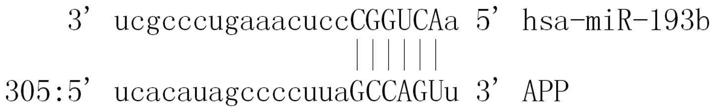
A total of 34 miRs were found to be putative targets
on the 3′-UTR of APP. MiR-193b was a miR that may target the
3′-UTRs of APP (Fig. 1).
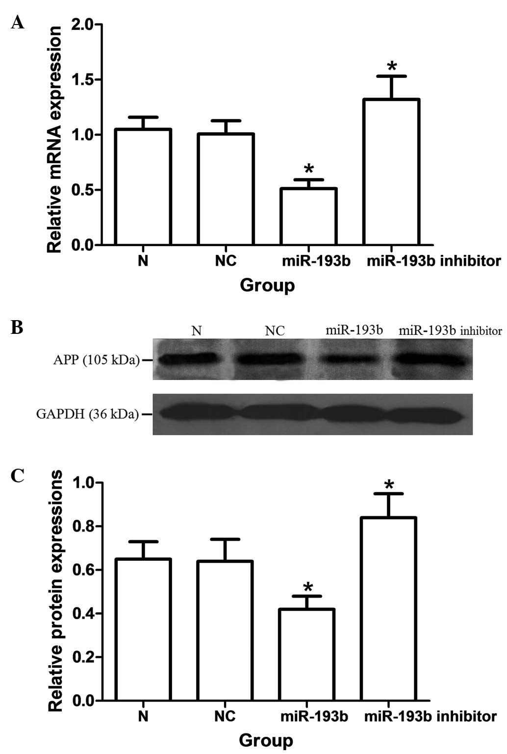
MiR-193b represses the mRNA and protein
expression of APP
As shown in Fig. 2,
the mRNA and protein expressions of APP were significantly
decreased by miRNA-193b in SH-SY5Y cells (P<0.05). The miR-193b
inhibitor oligonucleotide induced a significant upregulation of
mRNA and protein expression of APP as compared with the nonspecific
control groups, respectively (P<0.05). By TaqMan qPCR, a ~55%
downregulation of endogenous miR-193b was observed (data not
shown).
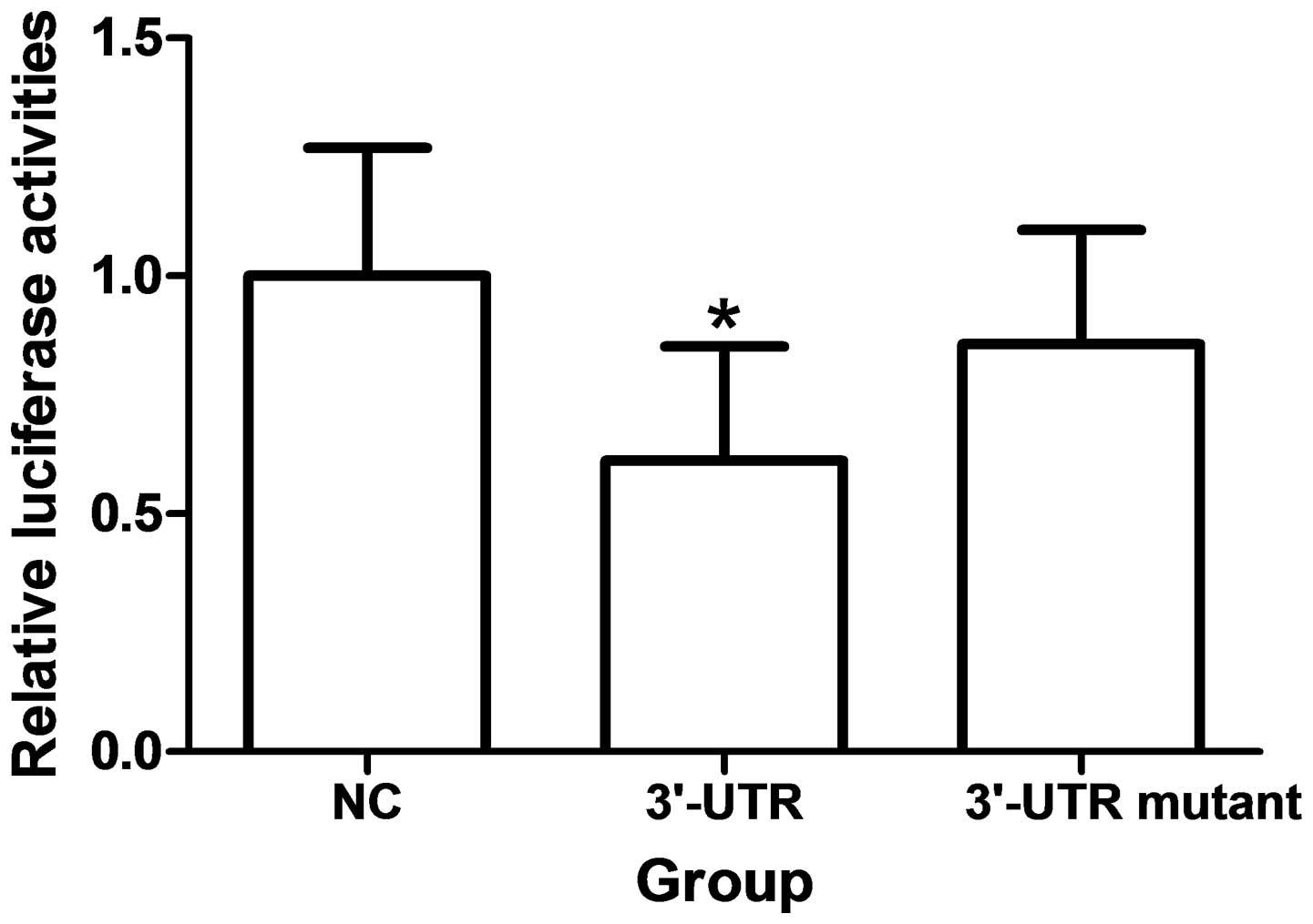
APP 3′-UTR is a miR-193b target
Over-expression of miR-193b significantly reduces
fluorescence from APP reporter vectors in HEK293 cells (P<0.05).
These reductions were not observed when mutations were made to the
3′UTR seed regions of APP or BACE-1 were generated (Fig. 3).
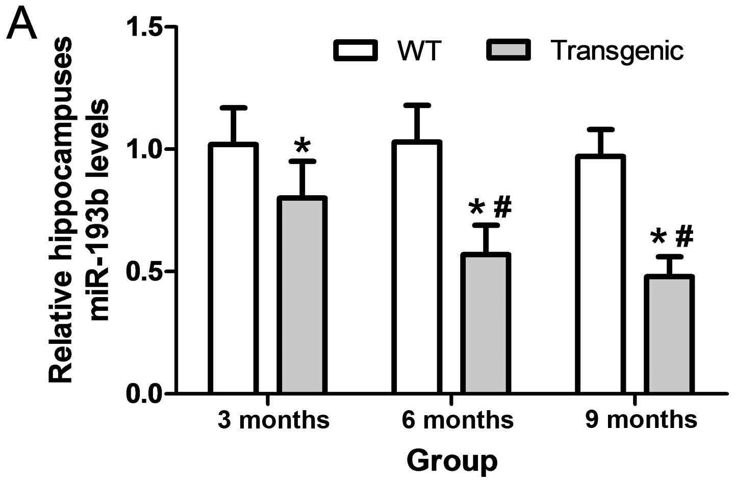
Exosomal miR-193b decreases in CSF-like
fluid and serum of transgenic mice
The levels of miR-193b were significantly
downregulated in the hippocampi of 3, 6 and 9 month APP/PS1
transgenic mice as compared with the WT mice (P<0.05). The
levels of exosomal miR-193b were significantly downregulated in the
CSF-like fluid and the serum of 3, 6 and 9 month APP/PS1 transgenic
mice, as compared with the WT mice (P<0.05). Levels of exosomal
miR-193b in the CSF-like fluid and serum of 6 and 9 month
transgenic mice were significantly lower than that of the 3 month
transgenic mice, respectively (P<0.05; Fig. 4).
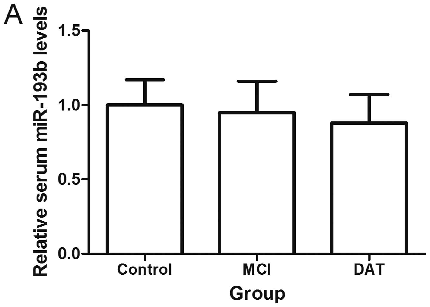
Exosomal miR-193b decreases in the CSF,
serum and plasma of AD patients
Compared with the control groups, patients with MCI
and DAT had lower levels of exosomal miR-193b in the serum and
plasma (P<0.05). Patients with DAT had lower exosomal miR-193b
levels in their serum and plasma compared with the MCI groups
(P<0.05). It was additionally found that exosomal miR-193b was
decreased in the CSF of patients with DAT as compared with the
control group (n=7; P<0.05; Fig.
5).
The level of exosomal miR-193b was lower in the CSF
compared with serum from a given individual (P<0.05; data not
shown). There was no correlation between exosomal miRNA-193b levels
and the CSF and serum from a given individual (data not shown).
When the cutoff value was set as the mean concentration - two
standard deviations of the controls, the positive rates of exosomal
miR-193b were 71.43% (5/7) and 58.82% (30/51) in the CSF and serum
of patients with DAT respectively, and 58.14% (25/43) in the serum
of patients with MCI.
Exosomal miR-193b is negatively
correlated with Aβ42 in the CSF but not in the serum
A weak, but significant, negative correlation was
found between the levels of exosomal miR-193b and Aβ42 in the CSF
of patients with DAT (r =−0.442, P<0.05), as well as the control
group (r =−0.503, P<0.05). MiR-193b and exosomal miR-193b had no
correlation with HCY, ApoE, tau and p-tau in the serum and CSF
(data not shown).
Discussion
The potential benefit in the analysis of miR in the
diagnosis and treatment of numerous diseases, including cancer,
infection and neurodegenerative disease has been previously
evaluated by numerous researchers (6,23,24).
The expression profiles of miRs are known to be altered in several
regions of the AD brain, however the cause or consequence in the
pathology of the disease is unknown. There has been no data to
suggest a direct genetic link between miRs or miR recognition
elements and neurodegenerative disease (6,9,10,23,24).
High expression of APP correlating with accelerated accumulation of
Aβ in the brain is a feature of AD. Previous research has
demonstrated miR-106a/b, -147, -160a, -520c and -655 function as
APP suppressors (5–8). In the present study, it was shown
that miR-193b could repress the expression of APP by binding its
3′-UTR and decay its mRNA. The deregulated miR-193b may play a role
in the development of AD and the downregulating effects of miR-193b
on APP may represent a study direction for AD therapy by miR.
Previous studies have demonstrated that miRs are
stably expressed in various bodily fluids, and their unique
expression patterns can serve as fingerprints of various diseases,
including AD (25). The CSF is in
direct contact with the extracellular space of the brain and can
reflect biochemical changes that occur in the latter. The CSF is
therefore the optimal source of AD biomarkers. CSF, however, is not
an appropriate sample for screening and routine testing due to its
invasive process of sample collection. From this perspective, blood
based biomarkers for AD screening and routine testing would be more
suitable (26). Circulating miRs
can derive from many sources, including cell death and lysed cells,
which are passive secretions, as well as active secretion from
cells by exosomes and microvesicles. Although endogenous plasma
miRNAs exist in a form that is resistant to plasma RNase activity
and other conditions such as extreme pH, the exosomes may provide
additional protection, which allows data to be obtained that
reflects the accurate expression level of miR actively secreted
from cells. The miRs can also be transported by other carriers such
as high and low density lipoprotein, both of which are highly
abundant in plasma and cannot be generated by brain (27). The total small RNAs isolated from
body fluid may contain more non-brain-derived miRs than that from
exosomes. Furthermore, exosomes are actively secreted from cells,
which can help to eliminate the interference from passively
secreted miRs.
APP/PS-1 double-transgenic mice contained insoluble
amyloid peptides at the age of 6–9 months, concomitant with the
formation of amyloid plaques (28). In the present study, it was shown
that the level of miR-193b was decreased in the hippocampi of 3
month transgenic mice, which suggested that the change of miR-193b
is earlier than the formation of amyloid plaques. The detection of
exosomal miR-193b in the CSF-like fluid and plasma of 3, 6 and 9
month transgenic mice demonstrated that exosomal miR-193b was a
potential AD biomarker, especially for the earlier stages of the
disease. This conjecture was further confirmed by clinical
detection, which showed that exosomal miR-193b levels in patients
with MCI were higher as compared with the control group and lower
as compared with the CSF and plasma samples from patients in the
DAT group. Although exosomes can be released by numerous organs,
the expression of miR-193b in the exosomes from CSF was ~73% of the
endogenous control, which indicated that it was abundant, and the
exosomal miR-193b levels in the CSF were correlated. Furthermore,
exosome-mediated secretion pathways exist in the blood brain
barrier (29,30). It is hypothesized that decreased
secretion of exosomal miR-193b may lead to the decreased level of
exosomal miR-193b in the plasma.
In conclusion, these findings showed that miR-193b
may function in the development of AD and exosomal miR-193b has
potential as a novel, noninvasive blood-based biomarkers of
patients with MCI and DAT.
Acknowledgements
This study was supported by the Natural Science
Foundation of China (no. 81271924) and Research Fund for the
Doctoral Program of Higher Education of China (no. 20121107110001).
The authors would like to thank Dr Shuang Meng of the Chinese
Center for Disease Control and Prevention, Beijing, People’s
Republic of China for the vector construction and fluorescence
detection.
References
|
1
|
Lewczuk P, Kamrowski-Kruck H, Peters O, et
al: Soluble amyloid precursor proteins in the cerebrospinal fluid
as novel potential biomarkers of Alzheimer’s disease: a multicenter
study. Mol Psychiatry. 15:138–145. 2010.
|
|
2
|
Weiner MW: Dementia in 2012: Further
insights into Alzheimer disease pathogenesis. Nat Rev Neurol.
2:65–66. 2013. View Article : Google Scholar : PubMed/NCBI
|
|
3
|
Singer O, Marr RA, Rockenstein E, et al:
Targeting BACE1 with siRNAs ameliorates Alzheimer disease
neuropathology in a transgenic model. Nat Neurosci. 8:1343–1349.
2005. View
Article : Google Scholar : PubMed/NCBI
|
|
4
|
Tan L, Yu JT, Hu N and Tan L: Non-coding
RNAs in Alzheimer’s disease. Mol Neurobiol. 47:382–393. 2013.
|
|
5
|
Junn E and Mouradian MM: MicroRNAs in
neurodegenerative diseases and their therapeutic potential.
Pharmacol Ther. 133:142–150. 2012. View Article : Google Scholar : PubMed/NCBI
|
|
6
|
Delay C, Mandemakers W and Hébert SS:
MicroRNAs in Alzheimer’s disease. Neurobiol Dis. 46:285–290.
2012.
|
|
7
|
Hébert SS and De Strooper B: Alterations
of the microRNA network cause neurodegenerative disease. Trends
Neurosci. 32:199–206. 2009.PubMed/NCBI
|
|
8
|
Long JM and Lahiri DK: MicroRNA-101
downregulates Alzheimer’s amyloid-β precursor protein levels in
human cell cultures and is differentially expressed. Biochem
Biophys Res Commun. 404:889–895. 2011.PubMed/NCBI
|
|
9
|
Delay C, Calon F, Mathews P and Hébert SS:
Alzheimer-specific variants in the 3′UTR of Amyloid precursor
protein affect microRNA function. Mol Neurodegener.
6:702011.PubMed/NCBI
|
|
10
|
Hébert SS, Papadopoulou AS, Smith P, et
al: Genetic ablation of Dicer in adult forebrain neurons results in
abnormal tau hyperphosphorylation and neurodegeneration. Hum Mol
Genet. 19:3959–3969. 2010.PubMed/NCBI
|
|
11
|
Blennow K, Hampel H, Weiner M and
Zetterberg H: Cerebrospinal fluid and plasma biomarkers in
Alzheimer disease. Nat Rev Neurol. 6:131–144. 2010. View Article : Google Scholar : PubMed/NCBI
|
|
12
|
Rembach A, Faux NG, Watt AD, et al: AIBL
research group, Changes in plasma amyloid beta in a longitudinal
study of aging and Alzheimer’s disease. Alzheimers Dement.
10:53–61. 2014.PubMed/NCBI
|
|
13
|
Lewczuk P, Kamrowski-Kruck H, Peters O, et
al: Soluble amyloid precursor proteins in the cerebrospinal fluid
as novel potential biomarkers of Alzheimer’s disease: a multicenter
study. Mol Psychiatry. 15:138–145. 2010.
|
|
14
|
Zetterberg H, Andreasson U, Hansson O, et
al: Elevated cerebrospinal fluid BACE1 activity in incipient
Alzheimer disease. Arch Neurol. 65:1102–1107. 2008. View Article : Google Scholar : PubMed/NCBI
|
|
15
|
Wang WX, Huang Q, Hu Y, Stromberg AJ and
Nelson PT: Patterns of microRNA expression in normal and early
Alzheimer’s disease human temporal cortex: white matter versus gray
matter. Acta Neuropathol. 2:193–205. 2011.PubMed/NCBI
|
|
16
|
Schonrock N, Ke YD, Humphreys D, et al:
Neuronal microRNA deregulation in response to Alzheimer’s disease
amyloid-beta. PLoS One. 5:e110702010.PubMed/NCBI
|
|
17
|
Hosseini HM, Fooladi AA, Nourani MR and
Ghanezadeh F: The role of exosomes in infectious diseases. Inflamm
Allergy Drug Targets. 1:29–37. 2013. View Article : Google Scholar : PubMed/NCBI
|
|
18
|
Pant S, Hilton H and Burczynski ME: The
multifaceted exosome: biogenesis, role in normal and aberrant
cellular function, and frontiers for pharmacological and biomarker
opportunities. Biochem Pharmacol. 11:1484–1494. 2012. View Article : Google Scholar : PubMed/NCBI
|
|
19
|
Boon RA and Vickers KC: Intercellular
transport of microRNAs. Arterioscler Thromb Vasc Biol. 33:186–192.
2013. View Article : Google Scholar : PubMed/NCBI
|
|
20
|
Wei PC, Tsai CH, Chiu PS and Lai SC:
Matrix metalloproteinase-12 leads to elastin degradation in BALB/c
mice with eosinophilic meningitis caused by Angiostrongylus
cantonensis. Int J Parasitol. 41:1175–1183. 2011. View Article : Google Scholar
|
|
21
|
Wang WX, Rajeev BW and Stromberg AJ: The
expression of microRNA miR-107 decreases early in Alzheimer’s
disease and may accelerate disease progression through regulation
of beta-site amyloid precursor protein-cleaving enzyme 1. J
Neurosci. 5:1213–1223. 2008.PubMed/NCBI
|
|
22
|
Liu CG, Xu KQ, Xu X, et al:
17Beta-oestradiol regulates the expression of
Na+/K+-ATPase beta1-subunit, sarcoplasmic
reticulum Ca2+-ATPase and carbonic anhydrase iv in H9C2
cells. Clin Exp Pharmacol Physiol. 34:998–1004. 2007.PubMed/NCBI
|
|
23
|
Dassow H and Aigner A: MicroRNAs (miRNAs)
in colorectal cancer: from aberrant expression towards therapy.
Curr Pharm Des. 19:1242–1252. 2013.PubMed/NCBI
|
|
24
|
Eulalio A, Schulte L and Vogel J: The
mammalian microRNA response to bacterial infections. RNA Biol.
9:742–750. 2012. View Article : Google Scholar : PubMed/NCBI
|
|
25
|
Mitchell PS, Parkin RK, Kroh EM, Fritz BR
and Wyman SK: Circulating microRNAs as stable blood-based markers
for cancer detection. Proc Natl Acad Sci USA. 105:10513–10518.
2008. View Article : Google Scholar : PubMed/NCBI
|
|
26
|
Giedraitis V, Sundelöf J, Irizarry MC, et
al: The normal equilibrium between CSF and plasma amyloid beta
levels is disrupted in Alzheimer’s disease. Neurosci Lett.
427:127–131. 2007.PubMed/NCBI
|
|
27
|
Vickers KC, Palmisano BT, Shoucri BM,
Shamburek RD and Remaley AT: MicroRNAs are transported in plasma
and delivered to recipient cells by high-density lipoproteins. Nat
Cell Biol. 4:423–433. 2011. View
Article : Google Scholar : PubMed/NCBI
|
|
28
|
van Leuven F: Single and multiple
transgenic mice as models for Alzheimer’s disease, Prog. Neurobiol.
61:305–312. 2000.
|
|
29
|
Ceruti S, Colombo L, Magni G, et al:
Oxygen-glucose deprivation increases the enzymatic activity and the
microvesicle-mediated release of ectonucleotidases in the cells
composing the blood-brain barrier. Neurochem Int. 59:259–271. 2011.
View Article : Google Scholar
|
|
30
|
Ma R, Jiang T and Kang X: Circulating
microRNAs in cancer: origin, function and application. J Exp Clin
Cancer Res. 31:382012. View Article : Google Scholar : PubMed/NCBI
|