Introduction
Angelicae dahuricae is a perennial plant that
grows naturally throughout large areas of China. It has a strong
scent, and its leaves are used to make incense (1). Angelicae dahuricae radix is
the dried root of Angelica dahurica Bentham et Hooker and
Angelica dahurica (Fisch. ex Hoffm). Benth. et Hook. f. var.
formosana (Boiss). Shan et Yuan, known as Bai Zhi in
Chinese, and is used in traditional Chinese medicine to treat
various diseases (2).
Angelicae dahuricae radix has been used for
the treatment of colds, headaches, rhinitis and psoriasis in
traditional medicine (3). Research
has been performed on the anti-inflammatory, analgesic,
antipyretic, antioxidant and cytochrome P450 activity of
Angelicae dahuricae radix (4–6).
Furthermore, Angelicae dahuricae radix has been suggested
for use in the treatment of oral diseases, including toothache
(3). Limited information is
currently available regarding the effects of Angelicae
dahuricae radix on dental tissue, and there is no information
on its effects on the mesenchymal stem cells derived from the
gingiva.
The aim of the present study was to evaluate the
effects of extracts of Angelicae dahuricae radix on the
morphology and viability of human stem cells derived from the
gingiva. To the best of our knowledge, this investigation is the
first to elucidate the effect of Angelicae dahuricae radix
on stem cells derived from the gingiva.
Materials and methods
Preparation of the materials
The dry roots of Angelica dahurica Bentham et
Hooker (500 g) were immersed in 2,000 ml distilled water for 2 h
and boiled under reflux for 2 h 30 min. The resulting extract was
centrifuged at 5,000 × g for 10 min. The supernatant was
concentrated to 300 ml using a rotary evaporator under reduced
pressure (Eyela NE-1001, Tokya Rikakikai Co. Ltd., Tokyo, Japan).
The concentrates were then freeze-dried in a lyophilizer (Labconco,
Kansas, MO, USA) to obtain 182.5 g of solid residue, resulting in a
yield of 36.5% (w/w).
Isolation and culture of the stem cells
derived from the gingival
Healthy gingival tissues were obtained from four
healthy patients undergoing crown-lengthening procedures. This
study was reviewed and approved by the Institutional Review Board
of Seoul St. Mary’s Hospital, College of Medicine, The Catholic
University of Korea (Seoul, Republic of Korea; KC11SISI0348), and
informed consent was obtained from all participants.
The tissues were immediately placed in sterile
phosphate-buffered saline (PBS, Welgene, Daegu, Korea) with 100
U/ml penicillin and 100 μg/ml streptomycin (Sigma-Aldrich,
St. Louis, MO, USA) at 4°C. The gingival tissue was
de-epithelialized, minced, digested with collagenase IV
(Sigma-Aldrich) and incubated at 37°C in a humidified incubator
with 5% CO2 and 95% O2. The non-adherent
cells were washed with PBS after 24 h, replaced with fresh medium,
and fed every 2–3 days.
Evaluation of stem cell morphology
The stem cells were plated at a density of
2.0×10³ cells/well in 96-well plates. The cells were
incubated in minimum Essential medium α (α-MEM, Gibco, Grand
Island, NY, USA) that was composed of 15% fetal bovine serum
(Gibco), 100 U/ml penicillin and 100 μg/ml streptomycin
(Sigma-Aldrich), 200 mM L-Glutamine (Sigma-Aldrich) and 10 mM
ascorbic acid 2-phosphate (Sigma-Aldric) in the presence of the
Angelicae dahuricae radix at final concentrations that
ranged from 0.001 to 100 μg/ml [0 (control), 0.001, 0.01,
0.1, 1, 10, and 100 μg/ml]. The morphology of the cells was
viewed under an inverted microscope (Leica DM IRM, Leica
Microsystems, Wetzlar, Germany) on days 1, 3 and 7. The images were
saved as JPEG fles.
Determination of cell proliferation
The analysis of cell proliferation was performed on
days 1, 3 and 7. Viable cells were identified using a cell counting
kit-8 (CCK-8, Dojindo, Tokyo, Japan) assay. The spectrophotometric
absorbance was measured with a microplate reader (BioTek, Winooski,
VT, USA), and the analysis was performed in triplicate.
Statistical analysis
The findings are represented as the mean ± standard
deviation of the experiments. Analysis of normality was performed,
and a one-way analysis of variance (ANOVA) with post hoc test was
performed to determine the differences between the groups using a
commercially available program (SPSS 12 for Windows, SPSS Inc.,
Chicago, IL, USA). P<0.05 was considered to indicate a
statistically significant difference.
Results
Evaluation of cell morphology
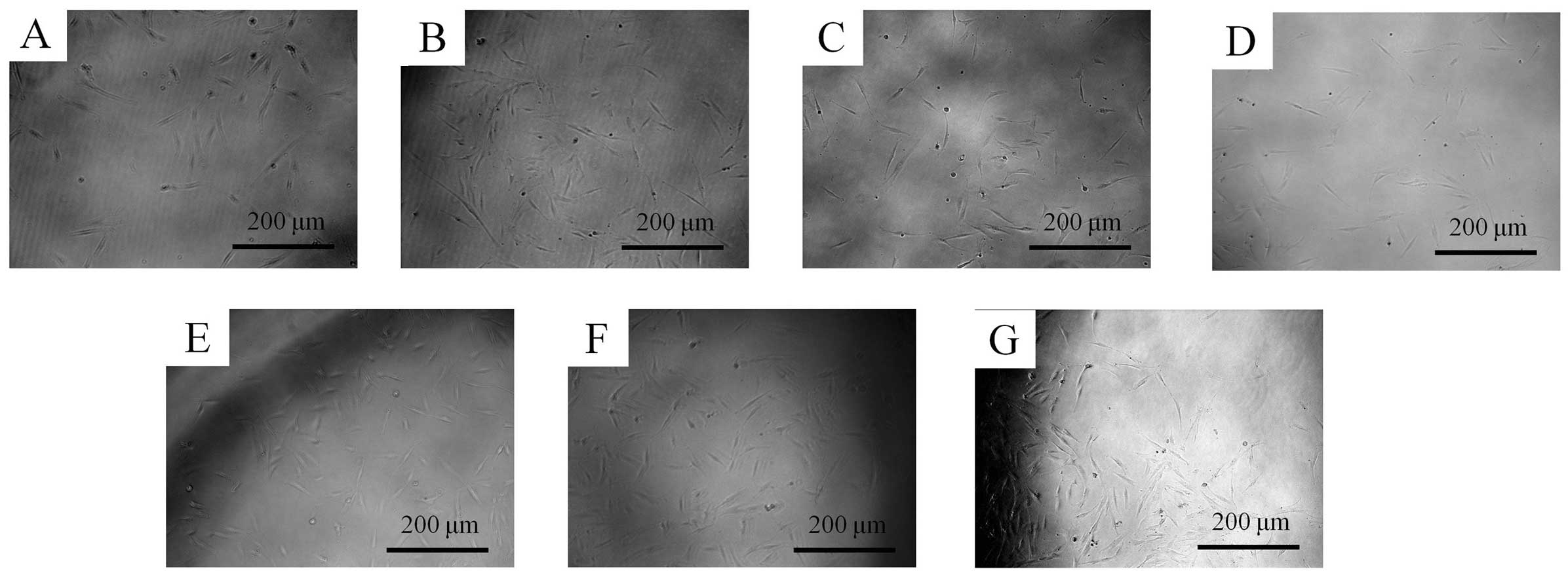
The morphology of the stem cells at day 1 is shown
in Fig. 1. Under optical
microscopy, the control group cells had a spindle-shaped,
fibroblast-like morphology. The shapes of the cells treated with
0.001, 0.01, 0.1, 1, 10, and 100 μg/ml Angelicae
dahuricae radix were similar to the shapes of the cells in the
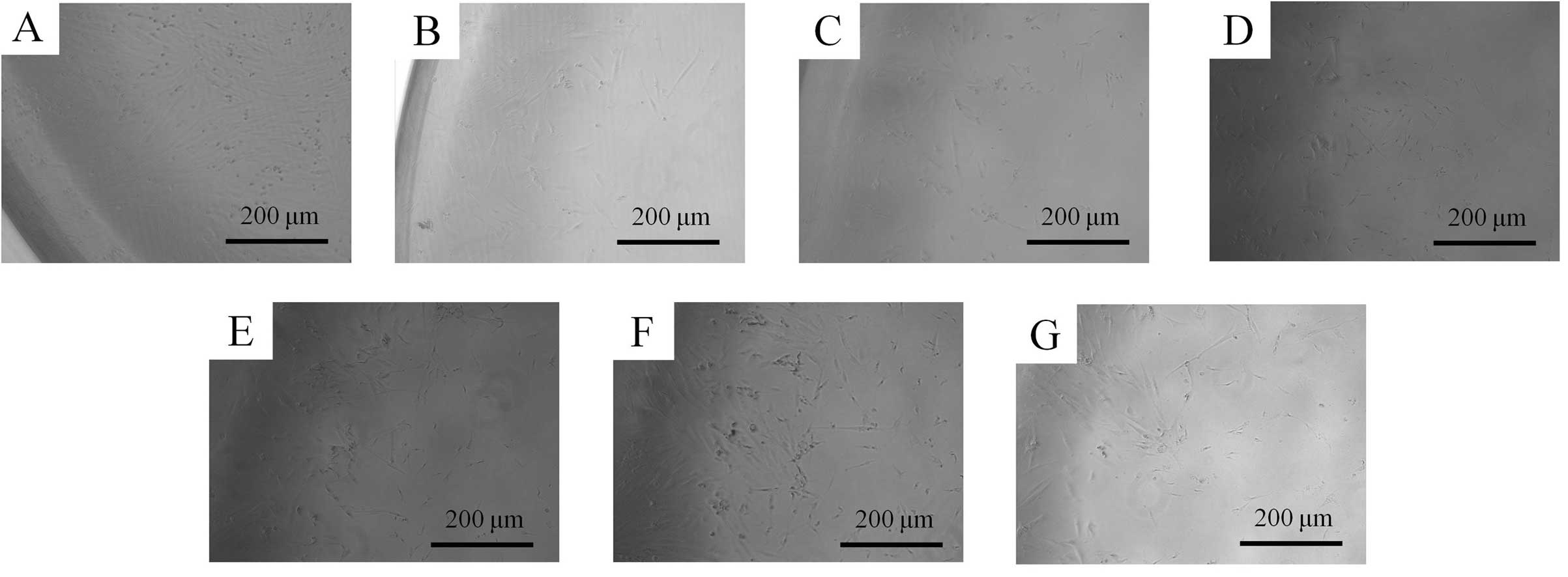
control group. The morphology of the cells on day 3 is shown in
Fig. 2. The shapes of the cells
treated with 0.001, 0.01, 0.1, 1, 10, and 100 μg/ml were
similar to those of cells in the control group. No significant
alterations were noted in the treated groups (0.001 to 100
μg/ml groups) when compared with the control group. The
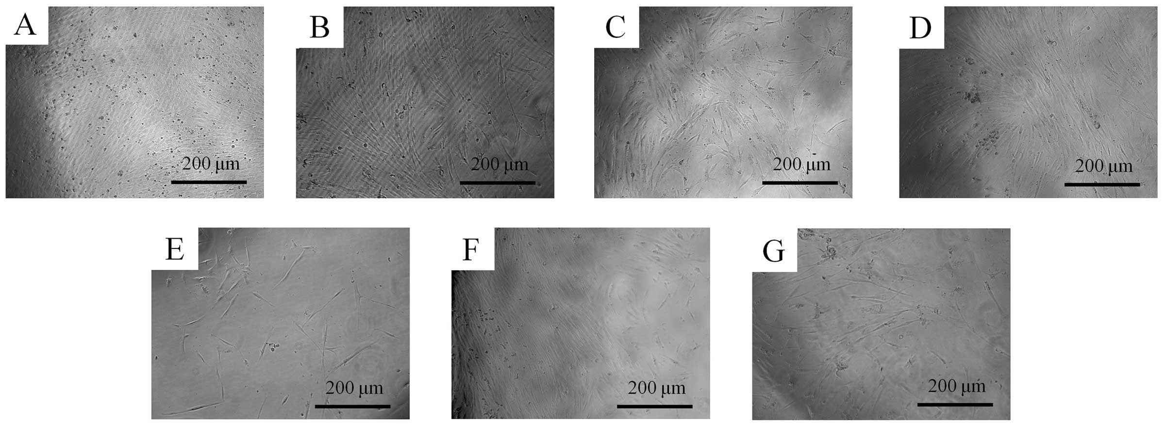
morphology of the cells on day 7 is shown in Fig. 3. The shapes of the cells treated
with 0.001, 0.01, 0.1, 1, 10, and 100 μg/ml groups were
similar to the shapes of the cells in the untreated control
group.
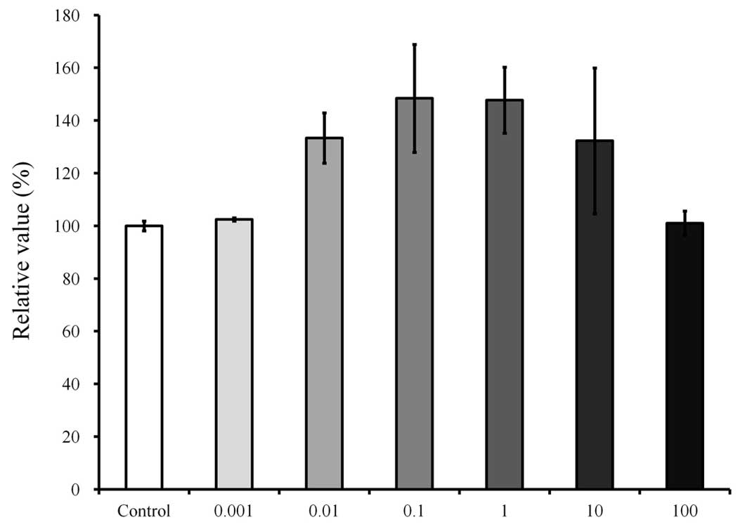
Cell proliferation
The results of cell proliferation at day 1 are shown
in Fig. 4, respectively. The
relative values of the CCK-8 assays of 0.001, 0.01, 0.1, 1, 10, and
100 μg/ml Angelicae dahuricae radix were 102.5±0.6,
133.3±9.6%, 148.4±20.5, 147.7±12.6, 132.3±277 and 101.1±4.6%,
respectively, when the CCK-8 result of the control group on day 1
was considered to be 100% (100.0±1.8). The proliferation rate of
the cultures that were growing in the presence of Angelicae
dahuricae radix increased marginally in the 0.1 and 1
μg/ml groups, but this was not indicated to be statistically
significant (P=0.052).
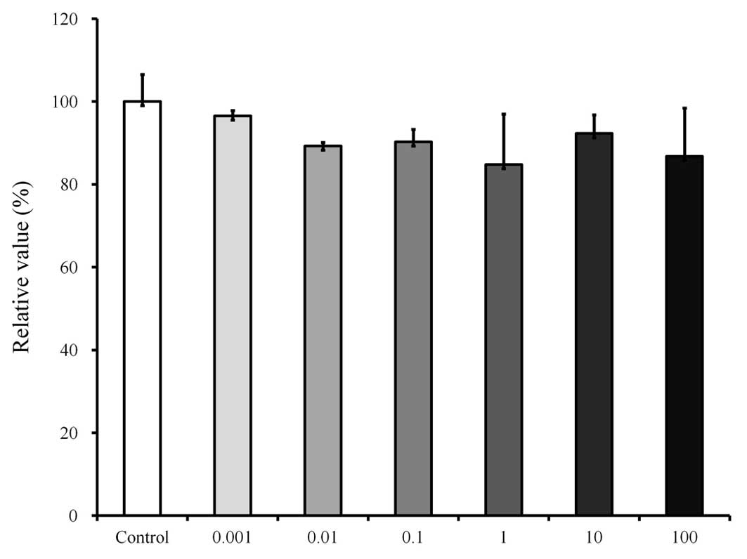
The results at day 3 are shown in Fig. 5. The relative values of the CCK-8
assays of 0.001, 0.01, 0.1, 1, 10, and 100 μg/ml of
Angelicae dahuricae radix were 96.5±1.3, 89.3±0.9, 90.3±3.0,
84.8±12.2, 92.3±4.5 and 86.8±11.7%, respectively, when the CCK-8
result of the control group on day 3 was considered to be 100%
(100.0±6.5). No significant differences were noted among the groups
(P>0.05).
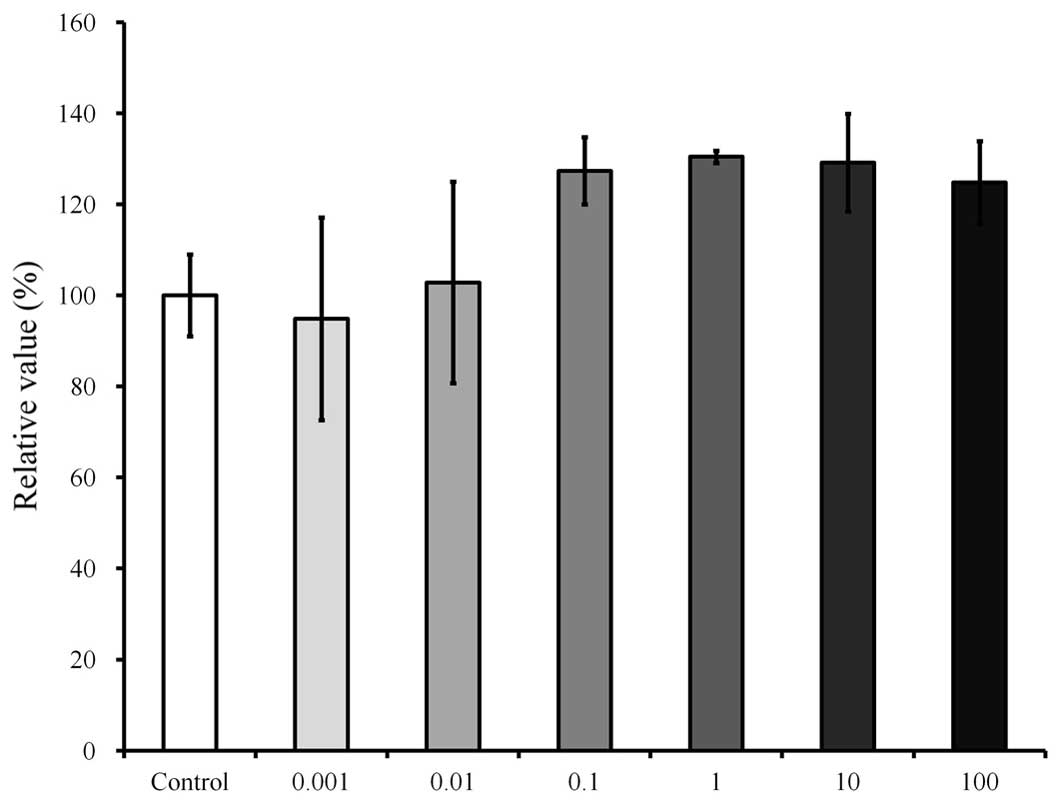
The results at day 7 are shown in Fig. 6. The relative values of the CCK-8
assays of 0.001, 0.01, 0.1, 1, 10, and 100 μg/ml of
Angelicae dahuricae radix were 94.9±22.3, 102.8±22.1,
1274±74, 130.4±1.3, 129.2±10.8 and 124.8±9.1%, respectively, when
the CCK-8 result of the control group on day 7 was considered to be
100% (100.0±9.0). The cultures that were growing in the presence of
Angelicae dahuricae radix exhibited marginally increased
proliferation in the 1 and 10 μg/ml groups; however, this
was not observed to achieve statistical significance
(P>0.05).
Discussion
This study discussed the effects of Angelicae
dahuricae radix on the morphology and proliferation of the
human mesenchymal stem cells derived from periodontal tissue. This
study showed that cell proliferation of the stem cells were
influenced by Angelicae dahuricae radix at day 1 and a
marginal increase in cell number was observed at certain
concentrations (0.1 to 1 μg/ml) (P=0.052); however, a
significant increase in cell proliferation was not achieved at day
3 and 7.
Angelicae dahuricae radix is a traditional
herbal medicine used to treat various diseases. More recently, it
was reported that Angelicae dahuricae radix suppressed the
development of atopic dermatitis-like skin lesions and could be
used for the treatment of hypertension and asthma (3,7,8). A
number of analytical methods have been reported for the
determination of several coumarins in Angelicae dahuricae
radix, including capillary electrochromatography, high-performance
liquid chromatography, liquid chromatography-mass spectrometry, gas
chromatography-mass spectrometry and high-speed countercurrent
chromatography (7,9–12).
It appears that the crude extract and essential oils of
Angelicae dahuricae radix may include multiple potentially
active chemical compounds. The main components of Angelicae
dahuricae radix are reported to be volatile oil and ingredients
of coumarin, including imperatorin, isoimperatorin and cnidilin,
and these active ingredients of Angelicae dahuricae radix
may explain its multiple functions (13). Previous studies have shown that
auraptenol, a major coumarin component from Angelicae
dahuricae radix, had a robust antinociceptive effect in a
chronic neuropathic pain mouse model (1).
Limited studies were performed to evaluate the
effects of Angelicae dahuricae radix on the cell viability
in vitro and in vivo (14,15),
and Angelicae dahuricae radix extract showed cytotoxicity
toward cancer cells, including mouse lymphocytic leukemia cells,
human promyelocytic leukemia cells, human myelogenous leukemia
cells, and mouse melanoma cells (14). Coumarins were isolated from
Angelicae dahuricae radix by silica gel column
chromatography, and the half maximal inhibitory concentration
(IC50) values varied from 8.6 to 14.6 μg/ml
cells. However, another study showed that extracts of Angelicae
dahuricae radix combined with methanol, water and ethyl acetate
at 100 μg/ml did not produce significant cytotoxic effects
on a murine macrophage cell line (15). Sprague Dawley rats were
administered 0.3, 1.0, and 3.0 g/kg Angelicae dahuricae
radix compound corresponding to the unprocessed drug of 2.78, 9.28,
and 27.8 g/kg by per os once a day for 12 weeks (16). The amounts were estimated to be
equal to 30, 100 and 300 times the clinical dose, and no apparent
chronic toxicity was reported during the experimental period.
Similarly, the present study showed that cultures growing in the
presence of Angelicae dahuricae radix showed marginal
increase in cell number in the 0.1 and 1 μg/ml groups;
however, statistically significant differences were not
identified.
There is increasing interest in mesenchymal stem
cells provide as they are an advantageous alternative therapeutic
option for tissue regeneration in comparison to current treatment
modalities (17). Previous reports
showed that stem cells derived from the gingiva exhibited
colony-forming abilities, plastic adherence and multilineage
differentiation (osteogenic, adipogenic, chondrogenic) potency, and
expressed CD44, CD73, CD90 and CD105 (18). Gingiva from the maxillofacial
region may be considered as a favorable source of mesenchymal stem
cells as the harvesting of stem cells from the mandible or maxilla
can be performed easily under local anesthesia (19). Furthermore, harvesting stem cells
from the gingiva may be more practical when compared with the bone
marrow of the maxilla and mandible as it is less invasive and has
less complications, including paresthesia and pain (20–22).
Within the limits of this study, Angelicae
dahuricae radix at the tested concentrations did not produce
statistically significant differences in the viability of stem
cells derived from the gingiva.
Acknowledgments
This study was supported by the Basic Science
Research Program through the National Research Foundation of Korea
(NRF) funded by the Ministry of Science, ICT & Future Planning
(grant no. NRF-2014R1A1A1003106).
References
|
1
|
Wang Y, Cao SE, Tian J, Liu G, Zhang X and
Li P: Auraptenol attenuates vincristine-induced mechanical
hyperalgesia through serotonin 5-HT1A receptors. Sci Rep.
3:33772013.PubMed/NCBI
|
|
2
|
Zhou RH: Resource science of Chinese
medicinal materials. China Medical & Pharmaceutical Sciences
Press; Beijing: pp. 19–32. 1993
|
|
3
|
Lee H, Lee JK, Ha H, Lee MY, Seo CS and
Shin HK: Angelicae dahuricae radix inhibits dust mite
extract-induced atopic dermatitis-like skin lesions in NC/Nga mice.
Evid Based Complement Alternat Med. 2012:7430752012.PubMed/NCBI
|
|
4
|
Li H, Dai Y, Zhang H and Xie C:
Pharmacological studies on the Chinese drug radix Angelicae
dahuricae. Zhongguo Zhong Yao Za Zhi. 16:560–562. 5761991.In
Chinese.
|
|
5
|
Kang OH, Chae HS, Oh YC, et al:
Anti-nociceptive and anti-inflammatory effects of Angelicae
dahuricae radix through inhibition of the expression of inducible
nitric oxide synthase and NO production. Am J Chin Med. 36:913–928.
2008. View Article : Google Scholar : PubMed/NCBI
|
|
6
|
Yi S, Cho JY, Lim KS, et al: Effects of
Angelicae tenuissima radix, Angelicae dahuricae radix and
Scutellariae radix extracts on cytochrome P450 activities in
healthy volunteers. Basic Clin Pharmacol Toxicol. 105:249–256.
2009. View Article : Google Scholar : PubMed/NCBI
|
|
7
|
Zhao G, Peng C, Du W and Wang S:
Pharmacokinetic study of eight coumarins of Radix Angelicae
dahuricae in rats by gas chromatography-mass spectrometry.
Fitoterapia. 89:250–256. 2013. View Article : Google Scholar : PubMed/NCBI
|
|
8
|
Tang W and Eisenbrand G: Angelica spp.
Chinese drugs of plant origin, chemistry, pharmacology and use in
traditional and modern medicine. Springer; Berlin: pp. 113–125.
1992, View Article : Google Scholar
|
|
9
|
Szucs V and Freitag R: Comparison of a
three-peptide separation by capillary electrochromatography,
voltage-assisted liquid chromatography and nano-high-performance
liquid chromatography. J Chromatogr A. 1044:201–210. 2004.
View Article : Google Scholar : PubMed/NCBI
|
|
10
|
Xie Y, Chen Y, Lin M, Wen J, Fan G and Wu
Y: High-performance liquid chromatographic method for the
determination and pharmacokinetic study of oxypeucedanin hydrate
and byak-angelicin after oral administration of Angelica dahurica
extracts in mongrel dog plasma. J Pharm Biomed Anal. 44:166–172.
2007. View Article : Google Scholar : PubMed/NCBI
|
|
11
|
Zheng X, Zhang X, Sheng X, et al:
Simultaneous characterization and quantitation of 11 coumarins in
Radix Angelicae dahuricae by high performance liquid chromatography
with electrospray tandem mass spectrometry. J Pharm Biomed Anal.
51:599–605. 2010. View Article : Google Scholar
|
|
12
|
Liu R, Li A and Sun A: Preparative
isolation and purification of coumarins from Angelica dahurica
(Fisch ex Hoffn) Benth, et Hook f (Chinese traditional medicinal
herb) by high-speed counter-current chromatography. J Chromatogr A.
1052:223–227. 2004. View Article : Google Scholar : PubMed/NCBI
|
|
13
|
Zhu M, Liang XL, Zhao LJ, et al:
Elucidation of the transport mechanism of baicalin and the
influence of a Radix Angelicae dahuricae extract on the absorption
of baicalin in a Caco-2 cell monolayer model. J Ethnopharmacol.
150:553–559. 2013. View Article : Google Scholar : PubMed/NCBI
|
|
14
|
Thanh PN, Jin W, Song G, Bae K and Kang
SS: Cytotoxic coumarins from the root of Angelica dahurica. Arch
Pharm Res. 27:1211–1215. 2004. View Article : Google Scholar
|
|
15
|
Kang OH, Lee GH, Choi HJ, et al: Ethyl
acetate extract from Angelica dahuricae Radix inhibits
lipopolysaccharide-induced production of nitric oxide,
prostaglandin E2 and tumor necrosis factor-alpha via
mitogen-activated protein kinases and nuclear factor-kappaB in
macrophages. Pharmacol Res. 55:263–270. 2007. View Article : Google Scholar : PubMed/NCBI
|
|
16
|
Zhao W, Cao Y and Liu J: Research on
chronic toxicology of Compound Radix Angelicae dahuricae capsule. J
Shaxi Med Univ. 37:160–165. 2006.
|
|
17
|
Moshaverinia A, Chen C, Xu X, et al: Bone
regeneration potential of stem cells derived from periodontal
ligament or gingival tissue sources encapsulated in RGD-modified
alginate scaffold. Tissue Eng Part A. 20:611–621. 2014.
|
|
18
|
Jeong SH, Lee JE, Jin SH, Ko Y and Park
JB: Effects of Asiasari radix on the morphology and viability of
mesenchymal stem cells derived from the gingiva. Mol Med Rep.
10:3315–3319. 2014.PubMed/NCBI
|
|
19
|
Tomar GB, Srivastava RK, Gupta N, et al:
Human gingiva-derived mesenchymal stem cells are superior to bone
marrow-derived mesenchymal stem cells for cell therapy in
regenerative medicine. Biochem Biophys Res Commun. 393:377–383.
2010. View Article : Google Scholar : PubMed/NCBI
|
|
20
|
Park JB, Kim YS, Lee G, Yun BG and Kim CH:
The effect of surface treatment of titanium with
sand-blasting/acid-etching or hydroxyapaptite-coating and
application of bone morphogenetic protein-2 on attachment,
proliferation and differentiation of stem cells derived from buccal
fat pad. Tissue Eng Regen Med. 10:115–121. 2013. View Article : Google Scholar
|
|
21
|
Fournier BP, Larjava H and Häkkinen L:
Gingiva as a source of stem cells with therapeutic potential. Stem
Cells Dev. 22:3157–3177. 2013. View Article : Google Scholar : PubMed/NCBI
|
|
22
|
Yang H, Gao LN, An Y, et al: Comparison of
mesenchymal stem cells derived from gingival tissue and periodontal
ligament in different incubation conditions. Biomaterials.
34:7033–7047. 2013. View Article : Google Scholar : PubMed/NCBI
|