Introduction
Methyl methanesulfonate (MMS) exerts serious
genotoxic effects, including induction of DNA damage, and
micronuclei and cell cycle alterations. MMS, a potent alkylating
agent, is a testicular toxicant that damages germ cell DNA, thereby
affecting sperm morphology in mice (1) and rats (2,3). MMS
also disrupts the differentiation of germ cells into sperm cells
(4,5). Oxidative stress is an imbalance
between the systemic manifestation of reactive oxygen species (ROS)
and the biological system's ability to detoxify reactive
intermediates or to repair the resulting damage efficiently
(6). The testis contains a high
level of polyunsaturated membrane lipids; thus, it is a target of
oxidative stress (7).
Nevertheless, organisms have developed numerous defense mechanisms
to protect themselves from damage caused by ROS. For example,
antioxidants, such as VE can scavenge free radicals (8,9) and
arrest the lipid peroxidation of lipoproteins that are present
within biological membranes (10).
VE is relatively abundant in the testis, where it protects sperm
from ROS and inhibits lipid peroxidation (11). Any deficiency in VE may result in
testicular damage, degenerated seminiferous tubules, and defective
germ cells (12). By contrast, an
increase in the level of VE has been shown to have an opposite
effect, that is, to increase total sperm output in rabbits
(13) and sheep (14). A previous study has shown VE to
have a protective effect on pesticide-induced oxidative stress
(13). Thus, the goal of this
study was to investigate the protective role of VE on MMS-induced
teratozoospermia in adult rats. Therefore, the effects of VE in
adult rats treated with MMS was compared with the effects of
treatment with MMS alone.
Materials and methods
Experimental animals
Eighteen adult male Sprague-Dawley (SD) outbred rats
at 8 weeks of age and weighing 210–230 g were obtained from a
closed random bred colony at the Animal Center of Nanjing Medical
University. Animals were housed under standard conditions of 21±2°C
and a 12 h light/dark cycle with access to food and water ad
libitum. Animals received humane care in compliance with the
guidelines of the National Institutes of Health. This study was
approved by the Institutional Animal Care and Use Committee of
Jiangsu University (Yixing, China).
Treatment of animals with MMS
Rats were randomly divided into three groups (n=6
per group) as follows: i) The control group, treated with distilled
water from days 1 to 5; ii) The MMS group, treated with MMS at a
dose of 40 mg·kg−1 (Sigma-Aldrich, St. Louis, MO, USA)
from day 1–5; and iii) the VE+MMS group, treated with MMS at a dose
of 40 mg·kg−1 from days 1–5, followed by VE
(Sigma-Aldrich, St. Louis, MO, USA) at a dose of 150
mg·kg−1 from day 6 for 6 weeks. All experimental agents
were delivered by gavage using a non-flexible stainless steel
feeding tube (Sangon Biotech Co., Ltd., Shanghai, China).
Thereafter, rats were anesthetized with 5% chloral hydrate (1.5
ml), and blood was collected by cardiac puncture into standard test
tubes after the rats had been anaesthetized. Samples were
centrifuged at 500 × g for 15 min at 4°C to obtain serum and then
stored at −40°C. Testes and epididymis were quickly excised and
suspended in ice-cold phosphate buffered saline (PBS). One testis
from one rat was fixed in 10% formaldehyde (w/v) and the other
testis was stored at −80°C. Sperm count, motility and morphology of
the three groups were examined following treatment with VE.
Counting of epididymal sperm
The epididymis was weighed prior to sperm counting.
Epididymal spermatozoids were counted as previously described with
minor modifications to the protocol (15). In brief, one of the epididymis was
minced in 5 ml PBS (pH 7.4), placed on an orbital shaker (Sangon
Biotech Co., Ltd.) for 10 min, and incubated at room temperature
for 2 min. Semen (25 μl) was transferred to 1 ml fixative,
and spermatozoids were counted under a light microscope (Olympus
BX41; Olympus Corporation, Tokyo, Japan) and using a hemocytometer
(Sangon Biotech Co., Ltd.). The other epididymis was stored at
−80°C.
Sperm motility
Approximately 200 spermatozoids with progressive
motility from each epididymis were evaluated under a light
microscope within 2–4 min after they were isolated as previously
described (16).
Detection of sperm abnormalities
Fixed spermatozoids were smeared onto a slide glass,
air-dried overnight and stained with the Diff-Quick kit (Baso
Diagnostics, Inc., Zhuhai, China) (17). Approximately 300 spermatozoids from
each treatment group were counted under a light microscope, and the
percentages of morphologically abnormal spermatozoids (detached
head and/or coiled tail) were recorded as previously described
(1).
Measurement of the serum testosterone
level
The serum testosterone level was measured as
previously described (18) using a
commercially available chemiluminescence-linked immunoassay
(Beckman-Coulter UniCel DxI 800, Beckman-Coulter, Brea, CA,
USA).
Antioxidant enzyme activity and oxidative
stress assays
Different oxidative stress indicators, including
free radicals (e.g., superoxide anion and hydroxyl radical) and
antioxidants [e.g., superoxide dismutase (SOD) and glutathione
peroxidase (GSH-Px)], were measured in sera from rats in each
treatment group. All assays were performed according to the
instructions provided in the kits (Jiancheng Bioengineering
Research Institute, Nanjing, China).
Immunohistochemistry
Testes were fixed in 10% formaldehyde (w/v) for at
least 24 h and then processed for paraffin embedding (19). Rabbit polyclonal anti-Vasa (cat.
no. ab13840), rabbit polyclonal anti-promyelocytic leukemia zinc
finger protein (Plzf; cat. no. ab39354), and rabbit polyclonal
anti-synaptonemal complex protein 3 (Sycp3; cat. no. ab150292)
antibodies were purchased from Abcam (Cambridge, UK). Anti-mouse
IgG (H+L, alkaline conjugate) was purchased from Promega
Corporation (Madison, WI, USA). Sections (5 μm) were
deparaffinized in xylene, rehydrated in a gradient series of
alcohol, and boiled in antigen retrieval solution (Sangon Biotech
Co., Ltd.) at 95°C for 10 min. The sections were treated with 3%
hydrogen peroxide (v/v) for 10 min, blocked with 3% bovine serum
albumin (v/v) (Sangon Biotech Co., Ltd.) and 10% normal donkey
serum (v/v) (Sangon Biotech Co., Ltd.) in Tris-buffered saline
(TBS), and then incubated with antibodies overnight at 4°C.
Thereafter, sections were washed with TBS and incubated with a
biotinylated and streptomycin-labeled goat anti-mouse antibody
(cat. no. KIT-5010; Maixin Bio, Ltd., Fujian, China) for 15 min at
room temperature. Immunoreactive proteins were visualized with
3,3′-diaminobenzidine tetrahydrochloride. Sections were examined
under a light microscope (Eclipse 80i; Nikon, Tokyo, Japan). The
expression levels of protein were measured and demonstrated by the
Integrated Optical Density (IOD) values using Image-Pro Plus 6.0
software (Media Cybernetics, Inc., Rockville, MD, USA).
Statistical analysis
SPSS 14.0 (SPSS Inc., Chicago, IL, USA) statistical
software for Windows was used. Results are presented as the mean ±
standard deviation. One-way analysis of variance (followed by
Tukey's post hoc test) or Mann-Whitney U-test was used to compare
the means across different treatment groups. P<0.05 was
considered to indicate a statistically significant difference.
Results
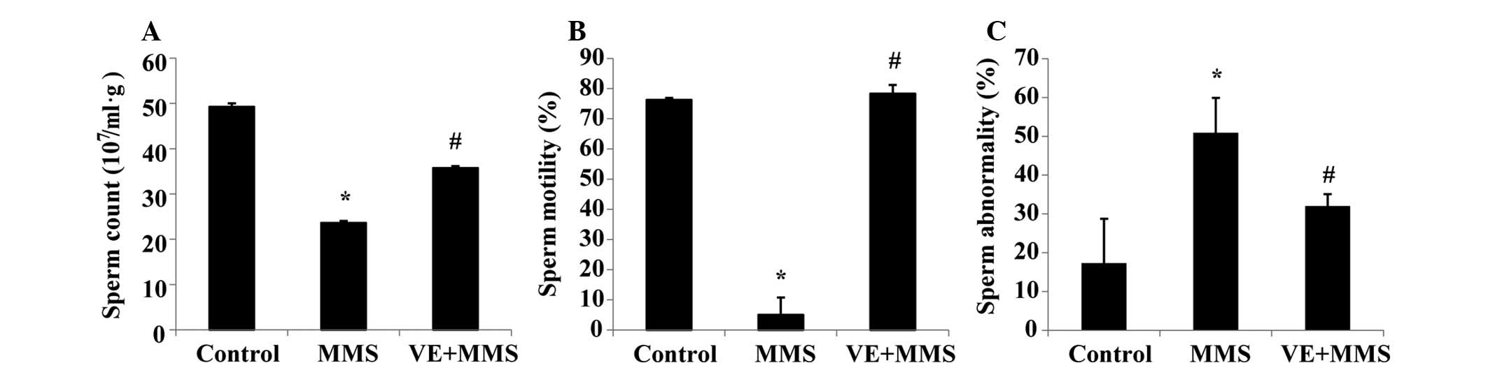
Sperm characteristics
Sperm count (2.36±0.04×10⁸) and motility
(5.0±5.8%) decreased significantly (P<0.05) in rats from the MMS
group compared with those from the control group
(4.92±0.07×10⁸ and 76.3±0.7%, respectively). However,
there was an increase in the percentage of abnormal sperm
(50.9±9.0%) compared with the control group (17.3±11.4%). After MMS
and VE treatment, sperm count (3.57±0.04×10⁸) and
motility (78.3±2.9%) were significantly higher (P<0.05) in rats
from the VE+MMS group than in the MMS group (Fig. 1A and B). However, there was a
significant decrease (P<0.05) in the percentage of abnormal
sperm in rats from the VE+MMS group (32.0±3.0%) compared with the
MMS group (Fig. 1C).
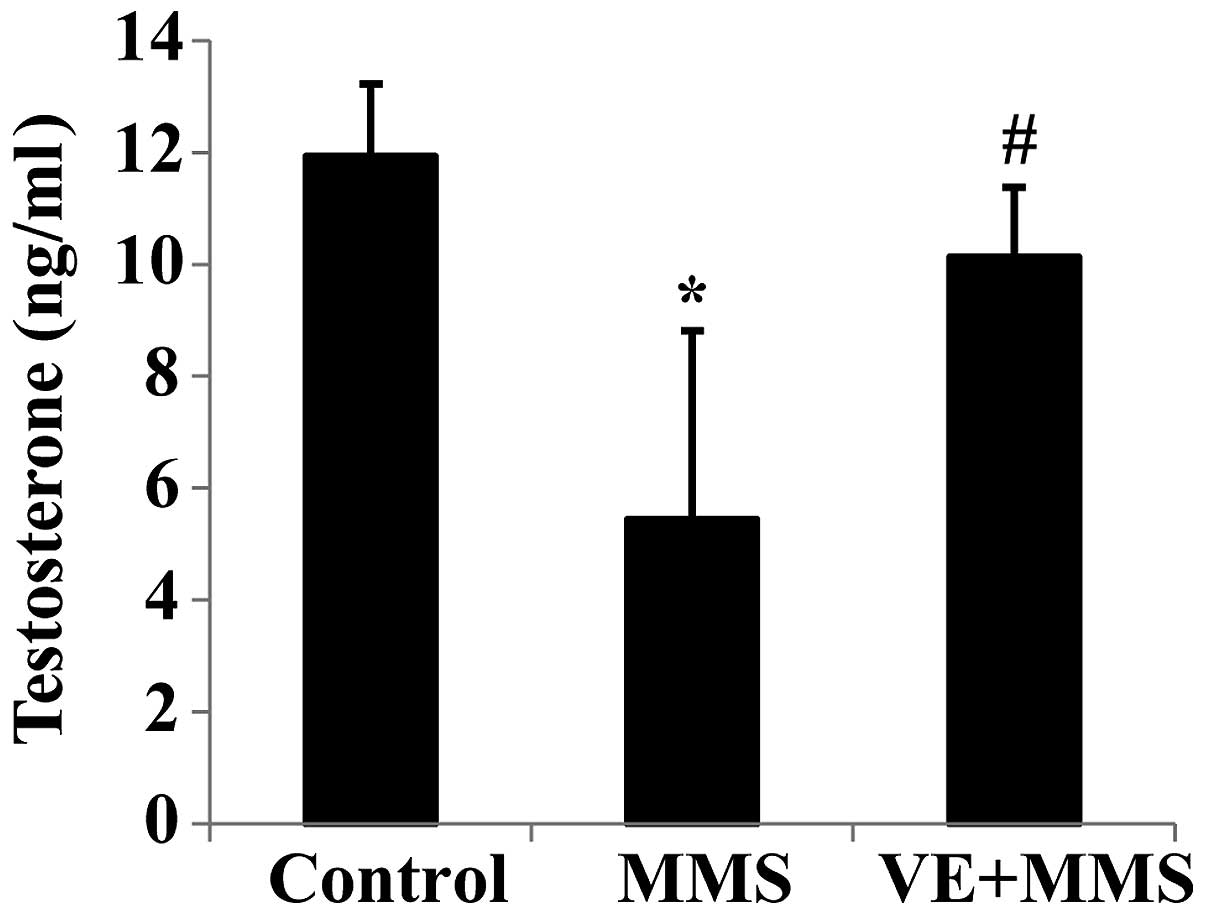
Levels of serum testosterone
There was a significant decrease (P<0.05) in the
serum testosterone level in the MMS group (5.4±3.4 ng/ml) compared
with the control (11.9±1.3 ng/ml) and a significant increase in the
VE+MMS group (10.1±1.3 ng/ml) compared with the MMS group (Fig. 2).
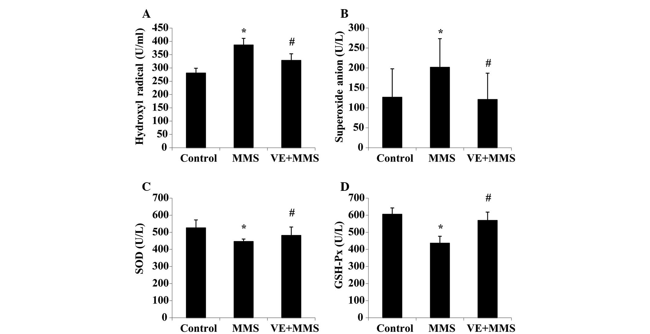
Serum oxidative stress indicators
The levels of hydroxyl and superoxide free radicals
were significantly higher in the MMS group, as compared with the
control group; and lower in the VE+MMS group, as compared with the
control group (Fig. 3A and B). By
contrast, the levels of antioxidants SOD and GSH-Px, were lower in
the MMS group than in the control and VE+MMS groups (Fig. 3C and D).
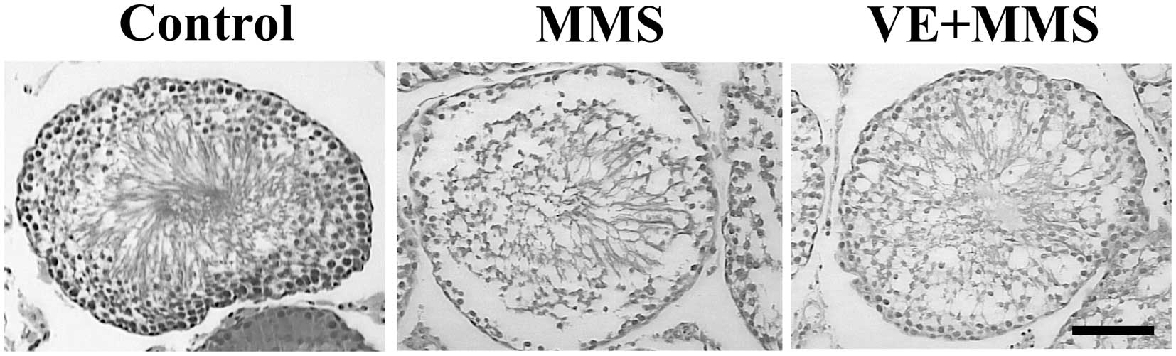
Immunohistochemical findings
The histo-architecture of the testes from control
rats was normal, consisting of uniform, well-organized seminiferous
tubules with complete spermatogenesis and normal interstitial
connective tissue (Fig. 4). By
contrast, testes from rats treated with MMS showed seminiferous
tubule degeneration manifested by shrunken, disorganized tubules
with irregular, buckled basement membranes and incomplete
spermatogenesis (Fig. 4).
Moreover, the seminiferous tubules were virtually devoid of
spermatids and sperm. After MMS+VE treatment, there was an
improvement in spermatogenesis, demonstrated by the presence of
elongated spermatids and sperm in the majority of the seminiferous
tubules (Fig. 4).
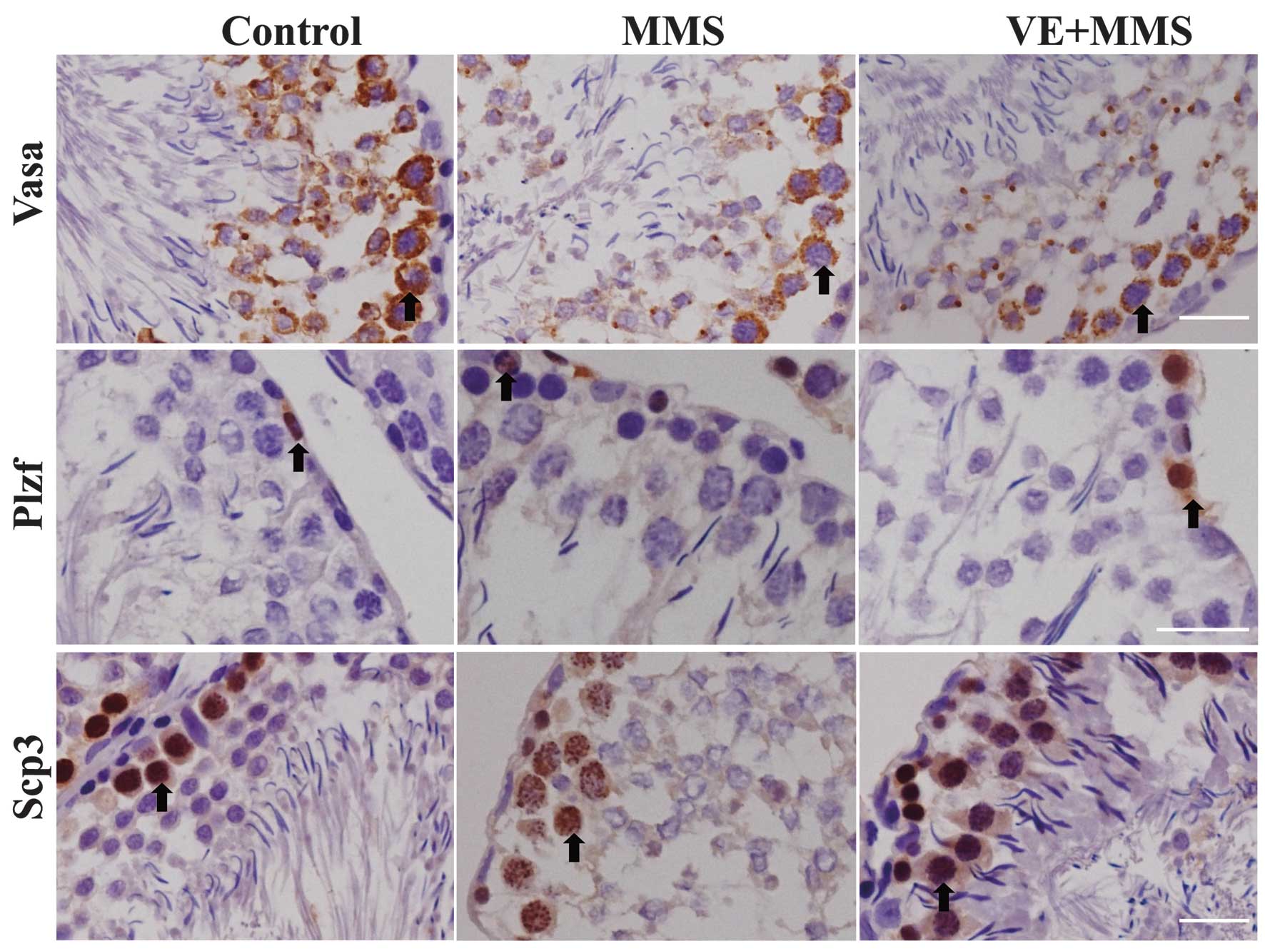
As observed using immunohistochemistry, the level of
Vasa, a biomarker of germ cells, decreased in the testis of rats
treated with MMS; however, its level increased in rats treated with
VE+MMS compared with those treated with MMS alone (Fig. 5). There were no changes in Plzf and
Sycp3, biomarkers of spermatogonial stem cells and spermatocytes,
respectively, following MMS and MSS+VE treatment (Table I).
 | Table IImmunohistochemical staining by
comparison of the average IOD values. |
Table I
Immunohistochemical staining by
comparison of the average IOD values.
| Group | Vasa | Plzf | Scp3 |
|---|
| Control | 0.18±0.04 | 0.18±0.05 | 0.18±0.06 |
| MMS | 0.12±0.03a | 0.20±0.04 | 0.17±0.07 |
| MMS+VE | 0.21±0.04b | 0.22±0.02 | 0.19±0.05 |
Discussion
Previous studies have shown MMS to decrease sperm
number and affect sperm head morphology in mice and rats (1–3).
Although VE can protect cells from damage caused by free radicals
and oxidative products, there are few studies on the protective
effect of VE in animals previously exposed to MMS (8,10,12–14).
The present study demonstrated that VE partially alleviated the
damage caused by MMS in the testis.
Ozawa et al (2) demonstrated that body, testis and
epididymis weights, as well as food consumption decrease following
administration of MMS. Germ cell exfoliation, Sertoli cell
vacuolization, and epididymal duct cell debris were also observed
(2). Although VE does not protect
the testes from acrylamide toxicity, treatment with VE following
cessation of acrylamide treatment improves recovery (20). In the present study, MMS treatment
resulted in seminiferous tubule degeneration, and VE treatment
improved the recovery of spermatogenesis.
VE can protect critical cellular structures from
free radical and oxidative damage (21). As a soluble, lipid-based
antioxidant, VE is protective against oxidative stress by
preventing the production of lipid peroxides and scavenging free
radicals (11). VE is also
important in maintaining the function of the testis, epididymis and
accessory glands (22), possibly
by improving sperm quality and quantity. For example, VE improved
semen quality and quantity in humans (11), sheep (12), and chickens (22). VE has also been demonstrated to
protect rat testis against experimental cryptorchidism (23,24).
Furthermore, the increased free radicals generated by acrylamide
exposure in the testes may have been scavenged in the testes during
recovery period (20). MMS at
doses of 20–40 mg/kg for two consecutive weeks increased heme
oxygenase-1 (HO-1) mRNA and protein in the rat testis (25). In this study, MMS increased free
radical formation, which decreased following VE treatment, while
the levels of two endogenous antioxidants indicators (SOD and
GSH-Px) showed an opposite tendency. These results indicate that
MMS induces oxidative stress in adult rats, and VE has the
capability of scavenge them.
MMS damages germ cell DNA by creating lesions within
the DNA, which can disrupt the differentiation of germ cells into
spermatozoa (5). This is in
agreement with the results of the present study, which showed an
increase in the percentage of defective spermatozoids. Moreover,
the level of Vasa decreased after MMS treatment, while the levels
of Plzf and Sycp3 remained unchanged, suggesting that MMS has
little influence on spermatogonial stem cells and spermatocytes.
Additional experiments are requried to address the mechanism of MMS
action in the adult testis.
In conclusion, MMS treatment of adult rats affects
germ cells in the testis, and VE may aid in repairing the damage
caused by oxidative stress.
Acknowledgments
This study was supported by the Fund of Science and
Technology of Yixing (grant no. 2013–15) and Development Fund of
Clinical Science and Technology (grant no. JLY20120070, 2012,
Jiangsu University).
References
|
1
|
Wyrobek AJ and Bruce WR: Chemical
induction of sperm abnormalities in mice. Proc Natl Acad Sci USA.
72:4425–4429. 1975. View Article : Google Scholar : PubMed/NCBI
|
|
2
|
Ozawa S, Yokoi R, Kitamura T, Kuriyama K,
Kobayashi K and Shibata N: Collaborative work to evaluate toxicity
on male reproductive organs by repeated dose studies in rats 15).
Two-week and 4-week administration study of methyl methanesulfonate
(MMS). J Toxicol Sci. 25:155–162. 2000. View Article : Google Scholar
|
|
3
|
Cassidy SL, Dix KM and Jenkins T:
Evaluation of a testicular sperm head counting technique using rats
exposed to dimethoxyethyl phthalate (DMEP), glycerol
alpha-monochlorohydrin (GMCH), epichlorohydrin (ECH), formaldehyde
(FA), or methyl methanesulphonate (MMS). Arch Toxicol. 53:71–s78.
1983. View Article : Google Scholar : PubMed/NCBI
|
|
4
|
Bruce WR and Heddle JA: The mutagenic
activity of 61 agents as determined by the micronucleus,
Salmonella, and sperm abnormality assays. Can J Genet Cytol.
21:319–334. 1979. View
Article : Google Scholar : PubMed/NCBI
|
|
5
|
Inoue M, Kurihara T, Yamashita M and
Tatsumi K: Effects of treatment with methyl methanesulfonate during
meiotic and postmeiotic stages and maturation of spermatozoa in
mice. Mutat Res. 294:179–186. 1993. View Article : Google Scholar : PubMed/NCBI
|
|
6
|
Ochsendorf FR: Infections in the male
genital tract and reactive oxygen species. Hum Reprod Update.
5:399–420. 1999. View Article : Google Scholar : PubMed/NCBI
|
|
7
|
Aitken RJ, Harkiss D and Buckingham DW:
Analysis of lipid peroxidation mechanisms in human spermatozoa. Mol
Reprod Dev. 35:302–315. 1993. View Article : Google Scholar : PubMed/NCBI
|
|
8
|
Ingold KU, Burton GW, Foster DO, et al: A
new vitamin E analogue more active than alpha-tocopherol in the rat
curative myopathy bioassay. FEBS Lett. 205:117–120. 1986.
View Article : Google Scholar : PubMed/NCBI
|
|
9
|
Jones DP, Kagan VE, Aust SD, Reed DJ and
Omaye ST: Impact of nutrients on cellular lipid peroxidation and
antioxidant defense system. Fundam Appl Toxicol. 26:1–7. 1995.
View Article : Google Scholar : PubMed/NCBI
|
|
10
|
Sheridan PA and Beck MA: The immune
response to herpes simplex virus encephalitis in mice is modulated
by dietary vitamin E. J Nutr. 138:130–137. 2008.
|
|
11
|
Akiyama M: In vivo scavenging effect of
ethylcysteine on reactive oxygen species in human semen. Nihon
Hinyokika Gakkai Zasshi. 90:421–428. 1999.In Japanese. PubMed/NCBI
|
|
12
|
Wilson MJ, Kaye D, Smith WE, Quach HT,
Sinha AA and Vatassery GT: Effect of vitamin E deficiency on the
growth and secretory function of the rat prostatic complex. Exp Mol
Pathol. 74:267–275. 2003. View Article : Google Scholar : PubMed/NCBI
|
|
13
|
Yousef MI: Vitamin E modulates
reproductive toxicity of pyrethroid lambda-cyhalothrin in male
rabbits. Food Chem Toxicol. 48:1152–1159. 2010. View Article : Google Scholar : PubMed/NCBI
|
|
14
|
Yue D, Yan L, Luo H, Xu X and Jin X:
Effect of Vitamin E supplementation on semen quality and the
testicular cell membranal and mitochondrial antioxidant abilities
in Aohan fine-wool sheep. Anim Reprod Sci. 118:217–222. 2010.
View Article : Google Scholar
|
|
15
|
Yokoi K, Uthus EO and Nielsen FH: Nickel
deficiency diminishes sperm quantity and movement in rats. Biol
Trace Elem Res. 93:141–154. 2003. View Article : Google Scholar : PubMed/NCBI
|
|
16
|
Sönmez M, Türk G and Yüce A: The effect of
ascorbic acid supplementation on sperm quality, lipid peroxidation
and testosterone levels of male Wistar rats. Theriogenology.
63:2063–2072. 2005. View Article : Google Scholar : PubMed/NCBI
|
|
17
|
Enginsu ME, Dumoulin JC, Pieters MH, Bras
M, Evers JL and Geraedts JP: Evaluation of human sperm morphology
using strict criteria after Diff-Quik staining: correlation of
morphology with fertilization in vitro. Hum Reprod. 6:854–858.
1991.PubMed/NCBI
|
|
18
|
Pratt JJ, Woldring MG and Villerius L:
Chemiluminescence-linked immunoassay. J Immunol Methods.
21:179–184. 1978. View Article : Google Scholar : PubMed/NCBI
|
|
19
|
Davidoff MS, Middendorff R, Pusch W,
Müller D, Wichers S and Holstein AF: Sertoli and Leydig cells of
the human testis express neurofilament triplet proteins. Histochem
Cell Biol. 111:173–187. 1999. View Article : Google Scholar : PubMed/NCBI
|
|
20
|
Rahangadale S, Jangir BL, Patil M, et al:
Evaluation of protective effect of vitamin e on acrylamide induced
testicular toxicity in wister rats. Toxicol Int. 19:158–161. 2012.
View Article : Google Scholar : PubMed/NCBI
|
|
21
|
Erin AN, Spirin MM, Tabidze LV and Kagan
VE: Formation of alpha-tocopherol complexes with fatty acids. A
hypothetical mechanism of stabilization of biomembranes by vitamin
E. Biochim Biophys Acta. 774:96–102. 1984. View Article : Google Scholar : PubMed/NCBI
|
|
22
|
Cerolini S, Zaniboni L, Maldjian A and
Gliozzi T: Effect of docosahexaenoic acid and alpha-tocopherol
enrichment in chicken sperm on semen quality, sperm lipid
composition and susceptibility to peroxidation. Theriogenology.
66:877–886. 2006. View Article : Google Scholar : PubMed/NCBI
|
|
23
|
Vigueras-Villaseñor RM, Ojeda I,
Gutierrez-Pérez O, et al: Protective effect of α-tocopherol on
damage to rat testes by experimental cryptorchidism. Int J Exp
Pathol. 92:131–139. 2011. View Article : Google Scholar
|
|
24
|
Rahangadale S, Kurkure N, Prajapati B,
Hedaoo V and Bhandarkar AG: Neuroprotective effect of vitamin e
supplementation in wistar rat treated with acrylamide. Toxicol Int.
19:1–8. 2012. View Article : Google Scholar : PubMed/NCBI
|
|
25
|
Ashino T, Ozawa S, Numazawa S and Yoshida
T: Tissue-dependent induction of heme oxygenase-1 and
metallothionein-1/2 by methyl methanesulfonate. J Toxicol Sci.
28:181–189. 2003. View Article : Google Scholar : PubMed/NCBI
|