Introduction
Bronchial asthma is a chronic allergic disease
characterized by airway inflammation and remodeling (1). The chronic airway inflammation can
induce several structural alterations of the airway, eventually
leading to airway remodeling (2,3).
Remodeling is frequently observed in patients with refractory
asthma, and can arise as a result of excessive repair or failure to
resolve the inflammation (4).
Eventually, remodeling can contribute to airway high reactivity and
progressive decline of lung function (5).
Transforming growth factor-β (TGF-β) is the critical
mediator in the regulation of airway inflammation and remodeling in
asthma (6). Increased TGF-β1
levels can be detected in asthmatic bronchoalveolar lavage fluids
(7), whilst increased levels of
TGF-β1 in the asthmatic airway mucosa correlates with the thickness
of the reticular basement membrane (8). Downstream of TGF-β, connective tissue
growth factor (CTGF) modulates the cellular response to TGF-β
(9). Furthermore, CTGF can
upregulate the production of collagen type I and fibronectin in
human lung fibroblasts (10). In
asthma, TGF-β upregulates the levels of CTGF in airway smooth
muscle cells (11). Thus, this
pathway is important in the regulation of airway remodeling in
asthma. Additionally, oxidative stress is of importance in airway
inflammation, and determines the severity of asthma, whilst reduced
antioxidant defense is also associated with asthma (12). Oxidative stress increases in the
lungs following antigen stimulation, and antioxidant administration
has been demonstrated to attenuate the regeneration of reactive
oxidative stress, and inflammation in vivo (13).
Glucocorticoids are the current gold standard in
treatment for asthma, however, their ability to modulate airway
remodeling is limited (14). The
combination of glucocorticoids and bronchodilators is also unable
to revert asthmatic features (14), and results in significant adverse
side-effects. Therefore, there is an urgent requirement for the
identification of novel therapeutic agents for the treatment of
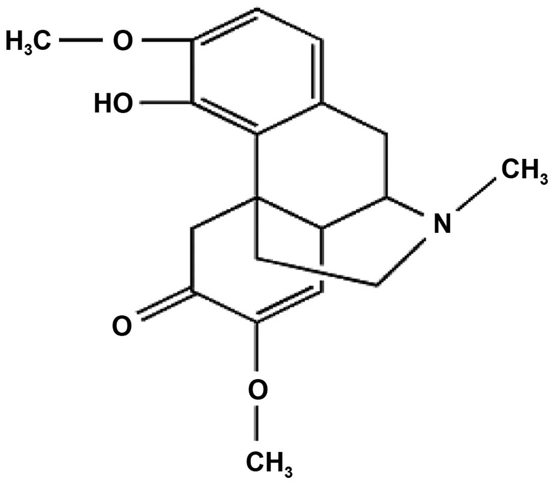
asthma. Sinomenine (SIN) is an alkaloid that is isolated from the
root and stem of the climbing plant Sinomenium acutum
(chemical structure presented in Fig.
1), and demonstrates immunosuppressive, anti-inflammatory,
anti-arrhythmic, analgesic and anti-arthritic properties (15,16).
SIN has been observed to significantly suppress the production of
inflammatory mediators in rats (17). However, the effects of SIN on
asthma remain to be established. The aim of the present study was
to evaluate the potential anti-inflammatory, anti-airway remodeling
and antioxidant effects of SIN in an asthma animal model.
Materials and methods
Animals
A total of 60 female BALB/c mice aged 6–8 weeks
(18–22 g) were purchased from the animal medical center of Lanzhou
University (Lanzhou, China). All animals received humane care and
were maintained with a 12 h light/dark cycle in a
temperature-controlled room (21–23°C), with free access to food and
water. All procedures described in the present study were approved
by the internal ethical committee of the First Hospital of Lanzhou
University (LDYYLL2014-0058).
Experimental asthma models and
intervention
Mice were divided randomly into six groups (ten
animals per group): i) Blank control group; ii) asthma model; iii)
asthma treated with dexamethasone (DEX; 2 mg/kg; Lianshui
Pharmaceutical Co., Ltd., Jiangsu, China); iv) asthma model with
low-dose SIN (25 mg/kg; Zelang Medical Technology Co., Ltd.
Nanjing, China); v) asthma treated with moderate-dose SIN (50
mg/kg); and vi) asthma treated with high-dose SIN (75 mg/kg). DEX
and SIN were administered by gavage.
Asthma was induced by ovalbumin (OVA; Sigma-Aldrich,
St. Louis, MO, USA) using a previously described method (18). Animals were immunized by
intraperitoneal injection (i.p.) of 20 µg OVA in the
presence of 2 mg of Al(OH)3 adjuvant (Pierce
Biotechnology, Inc., Rockford, IL, USA) diluted in 0.2 ml saline
solution on days 0, 7 and 14. Mice were exposed to 2.5% (w/v) OVA
solution in phosphate-buffered saline (PBS) for 30 min, using an
pump nebulizer (Pari TurboBoy N; Pari GmbH, Starnberg, Germany) on
days 21–28 subsequent to initial sensitization. Subsequently, mice
were exposed to aerosolized 2.5% OVA for 30 min, once every 2 days,
from days 29–70. DEX and SIN were administered for 1 h prior to the
OVA inhalation. Control mice received the adjuvant (i.p.) and were
exposed to nebulized aerosol of 0.9% NaCl (Gansu Fuzheng
Pharmaceutical Technology Co., Ltd., Lanzhou, China) at the same
time points. Mice were sacrificed via cervical dislocation at 24 h
subsequent to the final challenge.
Histochemical analyses
The left lobe of the lung (which was fixed in 10%
formalin (Shanghai Jianxin Chemical Co., Ltd., Shanghai, China) and
embedded in paraffin (Shanghai Yongye Biological Technology Co.,
Ltd., Shanghai, China) was cut into 3-µm sections using a
Leica RM 2135 microtome (Leica Microsystems GmbH, Wetzlar, Germany)
for histological and immunohistochemical analysis. The remainder of
the lung was triturated using a mortar and pestle for reverse
transcription-quantitative polymerase chain reaction (RT-qPCR) and
oxidative stress detection.
The sections were stained with hematoxylin and eosin
(HE) in order to observe the infiltration of cells. The degree of
inflammatory cell infiltration was determined using the established
scoring system (19). The
point-counting technique was used to evaluate levels of eosinophil
(20). To determine the
eosinophils/unit area (mm2) (21), the number of points of the
integrating eyepiece falling on areas of peribronchiolar
inflammation in three areas of each airway wall and the number of
eosinophils in the same area were counted using a Olympus DP71
microscope (Olympus Corporation, Tokyo, Japan). Goblet cells and
mucus expression were determined by staining the sections with
periodic acid Schiff's reagent (PAS). PAS-positive cells were
counted as goblet cells in four airways from one section. The
degree of mucus plugging of the airways (0.5–0.8 mm in diameter)
was classified by a semiquantitative system (22). Airway smooth muscle thickness was
determined in HE-stained lung sections using MetaMorph 6.1 image
software (Universal Imaging Corporation, West Chester, PA, USA) by
measuring the thickness of the smooth muscle cell layer per
micrometer perimeter of basement membrane (Wam/Pbm). Masson's
trichrome (Sigma-Aldrich) was used to evaluate collagen deposition
by analyzing the positive area per micrometer perimeter of basement
membrane (Wac/Pbm) by MetaMorph 6.1 image software (Universal
Imaging Corporation) (23).
RT-qPCR
Total RNA was prepared using the RNAiso Plus reagent
(Takara Bio, Inc., Otsu, Japan) according to the manufacturer's
instructions. RNA (2 µg per sample) was reverse-transcribed
into cDNA using PrimeScript™ Reverse Transcriptase (Takara Bio,
Inc.). qPCR was performed with a Rotor-gene 6000 Thermal Cycler
(Corbett Research Australia, Mortlake, Australia) and the SYBR
Premix Ex Taq II kit (Takara Bio, Inc.) in accordance with the
manufacturer's protocol. The sequences for the primers were as
follows: TGF-β1, F 5′-GTGTGGAGCAACATGTGGAACTCTA-3′ and R
5′-TTGGTTCAGCCACTGCCGTA-3′; CTGF, F 5′-CCTGTGCCTGCCATTA-3′ and R
5′-TTCCTCCCACGGTAGTT-3′; β-actin, F 5′-CATCCGTAAAGACCTCTATGCCAAC-3′
and R 5′-ATGGAGCCACCGATCCACA-3′. Gene expression was normalized to
β-actin. The experiments were repeated twice. Data are expressed as
the fold increase in RNA expression compared with control
animals.
Immunohistochemistry (IHC)
TGF-β1 and CTGF-induced protein expression was
determined by immunohistochemical staining with a Strept
Actividin-Biotin Complex kit (Zhongshan Biological Technology,
Beijing, China). IHC was performed using the specific rabbit
polyclonal anti-TGF-β1 (sc-146) and anti-CTGF immunoglobulin G
(sc-25440) antibodies (1:150; Santa Cruz Biotechnology, Inc., Santa
Cruz, CA, USA). Images were obtained using a Leica DM1000 optical
microscope (Leica Microsystems GmbH) and were analyzed using
Image-Pro Plus software, version 6.0 (Media Cybernetics, Inc.,
Silver Spring, MD, USA). Five unduplicated fields were selected in
one slide.
Measurement of the levels of oxidative
stress
Subsequent to thawing, the lung tissues were weighed
and homogenized in ice-cold PBS (20 mM, pH 7.4, w/v=1:9) using a
glass homogenizer on ice. The homogenate was centrifuged at 3,000 ×
g for 20 min at 4°C. The obtained supernatant was used for
measurement of the total antioxidant capacity (TAC),
malondialdehyde (MDA) content and myeloperoxidase (MPO) activities.
Detection of TAC, MDA and MPO were measured using commercially
available assay kits (Nanjing Jiancheng Bioengineering Institute,
Nanjing, China) following the manufacturer's protocol.
Statistical analysis
SPSS software, version 19.0 (IBM SPSS, Armonk, NY,
USA) was used for the statistical analysis. All data are expressed
as the mean ± standard deviation. The measurements were subjected
to one-way analysis of variance. Differences between experimental
groups were determined by Fisher's least significant differences
post-hoc test. P<0.05 was considered to indicate a statistically
significant difference.
Results
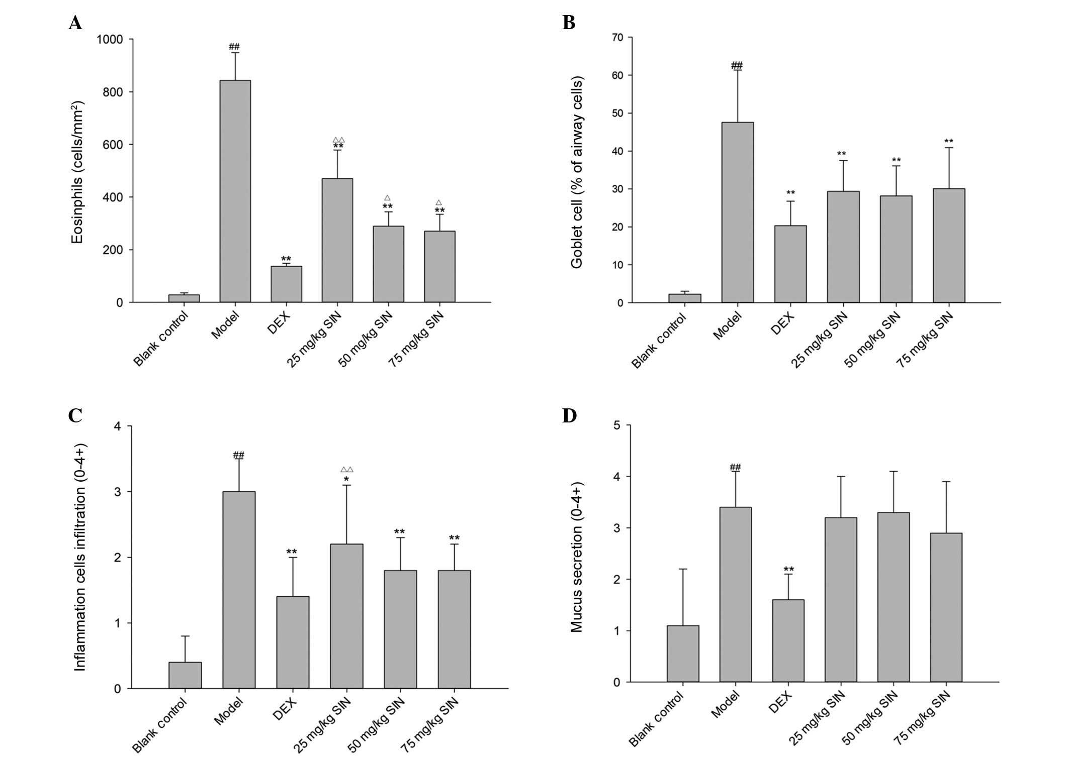
SIN reduced airway inflammation and
remodeling
Compared with the control, the OVA asthma group
presented significantly increased airway eosinophil infiltration
(P<0.01; Fig. 2A). DEX and SIN
treatment reduced the number of eosinophils in sensitized animals
compared with the model group (P<0.01), and the inhibitory
effects of SIN (25, 50 and 75 mg/kg) were not as effective as DEX
treatment (Fig. 2A). The OVA
challenge resulted in an increase in the number of goblet cells.
While compared with the control (P<0.01), therapeutic
administration of DEX and SIN significantly reduced the number of
goblet cells compared with model mice (Fig. 2B). Additionally, OVA challenge led
to an increase in the peribronchial and perivascular inflammatory
cell infiltration score, compared with that of the blank control.
DEX and various doses of SIN significantly reduced inflammation
score. (P<0.05 compared with model) Compared to the DEX group,
the inflammation scores were higher in the low-dose SIN group (25
mg/kg), however, no significant differences between moderate (50
mg/kg) and high-dose (75 mg/kg) SIN, and the DEX group were
identified (Fig. 2C). A marked
increase in airway mucus secretion was observed in OVA-treated mice
compared with the control animals. The DEX treatment group
displayed airway occlusion mucus scores which were significantly
lower than those of the model mice. However, the SIN (25, 50 and 75
mg/kg) groups demonstrated no alterations in mucus secretion
compared with the model group (Fig.
2D).
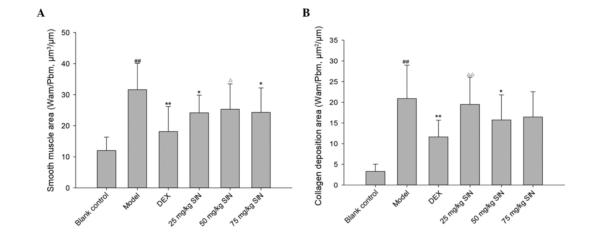
The OVA-challenged mice presented with a thicker
smooth muscle layer compared with the control group subsequent to
correction for the airway basement perimeter. DEX and SIN (25, 75
mg/kg) were effective in reducing myocyte hyperplasia (Fig. 3A). OVA challenge led to an increase
in collagen deposition compared with control mice, whilst DEX and
SIN (50 mg/kg) inhibited this collagen deposition (Fig. 3B).
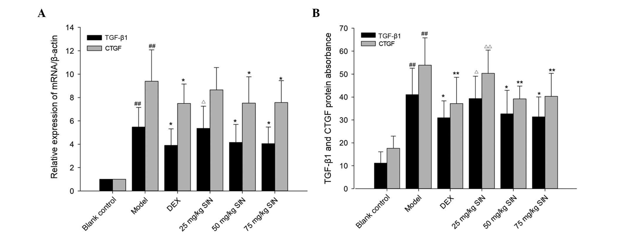
SIN modulated the TGF-β1 pathway
As demonstrated in Fig.
4, following the OVA-challenge, TGF-β1 and CTGF mRNA expression
was observed to be significantly increased compared with the blank
control group (P<0.01). TGF-β1 and CTGF mRNA expression levels
in the SIN (50 and 75 mg/kg) and DEX groups was significantly
reduced compared with those in the OVA group (P<0.05). No
significant differences in TGF-β1 and CTGF mRNA expression between
mice treated with SIN (50 and 75 mg/kg), and DEX were identified
(Fig. 4).
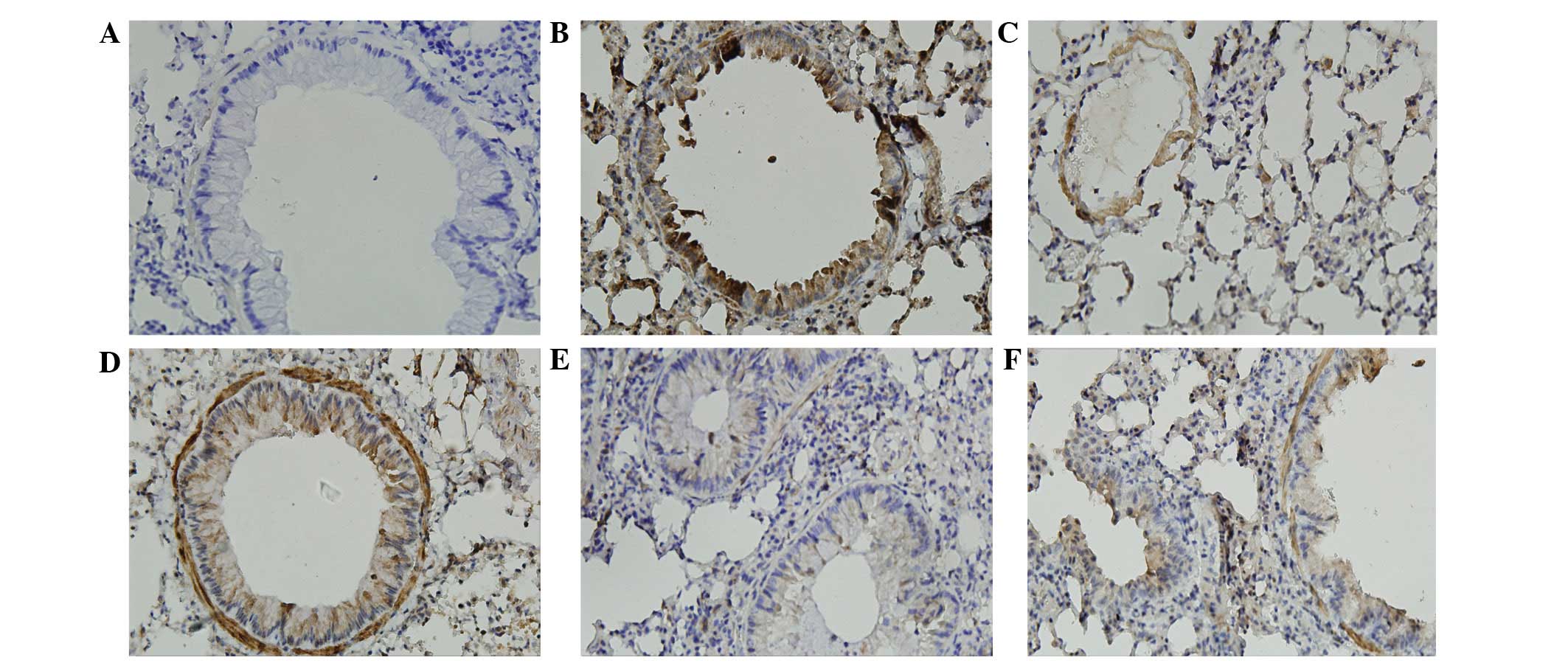
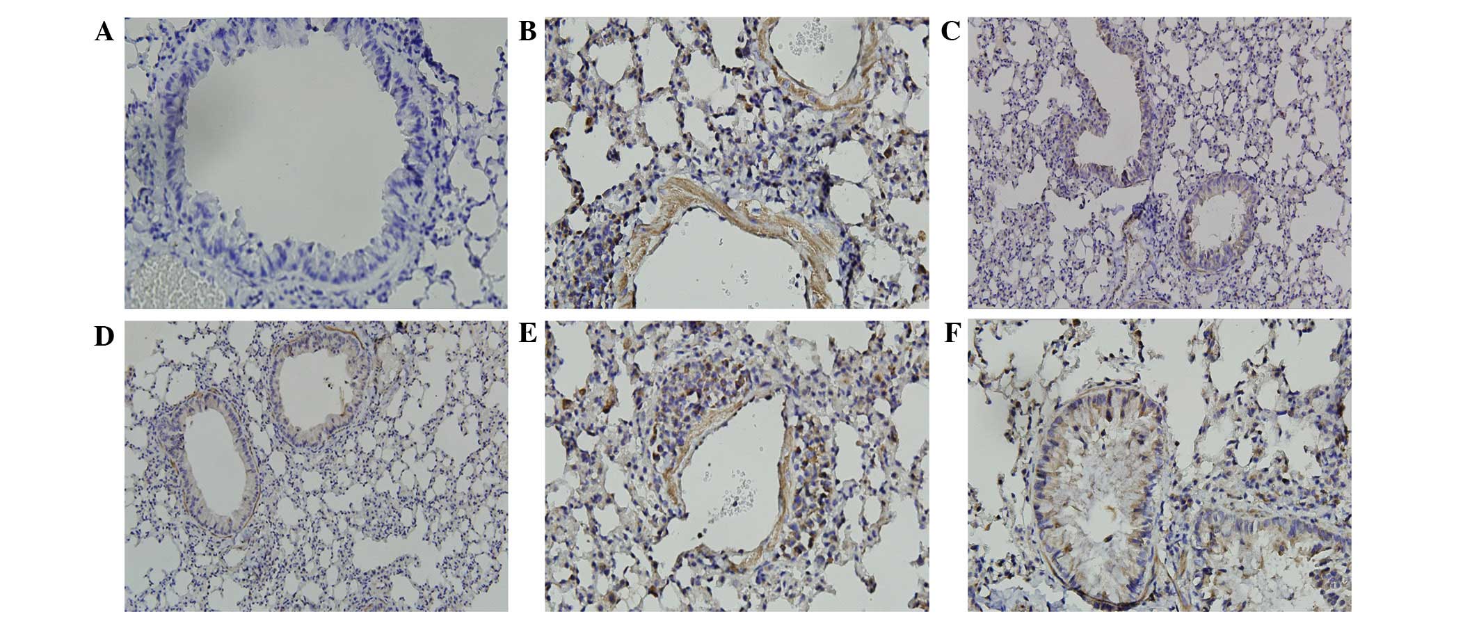
The immunostained areas of peribronchial TGF-β1 and
CTGF in the OVA group were greater than those in the control group
(Figs. 5 and 6). Administration of SIN (50 and 75
mg/kg) and DEX in OVA-challenged mice reduced the immunostained
area of TGF-β1 and CTGF compared with the OVA group. There were no
differences in TGF-β1 and CTGF expression levels between mice
treated with SIN (50 and 75 mg/kg) and DEX (Figs. 5 and 6).
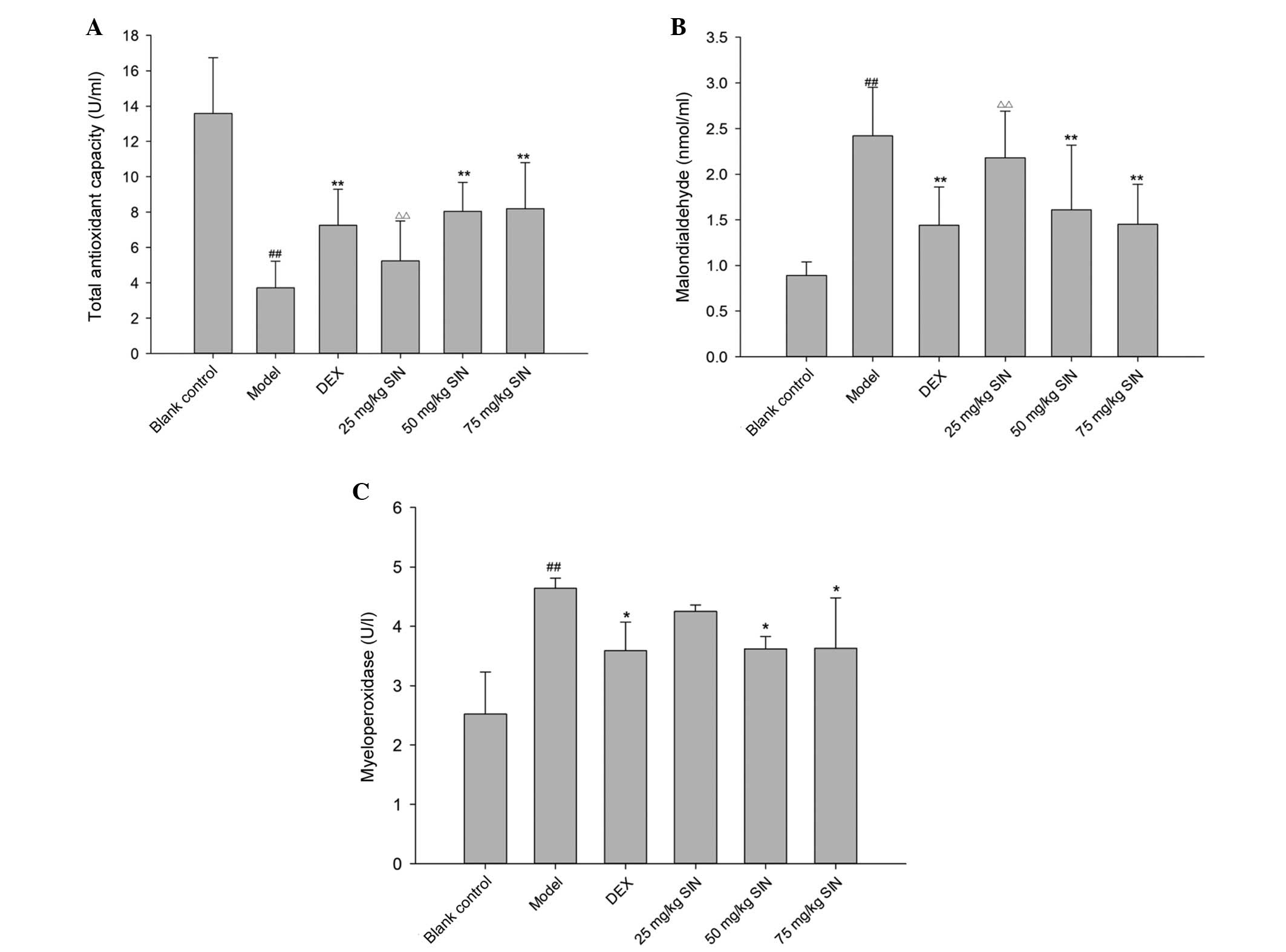
SIN attenuated pulmonary oxidative
stress
The model group presented lower TAC compared with
the blank control group (P<0.01; Fig. 7A). TAC was increased significantly
in the SIN (50 and 75 mg/kg) and DEX groups, compared with the
model group (Fig. 7A). The
concentration of lung MDA increased in the model group compared
with the blank control. (P<0.01; Fig. 7B) However, the increase was
significantly attenuated in the DEX and SIN (50 and 75 mg/kg)
treatment groups (P<0.01; Fig.
7B). Lung MPO activity was increased in the model group
compared with the blank control group, and reduced in the SIN (50
and 75 mg/kg) and DEX treatment groups compared with the model
group (Fig. 7C).
Discussion
The present study investigated the effects of SIN on
airway inflammation and remodeling in a mouse model of asthma. The
principle observation of the current study was that SIN was able to
inhibit airway inflammation and remodeling in an asthma mouse
model, however, there remains a limited amount of data regarding
the treatment of human patients. The results of the present study
do however demonstrate that SIN is able to reduce airway
inflammation and remodeling, perhaps by modulation of TGF-β1
pathways and pulmonary oxidative stress. Thus, SIN treatment may
have the potential to treat asthma.
Allergic asthma is characterized by allergen-induced
airway bronchoconstriction, inflammation involving eosinophils and
mucus hypersecretion and airway remodeling (24). Infiltrating inflammatory cells
entering the lung strongly contribute to the development of
allergic airway inflammation and remodeling (25). SIN, which is an alkaloid and a
morphinan derivative, has anti-inflammatory, antiarrhythmic,
antirheumatic, analgesic and immunosuppressive properties (16,26,27).
However, the effects of SIN on airway inflammation and remodeling
remain unclear. Although SIN is known to trigger release of
histamine, none of the mice with asthma died following its
administration in the present study. Utilizing this safe dose of
SIN, the present study demonstrated that models of OVA exposure for
6 weeks induced leukocyte infiltration to the lungs in mice.
Additionally, following exposure to OVA, mice displayed greater
eosinophil infiltration in the airway compared with mice in the
control group, a significant feature of airway inflammation in
asthma (28). SIN significantly
reduced the size and extent of leukocyte infiltration to the lung,
and also reduced the number of eosinophils surrounding airways in
the model. These effects were similar to those of mice treated with
DEX, suggesting that SIN was able to inhibit eosinophilic
inflammation.
Distinctive features of airway remodeling include
the thickening of the airway wall, increased smooth muscle mass,
goblet cell hyperplasia and mucous gland hypertrophy (29). The experimental model in the
present study reproduced several of the features observed in
asthmatic patients. It was indicated that OVA-sensitized animals
presented eosinophilic inflammation and airway remodeling in lungs,
including airway mucus expression, goblet cell hyperplasia,
collagen deposition and airway smooth muscle layer thickening.
Furthermore, it was observed that treatment with SIN reduced
collagen deposition and smooth muscle layer thickening, in addition
to mucus hypersecretion in a mouse model. It was demonstrated that
SIN has potential as a therapeutic agent for asthma through its
anti-remodeling properties. Compared with DEX, SIN has a similar
ability to prevent asthma airway remodeling. These observations
indicate potential for the use of this small molecular compound in
the treatment of asthma airway remodeling.
TGF-β is a profibrotic cytokine important in
promoting the structural alterations of airway remodeling, and is
highly expressed in asthma (30).
It has been demonstrated that airway wall remodeling is closely
associated with TGF-β in asthma (27,31).
CTGF is responsible for modulating the cellular response to TGF-β1,
and contributes to airway remodeling and the production of collagen
smooth muscle cells (11). The
present study demonstrated that OVA-challenge resulted in a
significant increase in TGF-β1 and CTGF levels, however, SIN was
able to block the TGF-β1 pathway by inhibiting TGF-β1 and CTGF
expression. No statistically significant differences between SIN
and DEX in TGF-β1 and CTGF expression levels were identified. These
observations suggest that the therapeutic action of SIN against
OVA-induced asthma may be associated with the inhibition of the
TGF-β1 pathway.
Oxidative stress is characterized by the excessive
production of reactive oxygen species (ROS) or reactive nitrogen
species (RNS) and the imbalance of oxidant/antioxidant response
(32). A large number of studies
have confirmed that ROS and RNS may lead to airway inflammation and
remodeling (33). Excessive ROS
and oxidative stress-mediated expression of TGF-β1 promote airway
remodeling in asthma (34). TAC is
an indicator possessing enzymatic and non-enzymatic properties,
allowing it to reflect the antioxidant status (35). MDA is the lipid peroxidation
product of a chain reaction of oxygen free radicals, and is
involved in amplification of injury by ROS (36). MPO can activate the matrix
metalloproteinases and is involved in asthmatic airway remodeling
(37). It has been elucidated that
increased MDA and MPO levels and reduced levels of TAC are present
in the peripheral blood, induced sputum and bronchoalveolar lavage
fluid of patients with asthma. These features are closely
associated with the severity of asthma (12,35).
The current study demonstrated that increased levels of TAC and
reduced levels of MDA or MPO were observed subsequent to SIN
administration, suggesting that SIN may inhibit oxidative stress in
asthma. This antioxidant effect of SIN may be associated with the
TGF-β1 pathway.
In conclusion, the current study reports that SIN
ameliorates airway inflammation and remodeling in vivo,
which may be associated with an ability to modulate the TGF-β1/CTGF
pathway and oxidative stress. These observations support the
hypothesis that SIN has therapeutic value for the treatment of
asthma.
References
|
1
|
Manuyakorn W, Howarth PH and Holgate ST:
Airway remodelling in asthma and novel therapy. Asian Pac J Allergy
Immunol. 31:3–10. 2013.PubMed/NCBI
|
|
2
|
Al-Muhsen S, Johnson JR and Hamid Q:
Remodeling in asthma. J Allergy Clin Immunol. 128:451–462; quiz
463–464. 2011. View Article : Google Scholar : PubMed/NCBI
|
|
3
|
Lederlin M, Ozier A, Montaudon M, Begueret
H, Ousova O, Marthan R, Berger P and Laurent F: Airway remodeling
in a mouse asthma model assessed by in-vivo respiratory-gated
micro-computed tomography. Eur Radiol. 20:128–137. 2010. View Article : Google Scholar
|
|
4
|
Kearley J, Buckland KF, Mathie SA and
Lloyd CM: Resolution of allergic inflammation and airway
hyperreactivity is dependent upon disruption of the T1/ST2-IL-33
pathway. Am J Respir Crit Care Med. 179:772–781. 2009. View Article : Google Scholar : PubMed/NCBI
|
|
5
|
King GG and Farah CS: Targeting airway
remodelling in asthma: The how, what and where? Respirology.
17:585–587. 2012. View Article : Google Scholar : PubMed/NCBI
|
|
6
|
Schmidt-Weber CB and Blaser K: The role of
TGF-beta in allergic inflammation. Immunol Allergy Clin North Am.
26:233–244. 2006. View Article : Google Scholar : PubMed/NCBI
|
|
7
|
Redington AE, Madden J, Frew AJ,
Djukanovic R, Roche WR, Holgate ST and Howarth PH: Transforming
growth factor-beta 1 in asthma. Measurement in bronchoalveolar
lavage fluid. Am Respir Crit Care Med. 156:642–647. 1997.
View Article : Google Scholar
|
|
8
|
Vignola AM, Chanez P, Chiappara G,
Merendino A, Pace E, Rizzo A, la Rocca AM, Bellia V, Bonsignore G
and Bousquet J: Transforming growth factor-beta expression in
mucosal biopsies in asthma and chronic bronchitis. Am J Respir Crit
Care Med. 156:591–599. 1997. View Article : Google Scholar : PubMed/NCBI
|
|
9
|
Grotendorst GR: Connective tissue growth
factor: A mediator of TGF-beta action on fibroblasts. Cytokine
Growth Factor Rev. 8:171–179. 1997. View Article : Google Scholar
|
|
10
|
Frazier K, Williams S, Kothapalli D,
Klapper H and Grotendorst GR: Stimulation of fibroblast cell
growth, matrix production, and granulation tissue formation by
connective tissue growth factor. J Invest Dermatol. 107:404–411.
1996. View Article : Google Scholar : PubMed/NCBI
|
|
11
|
Johnson PR, Burgess JK, Ge Q, Poniris M,
Boustany S, Twigg SM and Black JL: Connective tissue growth factor
induces extracellular matrix in asthmatic airway smooth muscle. Am
J Respir Crit Care Med. 173:32–41. 2006. View Article : Google Scholar
|
|
12
|
Sahiner UM, Birben E, Erzurum S, Sackesen
C and Kalayci O: Oxidative stress in asthma. World Allergy Organ J.
4:151–158. 2011. View Article : Google Scholar : PubMed/NCBI
|
|
13
|
Park CS, Kim TB, Lee KY, Moon KA, Bae YJ,
Jang MK, Cho YS and Moon HB: Increased oxidative stress in the
airway and development of allergic inflammation in a mouse model of
asthma. Ann Allergy Asthma Immunol. 103:238–247. 2009. View Article : Google Scholar : PubMed/NCBI
|
|
14
|
Baraket M, Oliver BG, Burgess JK, Lim S,
King GG and Black JL: Is low dose inhaled corticosteroid therapy as
effective for inflammation and remodeling in asthma? A randomized,
parallel group study. Respir Res. 13:112012. View Article : Google Scholar : PubMed/NCBI
|
|
15
|
Gao T, Hao J, Wiesenfeld-Hallin Z, Wang DQ
and Xu XJ: Analgesic effect of sinomenine in rodents after
inflammation and nerve injury. Eur J Pharmacol. 721:5–11. 2013.
View Article : Google Scholar : PubMed/NCBI
|
|
16
|
Wang Q and Li XK: Immunosuppressive and
anti-inflammatory activities of sinomenine. Int Immunopharmacol.
11:373–376. 2011. View Article : Google Scholar
|
|
17
|
Zhou H, Wong YF, Wang J, Cai X and Liu L:
Sinomenine ameliorates arthritis via MMPs, TIMPs, and cytokines in
rats. Biochem Biophys Res Commun. 376:352–357. 2008. View Article : Google Scholar : PubMed/NCBI
|
|
18
|
Temelkovski J, Hogan SP, Shepherd DP,
Foster PS and Kumar RK: An improved murine model of asthma:
Selective airway inflammation, epithelial lesions and increased
methacholine responsiveness following chronic exposure to
aerosolised allergen. Thorax. 3:849–856. 1998. View Article : Google Scholar
|
|
19
|
Duan W, Chan JH, Wong CH, Leung BP and
Wong WS: Anti-inflammatory effects of mitogen-activated protein
kinase kinase inhibitor U0126 in an asthma mouse model. J Immunol.
172:7053–7059. 2004. View Article : Google Scholar : PubMed/NCBI
|
|
20
|
Weibel ER: Principles and methods for the
morphometric study of the lung and other organs. Lab Invest.
12:131–155. 1963.PubMed/NCBI
|
|
21
|
Angeli P, Prado CM, Xisto DG, Silva PL,
Pássaro CP, Nakazato HD, Leick-Maldonado EA, Martins MA, Rocco PR
and Tibério IF: Effects of chronic L-NAME treatment lung tissue
mechanics, eosinophilic and extracellular matrix responses induced
by chronic pulmonary inflammation. Am J Physiol Lung Cell Mol
Physiol. 294:L1197–L1205. 2008. View Article : Google Scholar : PubMed/NCBI
|
|
22
|
Henderson WR Jr, Tang LO, Chu SJ, Tsao SM,
Chiang GK, Jones F, Jonas M, Pae C, Wang H and Chi EY: A role for
cysteinyl leukotrienes in airway remodeling in a mouse asthma
model. Am J Respir Crit Care Med. 165:108–116. 2002. View Article : Google Scholar : PubMed/NCBI
|
|
23
|
Zhou L, Qian ZX, Li F and Liu RY: The
effect of melatonin on the regulation of collagen accumulation and
matrix metal-loproteinase-9 and tissue inhibitor of matrix
metalloproteinase-1 mRNA and protein in a murine model of chronic
asthma. Zhonghua Jie He He Hu Xi Za Zhi. 30:527–532. 2007.In
Chinese. PubMed/NCBI
|
|
24
|
Toledo AC, Sakoda CP, Perini A, Pinheiro
NM, Magalhães RM, Grecco S, Tibério IF, Câmara NO, Martins MA, Lago
JH and Prado CM: Flavonone treatment reverses airway inflammation
and remodelling in an asthma murine model. Br J Pharmacol.
168:1736–1749. 2013. View Article : Google Scholar :
|
|
25
|
Oda H, Kawayama T, Imaoka H, Sakazaki Y,
Kaku Y, Okamoto M, Kitasato Y, Edakuni N, Takenaka S, Yoshida M, et
al: Interleukin-18 expression, CD8(+) T cells, and eosinophils in
lungs of nonsmokers with fatal asthma. Ann Allergy Asthma Immunol.
112:23–28. 2014. View Article : Google Scholar
|
|
26
|
Kok TW, Yue PY, Mak NK, Fan TP, Liu L and
Wong RN: The anti-angiogenic effect of sinomenine. Angiogenesis.
8:3–12. 2005. View Article : Google Scholar : PubMed/NCBI
|
|
27
|
Chen DP, Wong CK, Leung PC, Fung KP, Lau
CB, Lau CP, Li EK, Tam LS and Lam CW: Anti-inflammatory activities
of Chinese herbal medicine sinomenine and Liang Miao San on tumor
necrosis factor-α-activated human fibroblast-like synoviocytes in
rheumatoid arthritis. J Ethnopharmacol. 137:457–468. 2011.
View Article : Google Scholar : PubMed/NCBI
|
|
28
|
Busse WW and Lemanske RF Jr: Asthma. N
Engl J Med. 344:350–362. 2001. View Article : Google Scholar : PubMed/NCBI
|
|
29
|
Manuyakorn W: Airway remodelling in
asthma: Role for mechanical forces. Asia Pac Allergy. 4:19–24.
2014. View Article : Google Scholar : PubMed/NCBI
|
|
30
|
Holgate ST, Holloway J, Wilson S, Howarth
PH, Haitchi HM, Babu S and Davies DE: Understanding the
pathophysiology of severe asthma to generate new therapeutic
opportunities. J Allergy Clin Immunol. 117:496–506; quiz 507. 2006.
View Article : Google Scholar : PubMed/NCBI
|
|
31
|
Hardy CL, Nguyen HA, Mohamud R, Yao J, Oh
DY, Plebanski M, Loveland KL, Harrison CA, Rolland JM and O'Hehir
RE: The activin A antagonist follistatin inhibits asthmatic airway
remodelling. Thorax. 68:9–18. 2013. View Article : Google Scholar
|
|
32
|
Bowler RP and Crapo JD: Oxidative stress
in allergic respiratory diseases. J Allergy Clin Immunol.
110:349–356. 2002. View Article : Google Scholar : PubMed/NCBI
|
|
33
|
Sugiura H and Ichinose M: Oxidative and
nitrative stress in bronchial asthma. Antioxid Redox Signal.
10:785–797. 2008. View Article : Google Scholar : PubMed/NCBI
|
|
34
|
Brown SD, Baxter KM, Stephenson ST, Esper
AM, Brown LA and Fitzpatrick AM: Airway TGF-β1 and oxidant stress
in children with severe asthma: Association with airflow
limitation. J Allergy Clin Immunol. 129:388–396. 2012. View Article : Google Scholar
|
|
35
|
Fatani SH: Biomarkers of oxidative stress
in acute and chronic bronchial asthma. J Asthma. 51:578–584. 2014.
View Article : Google Scholar : PubMed/NCBI
|
|
36
|
Ayala A, Muñoz MF and Argüelles S: Lipid
peroxidation: production, metabolism, and signaling mechanisms of
malondialdehyde and 4-hydroxy-2-nonenal. Oxid Med Cell Longev.
2014:360–438. 2014. View Article : Google Scholar
|
|
37
|
Vignola AM, Riccobono L, Mirabella A,
Profita M, Chanez P, Bellia V, Mautino G, D'accardi P, Bousquet J
and Bonsignore G: Sputum metalloproteinase-9/tissue inhibitor of
metallopro-teinase-1 ratio correlates with airflow obstruction in
asthma and chronic bronchitis. Am J Respir Crit Care Med.
158:1945–1950. 1998. View Article : Google Scholar : PubMed/NCBI
|