Introduction
Clefts of the lip and palate (CLP) are common birth
defects. CLP can be corrected to varying degrees by surgery, dental
treatment, speech therapy and psychosocial intervention. CLP is not
a major cause of mortality in developed countries, however, it does
cause considerable morbidity in affected children and imposes a
societal and financial burden for families (1). Thus, preventing the occurrence of CLP
has important practical significance. In order for prevention
strategies to be effective, the etiology and pathogenesis of CLP
requires further elucidation.
Previous studies have demonstrated that various
environmental risk factors are associated with the incidence of a
cleft lip and palate, including exposure to dioxin-like chemicals
during pregnancy (2,3). In addition, mice exposed to
2,3,7,8-tetrachlorodibenzo-p-dioxin (TCDD) during
organo-genesis was shown to result in the formation of a cleft
palate (4). It has been
established that the transforming growth factor-β (TGF-β)
superfamily is important in palate development (5). TGF-β3 is considered to be an
important regulator of palatal fusion in mice, and TGF-β3-null mice
exhibited cleft palates without any other major deformities
(5). Congenital cleft palate
patients usually have aberrant expression of the TGF-β3 gene as a
result of epigenetic regulation rather than genetic mutations
(6,7). Our previous study determined that the
expression of TGF-β3 was gradually upregulated in TCDD-induced
cleft palates in fetal mice (8);
however, the underlying mechanism for elevated expression of TGF-β3
in the TCDD-induced cleft palate remains to be determined.
A previous study demonstrated that changes in DNA
methylation caused by environmental factors, such as maternal
exposure to all-trans-retinoic acid may be important in
cleft palate formation (9).
Additionally, treatment with lipopolysaccharide or TCDD may alter
DNA methylation in splenocytes (10). Histones are globular proteins that
undergo post-translational modifications and interact with DNA and
other nuclear proteins (11). H3
and H4 histones have long tails protruding from the nucleosome
which may be covalently modified by acetylation, methylation,
ubiquitination, phosphorylation, sumoylation, citrullination and
ADP-ribosylation, thus, modifying chromatin structure and gene
expression. Histone acetylation is a dynamic process regulated by
histone acetyltransferases (HATs) and histone deacetylases (HDACs).
HATs activate gene transcription via histone acetylation, while
HDACs inhibit gene transcription via histone deacetylation. Our
previous study revealed that the acetylation of histone H3 (H3) is
involved in TCDD-induced cleft palate development in C57BL/6J mice
(12). Therefore, it is possible
that an epigenetic mechanism may be involved in the TCDD-induced
cleft palate formation in fetal mice and may be one of the factors
contributing to the upregulation of TGF-β3 expression. The present
study aimed to elucidate the effects of epigenetic regulation,
including DNA methylation and histone acetylation, on TCDD-induced
cleft palate in fetal mice.
Materials and methods
Animals and drugs
The present study was approved by the Institutional
Review Board of the Children's Hospital of Chongqing Medical
University (Chongqing, China). C57BL/6J mice (46 female
experimental mice; 23 male mice to impregnate the females; age,
8–10 weeks) were obtained from the Experimental Animal Center of
Chongqing Medical University and treated in accordance with the
Guide for the Care and Use of Laboratory Animals, US National
Institutes of Health (13). Mice
were caged under controlled conditions of 22–24°C, 55±5% humidity,
a 12 h light/dark cycle and access to food and water ad
libitum. Two female mice were housed overnight with one male
mouse after one week of acclimation, and the presence of vaginal
plugs on the next morning was defined as gestation day 0 (GD
0).
TCDD and corn oil were purchased from Sigma-Aldrich
(St. Louis, MO, USA). TCDD was diluted with corn oil to 4
µg/ml.
Animal treatment and sample
collection
A total of 10 pregnant mice were randomly divided
into two groups (n=5). The TCDD group was administered a single
dose of 28 µg/kg TCDD by oral gavage on GD 10. The control
group was given an equal volume of corn oil by oral gavage on GD
10. All of these mice were sacrificed by cervical dislocation on GD
16.5. The body weight of the pregnant and fetal mice, and the
numbers of live and total fetal mice were recorded. The cleft
palate and other malformations were examined under a
stereomicroscope (SMZ1500; Nikon Corporation, Tokyo, Japan). The
palate was quickly isolated from each embryo and fixed overnight in
4% paraformaldehyde (Shanghai Chemical Reagent Co., Ltd., Shanghai,
China) for histological analysis. Fixed palates were dehydrated in
alcohol (80% ethanol for 60 min; 95% ethanol for 60 min, twice;
100% ethanol for 60 min, three times), embedded in paraffin
(Shanghai Chemical Reagent Co., Ltd.), and cut into 4-µm
sections. The palate sections were stained with hematoxylin and
eosin (Shanghai Chemical Reagent Co., Ltd.) and examined by light
microscopy (Eclipse 55i; Nikon Corporation).
An additional 36 pregnant mice were randomly divided
into TCDD and control groups (n=18) as described, and sacrificed on
GD 13.5 (pre-palate fusion), GD 14.5 (period of palate fusion), and
GD 15.5 (post-palate fusion). A total of 6 mice were sacrificed at
each time point. The palate was quickly removed from every embryo
following sacrifice of the mother and stored at −80°C for molecular
analysis.
Reverse transcription-quantitative
polymerase chain reaction (RT-qPCR)
The mRNA expression of TGF-β3 in the palates of
fetal mice was quantified by RT-qPCR. Total RNA was extracted from
the frozen palates using RNApure High-purity Total RNA Rapid
Extraction kit (BioTeke Corporation, Beijing, China) and 1,000 ng
was reverse transcribed into cDNA using PrimeScript™ RT reagent kit
(Takara Biotechnology Co., Ltd., Dalian, China). qPCR was performed
with a total reaction volume of 20 µl, containing 2
µl cDNA, 10 µl SYBR® Fast qPCR mix (Takara
Biotechnology Co., Ltd.), 0.8 µl of sense and antisense
primers, and 6.4 µl ddH2O by using StepOnePlus™
real-time PCR system (Applied Biosystems; Thermo Fisher Scientific,
Inc., Waltham, MA, USA). The primer sequences (Beijing SBS Genetech
Co., Ltd., Beijing, China) for TGF-β3 and β-actin were as follows:
Sense: 5′-CCTGGCCCTGCTGAACTTG-3′ and antisense:
5′-TTGATGTGGCCGAAGTCCAAC-3′ for TGF-β3; and sense:
5′-CCAGCCTTCCTTCTTGGGTAT-3′ and antisense:
5′-TTGGCATAGAGGTCTTTACGG-3′ for β-actin. The PCR was performed at
95°C for 30 sec, followed by 39 cycles of 95°C for 5 sec, 60°C for
30 sec, and 63°C for 30 sec. The quantification cycle indicated the
fractional cycle number at which the PCR product was first detected
above a fixed threshold. The relative mRNA expression levels were
determined by normalization to β-actin using the 2−ΔΔCq
method (14).
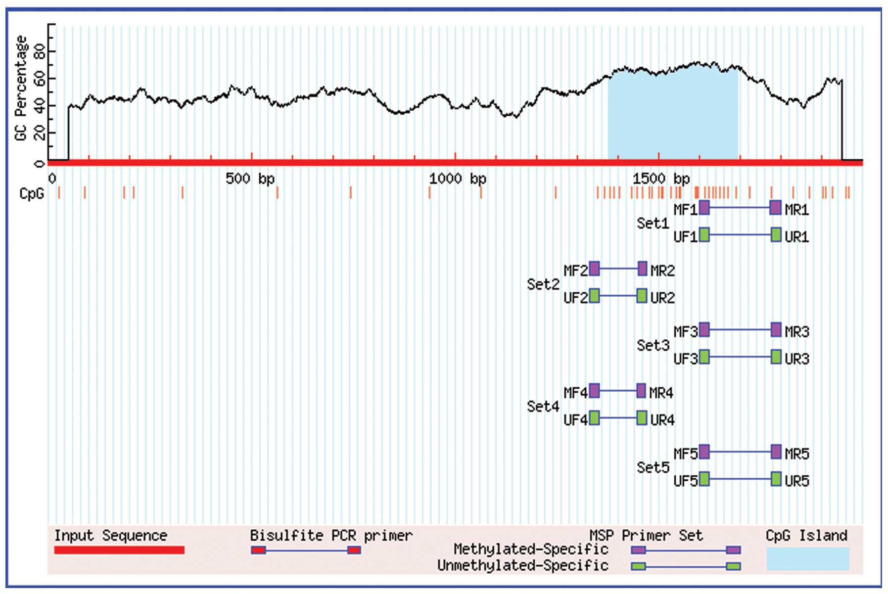
Methylation analysis
Methylation of the TGF-β3 gene promoter was examined
using a DNA methylation kit (Zymo Research Corporation, Irvine, CA,
USA). The TGF-β3 gene promoter sequences were obtained from the
UCSC Genome Browser Home and inputted into MethPrimer software
(15). The location of the CpG
island in the TGF-β3 gene promoter is shown in Fig. 1, and the primers for methylation
analysis were synthesized by Shanghai Biological Engineering Co.,
Ltd. (Shanghai, China) and are presented in Table I. Total DNA was extracted from the
frozen palates using a Cell/Tissue DNA Extraction kit (BioTeke
Corporation). PCR was performed in a reaction volume of 25
µl containing 2 µl cDNA, 12.5 µl Zymo Taq
PreMix (Zymo Research Corporation), 1 µl of forward and
reverse primers (Beijing SBS Genetech Co., Ltd.), and 8.5 µl
ddH2O. The PCR parameters were as follows: Denaturation
at 95°C for 10 min; 35 cycles of 95°C for 30 sec, 56°C for 40 sec,
and 72°C for 60 sec; and a final extension at 72°C for 7 min. Equal
quantities (5 µl) of PCR product from each group were
analyzed on a 1.5% agarose gel containing ethidium bromide
(Shanghai Chemical Reagent), and the target bands were analyzed
densitometrically on a Vistra Fluor Imager SI (Molecular Dynamics
Inc., Albuquerque, NM, USA).
 | Table ISequences of primers used for
methylation analysis. |
Table I
Sequences of primers used for
methylation analysis.
| Primer | Primer sequence | Product length
(bp) |
|---|
| Methylated TGF-β3
primer | F:
5′-TATTTAATTAGATTGGAAAGGAGCG-3′
R: 5′-AACCGAAAATACCCTCTAACGA-3′ | 138 |
| Non-methylated TGF-β3
primer | F:
5′-ATTTAATTAGATTGGAAAGGAGTGG-3′
F: 5′-AAAAAACCAAAAATACCCTCTAACAA-3′ | 141 |
Detection of HAT activity
Palatal tissues (0.05–0.1 g) were homogenized in 300
µl of ice-cold lysis buffer (50 mM NaCl, 0.5% Triton X-100,
10 mM HEPES, 1 mM EDTA, 0.05% 2-mercaptoethanol, 0.1 mM polymethyl
sulphonate and 10 µg/ml aprotinin). After centrifugation at
22 x g and 4°C for 10 min, the supernatant lysates were collected
and stored at −80°C. The palatal protein concentration was
quantified using the bicinchoninic acid assay reagent (BioTeke
Corporation) and adjusted to 1.25 µg/µl. Next, 68
µl lysate was mixed with HAT activity analysis liquid
(BioVision, Inc., Milpitas, CA, USA), added to a 96-well plate, and
incubated at 37°C for 4 h. The optical density was measured at a
wavelength of 440 nm using a microplate reader (Varioskan™ Flash;
Thermo Fisher Scientific, Inc.). The HAT activity was determined by
the optical density of the experimental group minus that of the
blank control.
Western blotting
A total of 50 µg of whole cell lysate was
separated by 10% sodium dodecyl sulfate-polyacrylamide gel
electrophoresis (Beyotime Institute of Biotechnology, Haimen,
China) at 60 V for 30 min and 110 V for 1 h, and transferred onto a
polyvinylidene difluoride membrane (Immobilon; EMD Millipore,
Billerica, MA, USA). After blocking in 5% non-fat dry milk, the
membranes were incubated with primary antibodies overnight at 4°C,
as follows: Rabbit polyclonal anti-acetylated H3 (Ac-H3, 1:10,000,
EMD Millipore; cat. no. 382158) and rat monoclonal anti-laminin β1
(1:500, Abcam, Cambridge, MA, USA; cat. no. ab44941) overnight at
4°C. Polyclonal horseradish peroxidase-conjugated goat anti-rabbit
IgG (1:5,000; OriGene Technologies, Beijing, China; cat. no.
TA130023) was used as the secondary antibody, and the bands were
detected using an Enhanced Chemiluminescence Detection kit (Nanjing
KeyGen Biotech Co., Ltd. Nanjing, China) and analyzed by Quantity
One Software (version 4.62; Bio-Rad Laboratories, Inc.). Protein
expression was determined relative to expression of laminin β1.
Statistical analysis
Data are expressed as the mean ± standard deviation
and analyzed using SPSS 17.0 (SPSS, Inc., Chicago, IL, USA).
Wilcoxon rank sum test was used to compare count data and the
Student's t-test was used to compare the data between two groups.
P<0.05 was considered to indicate a statistically significant
difference.
Results
Effects of TCDD on pregnant and fetal
mice
The appearance, feeding, drinking, and activity of
the mice were not affected by TCDD treatment. There was no
significant difference in the weight gain of pregnant mice, body
weights of live fetal mice, or the numbers of live fetuses per
litter in the TCDD group compared with the control group
(P>0.05, Table II). No cleft
palates were observed in fetal mice in the control group, while
93.55% of fetal mice in the TCDD group had a cleft palate. The
incidence of a cleft palate was significantly different between the
two groups (P<0.01, Table II).
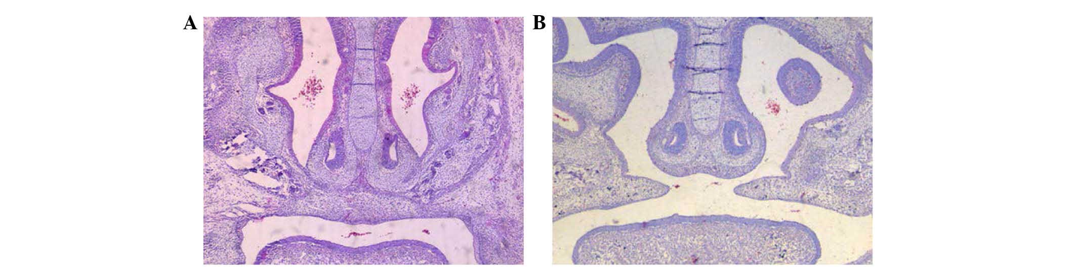
Additionally, on GD 16.5, the palatine process fused to the
opposite side and formed a complete palate in the control group,
whereas the palatine process in the TCDD group was not complete and
a cleft was observed in the middle (Fig. 2).
| Table IIDevelopment of pregnant mice and fetal
mice and the incidence of cleft palate in each group. |
Table II
Development of pregnant mice and fetal
mice and the incidence of cleft palate in each group.
| Group (n=5) | Pregnant mouse weight
gain (g) | Live fetus body
weight (g) | Number of fetal mice
|
|---|
| Total | Cleft palate | Stillbirth or
absorbed fetus | Incidence of cleft
palate (%) |
|---|
| Control | 2.05±0.43 | 1.08±0.10 | 33 | 0 | 0 | 0.00 |
| TCDD (28
µg/kg) | 1.79±0.57 | 1.06±0.11 | 31 | 29 | 1 | 93.55a |
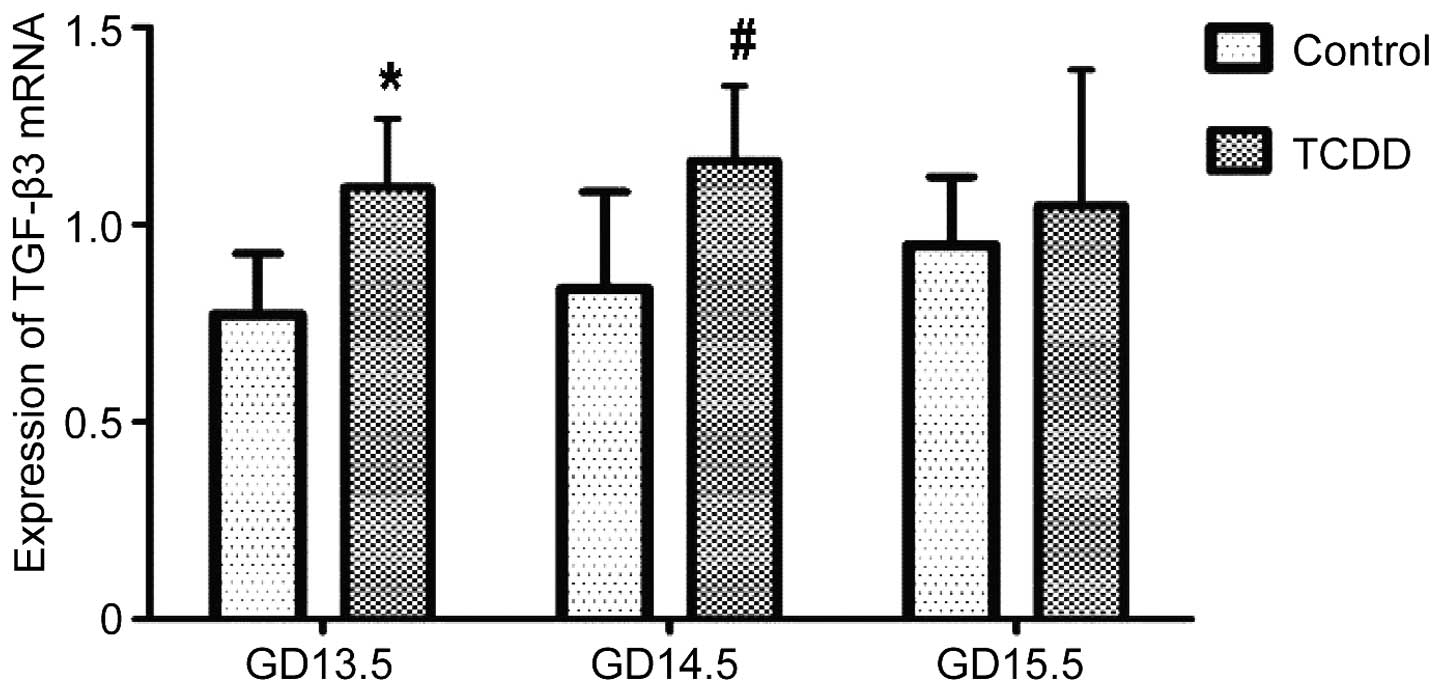
Effect of TCDD on TGF-β3 mRNA expression
levels in the palate and on methylation in the TGF-β3 gene
promoter
The relative expression of TGF-β3 mRNA in the palate
of fetal mice was normalized to that of β-actin. On GD 13.5 and GD
14.5, TGF-β3 mRNA expression was significantly greater in the TCDD
group compared with the control (P<0.01 and P<0.05,
respectively; Fig. 3); however,
the mRNA expression level was not significantly different on GD
15.5 between the two groups (P>0.05, Fig. 3). In addition, the effect of TCDD
on methylation of the TGF-β3 gene promoter was examined using PCR.
Notably, on GD 13.5, GD 14.5 and GD 15.5, only unmethylated bands
were observed in the TCDD and control groups (Fig. 4).
 | Figure 4Detection of methylation status in the
TGF-β3 gene promoter by methylation-specific polymerase chain
reaction. Marker, 100–600 bp; M, methylation band (138 bp); U,
nonmethylated band (141 bp); 1–3, GD13.5, GD14.5, and GD15.5,
respectively, in the control group; 4–6, GD13.5, GD14.5 and GD15.5,
respectively, in the TCDD group. TGF-β3, transforming growth
factor-β3; GD, gestation day; TCDD,
2,3,7,8-tetrachlorodibenzo-p-dioxin. |
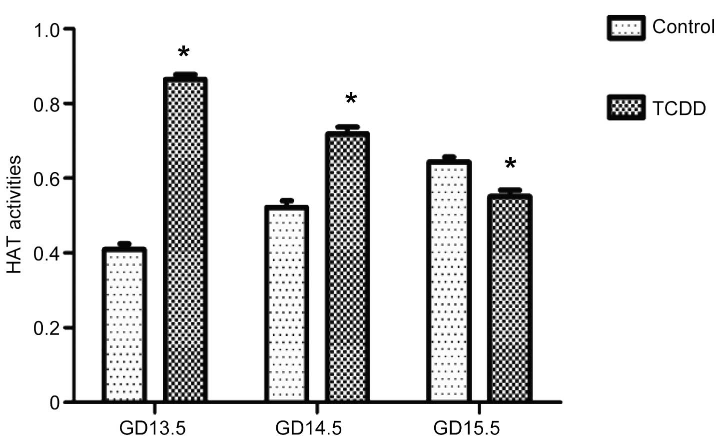
Effect of TCDD on HAT activity
On GD 13.5 and GD 14.5, HAT activity in the TCDD
group was significantly higher compared with the control group
(P<0.01). Conversely, on GD 15.5, it was significantly lower
than that in the control group (P<0.01, Fig. 5). HAT activity in the control group
was gradually increased from GD 13.5 to GD 15.5 by contrast, its
activity in the TCDD group was significantly reduced (P<0.05,
Fig. 5).
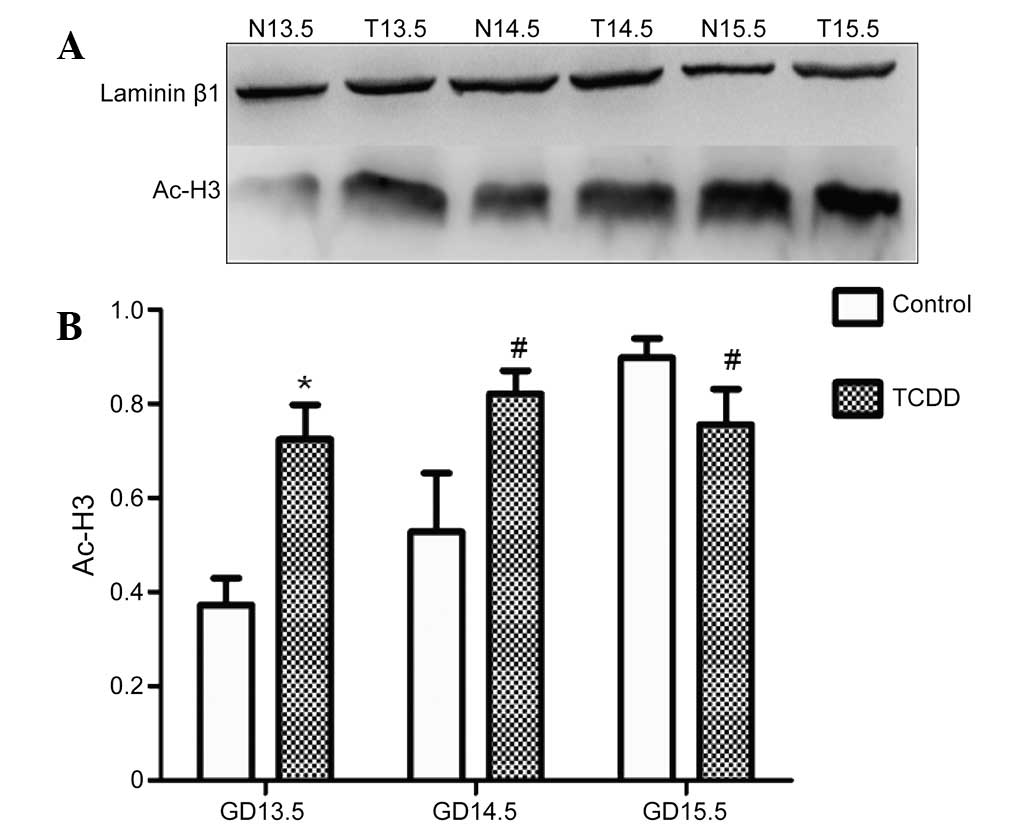
Effect of TCDD on H3 acetylation
The level of H3 acetylation in the TCDD group was
significantly increased on GD 13.5 and GD 14.5 compared with the
control group (P<0.01 and P<0.05, respectively; Fig. 6). On GD 15.5, it was significantly
reduced (P<0.05; Fig. 6). H3
acetylation level in the control group was increased from GD 13.5
to GD 15.5; however, in the TCDD group, it was not significantly
affected from GD 13.5 to GD 15.5 (Fig.
6).
Discussion
The combination of gene mutations and environmental
effects contribute to the complex etiology of cleft palate
formation. Although progress has been made in screening mutant
genes in the cells of the cleft palate, it is unclear how
environmental factors can result in this defect. Environmental
influences can adversely affect the development of the secondary
palate through genetic variants or epigenetic alterations. Single
nucleotide polymorphisms (SNPs) in specific genes confer an
increased risk for adverse developmental outcomes subsequent to
exposure to certain environmental hazards. For example, infants
with a specific transforming growth factor-α polymorphism whose
mothers smoked cigarettes during their pregnancy exhibit an
increased risk for cleft palate (16–18).
Although SNPs are easily identifiable due to the change in DNA
sequence, epigenetic alterations may result in gene expression
changes without altering the DNA sequence (19), and so cannot be identified through
genome-wide association studies that determine nucleotide changes.
Epigenetics include, but are not limited to, DNA methylation,
histone modification, and microRNA function (19). The pollutant TCDD, a highly toxic
halogenated aromatic hydrocarbon, has been implicated in the
etiology of various human diseases and birth defects, including the
cleft palate. However, the exact mechanisms of TCDD-induced cleft
palate formation remain to be fully elucidated.
TGF-β3 is considered to be an important regulator of
palatal fusion in mice as TGF-β3-null mice were shown to exhibit a
cleft palate but did not have any other major deformities (5). Exposure to dioxin in vivo and
in vitro, leads to a reduction in the number of filopodial
extensions at the medial epithelial edge of the developing palate,
similar to what was observed in TGF-β3-null mice (20). The addition of TGF-β3 to an in
vitro palate culture model prevented the dioxin-induced
reduction in filopodial density (20). TGF-β3 exposure completely prevented
the dioxin-induced block of palatal fusion (20). Our previous study revealed that the
expression of TGF-β3 was gradually upregulated in the TCDD-treated
cleft palate (32 and 64 µg/kg/day TCDD on GD 10) (8). In the present study, the optimal dose
of TCDD was reduced to 28 µg/kg to induce a cleft palate
(21) and the epigenetic
mechanisms were investigated. Consistent with our previous study,
TGF-β3 mRNA expression was significantly increased in the
TCDD-induced cleft palate on GD 13.5 and GD 14.5. However, when the
methylation status of the TGF-β3 promoter was examined, only
unmethylated bands were observed in the cleft palate from the TCDD
and control groups. In human and mouse genomes, ~50% of all CpG
islands are remote from annotated promoters but often have
promoter-like features (22). In a
previous study, dioxin exposure was associated with changes in DNA
methylation (23). TCDD may induce
CpG methylation of the breast cancer-1 (BRCA-1) gene and reduce
BRCA-1 expression in breast cancer cell lines (24). Additionally, when a pregnant female
was exposed to TCDD epigenetic transgenerational inheritance of
adult-onset disease and sperm epimutations through altering DNA
methylated regions in gene promoters occurred (25). Therefore, TCDD may affect the
methylation status of other gene promoters, global DNA methylation,
and promoter-like CpG island methylation in the TCDD-induced cleft
palate. However, these mechanisms require further
investigation.
Histones are globular proteins that undergo
posttranslational modifications and interact with DNA and other
nuclear proteins (11). H3 and H4
histones have long tails protruding from the nucleosome, which may
be covalently modified by acetylation, methylation, ubiquitination,
phosphorylation, sumoylation, citrullination and ADP-ribosylation,
thus influencing chromatin structure and gene expression. Histone
acetylation is a dynamic process regulated by HATs and HDACs. HATs
activate gene transcription through histone acetylation, while
HDACs inhibit gene transcription through histone deacetylation. The
key period for palate development is between GD 13.5 and GD 14.5;
therefore, TCDD may regulate HAT activity and H3 acetylation at the
critical period of palate fusion. HAT activity and the acetylated
H3 level in the cleft palates from the TCDD group was observed to
be significantly higher in the present study on GD 13.5 and GD 14.5
than those from the control group. Since palate fusion completes on
GD 15.5, the HAT activity and acetylated H3 levels in the TCDD
group were determined to be significantly lower than those in the
control group of the current study. The HAT activity and acetylated
H3 levels in the control group were increased from GD 13.5 to GD
15.5; however, in the TCDD group, the HAT activity was gradually
reduced and the acetylated histone H3 level was not significantly
affected. These data indicate that histone acetylation is important
in cleft palate development and that the TCDD-induced cleft palate
may be the result of the deregulation of HAT activity and histone
H3 acetylation. However, the underlying mechanisms for the
deregulation of histone acetylation remain to be determined. A
previous study reported that the exposure of pregnant women to
valproic acid, an antiepileptic drug and an established human
teratogen, led to neural tube defects and orofacial clefts,
including a cleft palate (26).
The teratogenicity of this drug was likely due to its ability to
inhibit histone deacetylases, thereby altering the chromatin
conformation and regulating gene transcription (27). In addition, the increased
deacetylation of histones was important in the TCDD-induced
reduction in luteinizing hormone β in the fetal pituitary (28). It has been determined that the
acetylation of H3 and H4 was markedly increased at the CYP1B1
promoter in the MCF-7 human breast cancer cell line following
dioxin treatment, which is dependent on p300 (29). In conjunction, these studies
indicate that histone acetylation is involved in dioxin-induced
teratogenic and pathogenic mechanisms.
In conclusion, 28 µg/kg TCDD successfully
induced cleft palate development in fetal mice, and upregulated
TGF-β3 mRNA expression, HAT activity and H3 levels in the cleft
palate. Therefore, TCDD-induced cleft palate development may be
associated with the deregulation of histone acetylation. Other
epigenetic mechanisms may be involved in TCDD-induced cleft palate
and require further research, which may contribute towards
strategies of cleft palate prevention.
Acknowledgments
The present study was funded by the National Natural
Science Youth Foundation (grant no. 81202167), the National Key
Clinical Specialist Construction Project Foundation [(2013) 544]
and the Yuzhong District Science and technology planning project
(grant nos. 20130121 and 20150112).
References
|
1
|
Wehby GL and Cassell CH: The impact of
orofacial clefts on quality of life and healthcare use and costs.
Oral Dis. 16:3–10. 2010. View Article : Google Scholar
|
|
2
|
Leite IC, Paumgartten FJ and Koifman S:
Chemical exposure during pregnancy and oral clefts in newborns. Cad
Saude Publica. 18:17–31. 2002. View Article : Google Scholar : PubMed/NCBI
|
|
3
|
Murray JC: Gene/environment causes of
cleft lip and/or palate. Clin Genet. 61:248–256. 2002. View Article : Google Scholar : PubMed/NCBI
|
|
4
|
Yamada T, Hirata A, Sasabe E, Yoshimura T,
Ohno S, Kitamura N and Yamamoto T: TCDD disrupts posterior
palato-genesis and causes cleft palate. J Craniomaxillofac Surg.
42:1–6. 2014. View Article : Google Scholar
|
|
5
|
Proetzel G, Pawlowski SA, Wiles MV, Yin M,
Boivin GP, Howles PN, Ding J, Ferguson MW and Doetschman T:
Transforming growth factor-beta 3 is required for secondary palate
fusion. Nat Genet. 11:409–414. 1995. View Article : Google Scholar : PubMed/NCBI
|
|
6
|
Marazita ML, Lidral AC, Murray JC, Field
LL, Maher BS, Goldstein McHenry T, Cooper ME, Govil M, Daack-Hirsch
S, Riley B, et al: Genome scan, fine-mapping and candidate gene
analysis of non-syndromic cleft lip with or without cleft palate
reveals phenotype-specific differences in linkage and association
results. Hum Hered. 68:151–170. 2009. View Article : Google Scholar :
|
|
7
|
Jenuwein T: The epigenetic magic of
histone lysine methylation. FEBS J. 273:3121–3135. 2006. View Article : Google Scholar : PubMed/NCBI
|
|
8
|
Gan LQ, Fu YX, Liu X, Qiu L, Wu SD, Tian
XF, Liu Y and Wei GH: Transforming growth factor-beta3 expression
up-regulates on cleft palates induced by
2,3,7,8-tetrachloro-dibenzo-p-dioxin in mice. Toxicol Ind Health.
25:473–478. 2009. View Article : Google Scholar : PubMed/NCBI
|
|
9
|
Kuriyama M, Udagawa A, Yoshimoto S,
Ichinose M, Sato K, Yamazaki K, Matsuno Y, Shiota K and Mori C: DNA
meth-ylation changes during cleft palate formation induced by
retinoic acid in mice. Cleft Palate Craniofac J. 45:545–551. 2008.
View Article : Google Scholar : PubMed/NCBI
|
|
10
|
McClure EA, North CM, Kaminski NE and
Goodman JI: Changes in DNA methylation and gene expression during
2,3,7,8-tetrachlorodibenzo-p-dioxin-induced suppression of the
lipopolysaccharide-stimulated IgM response in splenocytes. Toxicol
Sci. 120:339–348. 2011. View Article : Google Scholar : PubMed/NCBI
|
|
11
|
Kouzarides T: Chromatin modifications and
their function. Cell. 128:693–705. 2007. View Article : Google Scholar : PubMed/NCBI
|
|
12
|
Cuiping L, Xingang Y, Yuexian F, Lin Q,
Xiaofei T, Yan L and Guanghui W: The role of histone H3 acetylation
on cleft palate in mice induced by
2,3,7,8-tetrachlorodibenzopdioxin. Zhonghua Zheng Xing Wai Ke Za
Zhi. 30:369–372. 2014.In Chinese. PubMed/NCBI
|
|
13
|
National Research Council: Guide for the
Care and Use of Laboratory Animals. 8th edition. National Academy
Press; Washington, DC: 2011
|
|
14
|
Livak KJ and Schmittgen TD: Analysis of
relative gene expression data using real-time quantitative PCR and
the 2(-Delta Delta C(T)) method. Methods. 25:402–408. 2001.
View Article : Google Scholar
|
|
15
|
Li LC and Dahiya R: MethPrimer: Designing
primers for methylation PCRs. Bioinformatics. 18:1427–1431. 2002.
View Article : Google Scholar : PubMed/NCBI
|
|
16
|
Beaty TH, Maestri NE, Hetmanski JB,
Wyszynski DF, Vanderkolk CA, Simpson JC, McIntosh I, Smith EA,
Zeiger JS, Raymond GV, et al: Testing for interaction between
maternal smoking and TGFA genotype among oral cleft cases born in
Maryland 1992–1996. Cleft Palate Craniofac J. 34:447–454. 1997.
View Article : Google Scholar : PubMed/NCBI
|
|
17
|
Hwang SJ, Beaty TH, Panny SR, Street NA,
Joseph JM, Gordon S, McIntosh I and Francomano CA: Association
study of transforming growth factor alpha (TGF alpha) TaqI
polymorphism and oral clefts: Indication of gene-environment
interaction in a population-based sample of infants with birth
defects. Am J Epidemiol. 141:629–636. 1995.PubMed/NCBI
|
|
18
|
Shaw GM, Wasserman CR, Lammer EJ, O'Malley
CD, Murray JC, Basart AM and Tolarova MM: Orofacial clefts,
parental cigarette smoking, and transforming growth factor-alpha
gene variants. Am J Hum Genet. 58:551–561. 1996.PubMed/NCBI
|
|
19
|
Seelan RS, Mukhopadhyay P, Pisano MM and
Greene RM: Developmental epigenetics of the murine secondary
palate. ILAR J. 53:240–252. 2012. View Article : Google Scholar
|
|
20
|
Thomae TL, Stevens EA and Bradfield CA:
Transforming growth factor-beta3 restores fusion in palatal shelves
exposed to 2,3,7,8-tetrachlorodibenzo-p-dioxin. J Biol Chem.
280:12742–12746. 2005. View Article : Google Scholar : PubMed/NCBI
|
|
21
|
He X, Liu C, Pu Y, Gan L, Yuan X, Wei G
and Fu Y: Be based on the morphological and histological changes to
study optimal dose of TCDD induced cleft palate in mice embryo. Wei
Sheng Yan Jiu. 42:277–281. 2013.In Chinese. PubMed/NCBI
|
|
22
|
Deaton AM, Webb S, Kerr AR, Illingworth
RS, Guy J, Andrews R and Bird A: Cell type-specific DNA methylation
at intragenic CpG islands in the immune system. Genome Res.
21:1074–1086. 2011. View Article : Google Scholar : PubMed/NCBI
|
|
23
|
Lind L, Penell J, Luttropp K, Nordfors L,
Syvänen AC, Axelsson T, Salihovic S, van Bavel B, Fall T, Ingelsson
E and Lind PM: Global DNA hypermethylation is associated with high
serum levels of persistent organic pollutants in an elderly
population. Environ Int. 59:456–461. 2013. View Article : Google Scholar : PubMed/NCBI
|
|
24
|
Papoutsis AJ, Borg JL, Selmin OI and
Romagnolo DF: BRCA-1 promoter hypermethylation and silencing
induced by the aromatic hydrocarbon receptor-ligand TCDD are
prevented by resveratrol in MCF-7 cells. J Nutr Biochem.
23:1324–1332. 2012. View Article : Google Scholar
|
|
25
|
Manikkam M, Tracey R, Guerrero-Bosagna C
and Skinner MK: Dioxin (TCDD) induces epigenetic transgenerational
inheritance of adult onset disease and sperm epimutations. PLoS
One. 7:e462492012. View Article : Google Scholar : PubMed/NCBI
|
|
26
|
Ornoy A: Valproic acid in pregnancy: How
much are we endangering the embryo and fetus? Reprod Toxicol.
28:1–10. 2009. View Article : Google Scholar : PubMed/NCBI
|
|
27
|
Phiel CJ, Zhang F, Huang EY, Guenther MG,
Lazar MA and Klein PS: Histone deacetylase is a direct target of
valproic acid, a potent anticonvulsant, mood stabilizer and
teratogen. J Biol Chem. 276:36734–36741. 2001. View Article : Google Scholar : PubMed/NCBI
|
|
28
|
Takeda T, Fujii M, Taura J, Ishii Y and
Yamada H: Dioxin silences gonadotropin expression in perinatal pups
by inducing histone deacetylases: A new insight into the mechanism
for the imprinting of sexual immaturity by dioxin. J Biol Chem.
287:18440–18450. 2012. View Article : Google Scholar : PubMed/NCBI
|
|
29
|
Ingelman-Sundberg M, Zhong XB, Hankinson
O, Beedanagari S, Yu AM, Peng L and Osawa Y: Potential role of
epigenetic mechanisms in the regulation of drug metabolism and
transport. Drug Metab Dispos. 41:1725–1731. 2013. View Article : Google Scholar : PubMed/NCBI
|