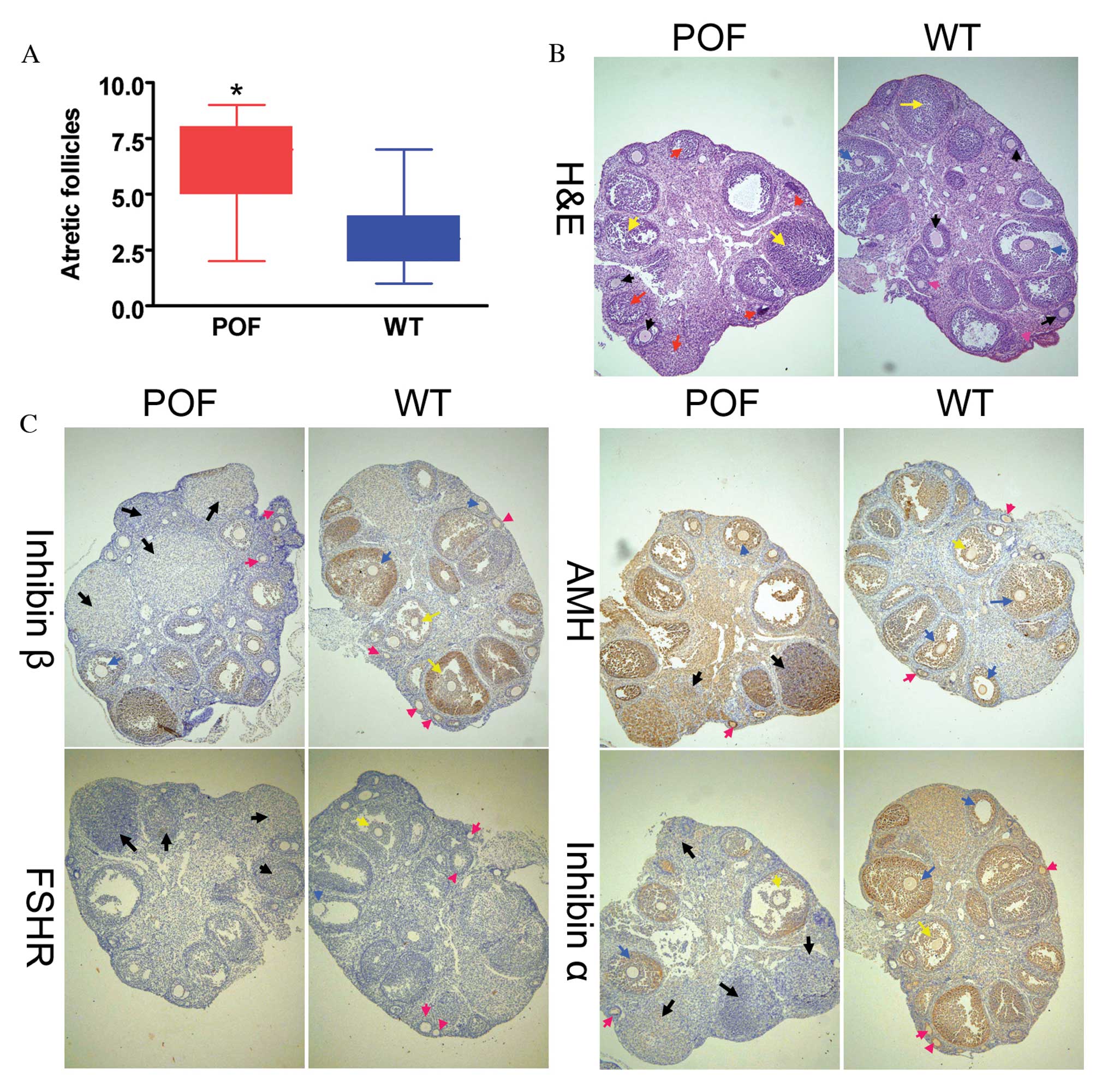
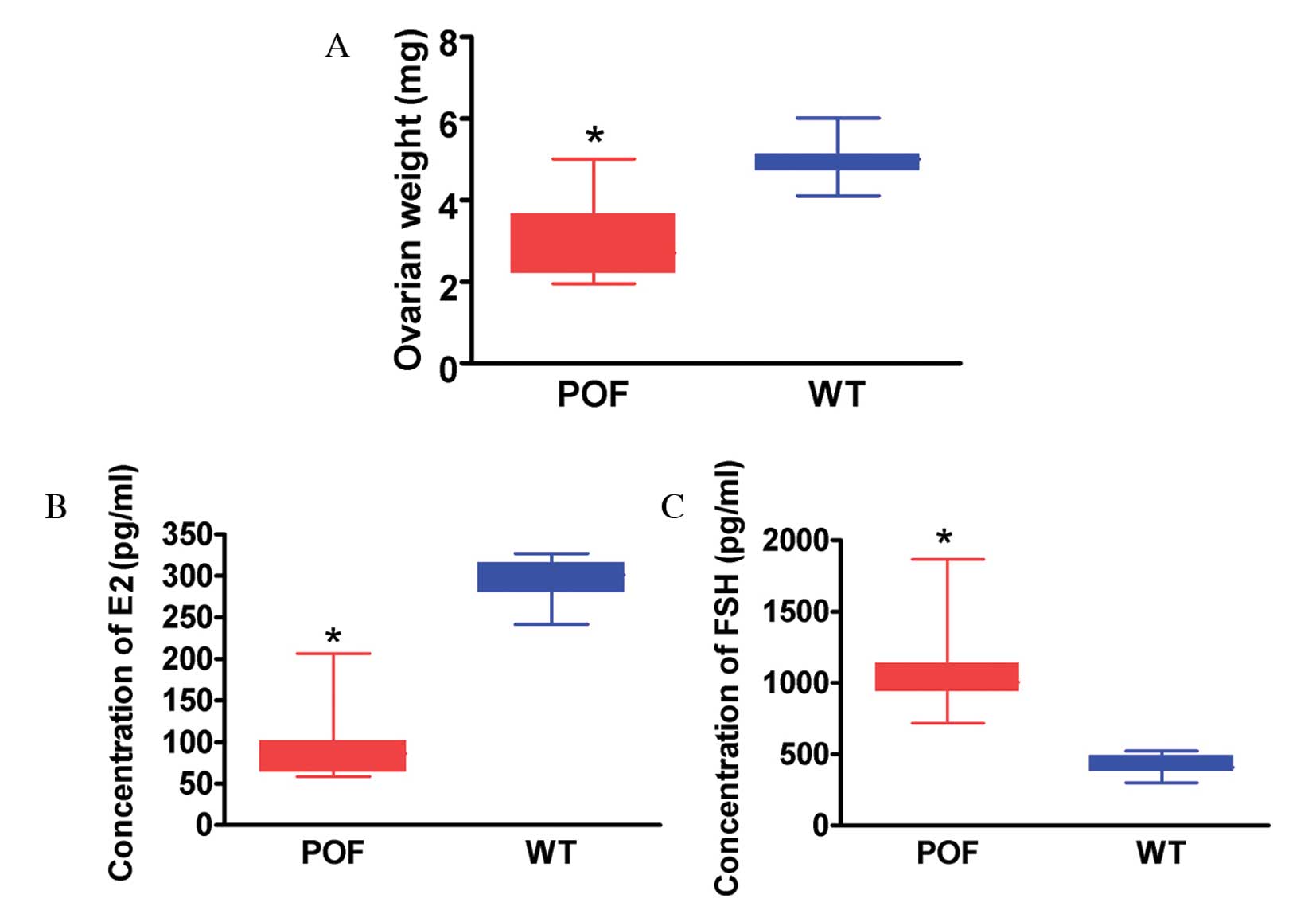
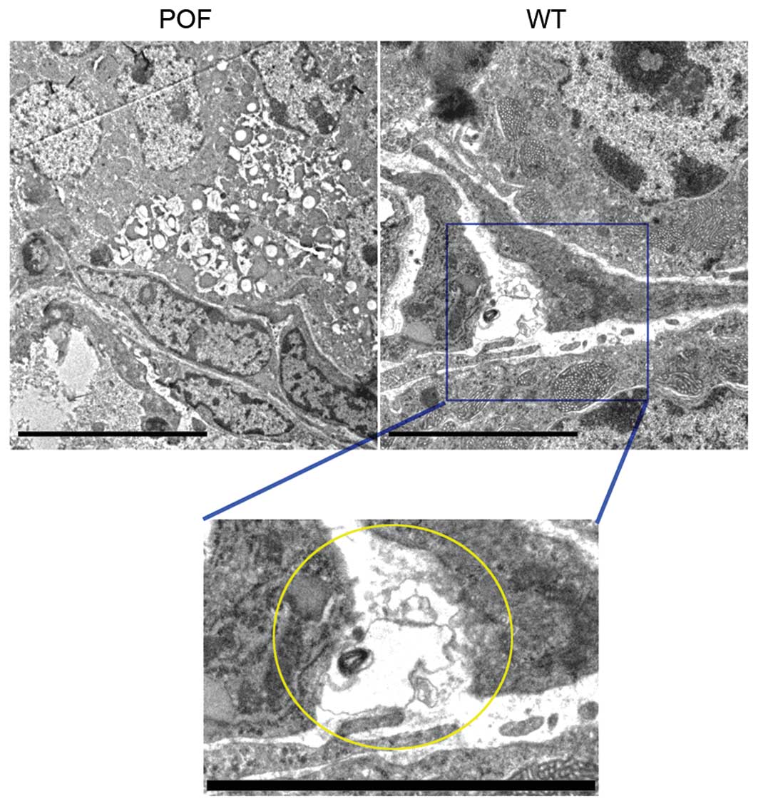
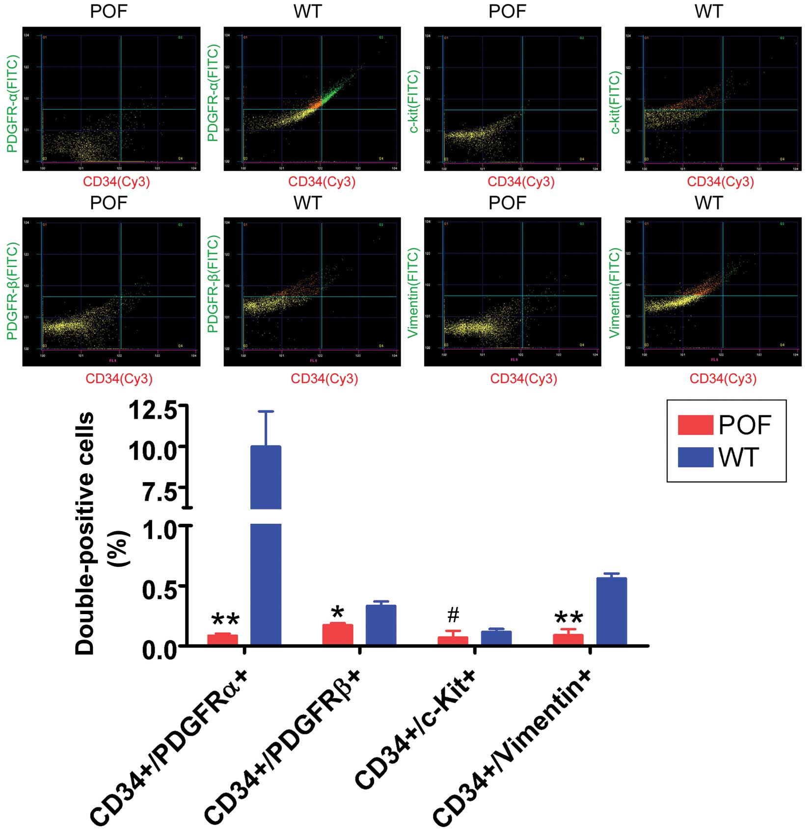
|
1
|
Beck-Peccoz P and Persani L: Premature
ovarian failure. Orphanet J Rare Dis. 1:92006. View Article : Google Scholar : PubMed/NCBI
|
|
2
|
Vujović S, Ivović M, Tancić-Gajić M,
Marina L, Barać M, Arizanović Z, Nenezić A, Ivanisević M, Micić J,
Sajić S and Micić D: Premature ovarian failure. Srp Arh Celok Lek.
140:806–811. 2012. View Article : Google Scholar
|
|
3
|
Liu T, Huang Y, Zhang J, Qin W, Chi H,
Chen J, Yu Z and Chen C: Transplantation of human menstrual blood
stem cells to treat premature ovarian failure in mouse model. Stem
Cells Dev. 23:1548–1557. 2014. View Article : Google Scholar : PubMed/NCBI
|
|
4
|
Liu T, Qin W, Huang Y, Zhao Y and Wang J:
Induction of estrogen-sensitive epithelial cells derived from
human-induced pluripotent stem cells to repair ovarian function in
a chemotherapy-induced mouse model of premature ovarian failure.
DNA Cell Biol. 32:685–698. 2013. View Article : Google Scholar : PubMed/NCBI
|
|
5
|
Liu T, Huang Y, Guo L, Cheng W and Zou G:
CD44+/CD105+ human amniotic fluid mesenchymal stem cells survive
and proliferate in the ovary long-term in a mouse model of
chemotherapy-induced premature ovarian failure. Int J Med Sci.
9:592–602. 2012. View Article : Google Scholar : PubMed/NCBI
|
|
6
|
Li H, Lu S, Liu H, Ge J and Zhang H:
Scanning electron microscope evidence of telocytes in vasculature.
J Cell Mol Med. 18:1486–1489. 2014. View Article : Google Scholar : PubMed/NCBI
|
|
7
|
Hatta K, Huang ML, Weisel RD and Li RK:
Culture of rat endometrial telocytes. J Cell Mol Med. 16:1392–1396.
2012. View Article : Google Scholar : PubMed/NCBI
|
|
8
|
Bei Y, Zhou Q, Fu S, Lv D, Chen P, Chen Y,
Wang F and Xiao J: Cardiac telocytes and fibroblasts in primary
culture: Different morphologies and immunophenotypes. PLoS One.
10:e01159912015. View Article : Google Scholar : PubMed/NCBI
|
|
9
|
Zheng Y, Cretoiu D, Yan G, Cretoiu SM,
Popescu LM, Fang H and Wang X: Protein profiling of human lung
telocytes and microvascular endothelial cells using iTRAQ
quantitative proteomics. J Cell Mol Med. 18:1035–1059. 2014.
View Article : Google Scholar : PubMed/NCBI
|
|
10
|
Li L, Lin M, Wang R, Zhang C, Qi G, Xu M,
Rong R and Zhu T: Renal telocytes contribute to the repair of
ischemically injured renal tubules. J Cell Mol Med. 18:1144–1156.
2014. View Article : Google Scholar : PubMed/NCBI
|
|
11
|
Cretoiu SM and Popescu LM: Telocytes
revisited. Biomol Concepts. 5:353–369. 2014. View Article : Google Scholar : PubMed/NCBI
|
|
12
|
Popescu LM, Curici A, Wang E, Zhang H, Hu
S and Gherghiceanu M: Telocytes and putative stem cells in ageing
human heart. J Cell Mol Med. 19:31–45. 2014. View Article : Google Scholar : PubMed/NCBI
|
|
13
|
Yang XJ, Yang J, Liu Z, Yang G and Shen
ZJ: Telocytes damage in endometriosis-affected rat oviduct and
potential impact on fertility. J Cell Mol Med. 19:452–462. 2014.
View Article : Google Scholar : PubMed/NCBI
|
|
14
|
Vannucchi MG, Traini C, Manetti M,
Ibba-Manneschi L and Faussone-Pellegrini MS: Telocytes express
PDGFRα in the human gastrointestinal tract. J Cell Mol Med.
17:1099–1108. 2013. View Article : Google Scholar : PubMed/NCBI
|
|
15
|
Cismasiu VB and Popescu LM: Telocytes
transfer extracellular vesicles loaded with microRNAs to stem
cells. J Cell Mol Med. 19:351–358. 2015. View Article : Google Scholar : PubMed/NCBI
|
|
16
|
Roatesi I, Radu BM, Cretoiu D and Cretoiu
SM: Uterine telocytes: A review of current knowledge. Biol Reprod.
93:102015. View Article : Google Scholar : PubMed/NCBI
|
|
17
|
Vannucchi MG, Traini C, Guasti D, Del
Popolo G and Faussone-Pellegrini MS: Telocytes subtypes in human
urinary bladder. J Cell Mol Med. 18:2000–2008. 2014. View Article : Google Scholar : PubMed/NCBI
|
|
18
|
Li H, Zhang H, Yang L, Lu S and Ge J:
Telocytes in mice bone marrow: Electron microscope evidence. J Cell
Mol Med. 18:975–978. 2014. View Article : Google Scholar : PubMed/NCBI
|
|
19
|
Fu S, Wang F, Cao Y, Huang Q, Xiao J, Yang
C and Popescu LM: Telocytes in human liver fibrosis. J Cell Mol
Med. 19:676–683. 2015. View Article : Google Scholar : PubMed/NCBI
|
|
20
|
Wang F, Song Y, Bei Y, Zhao Y, Xiao J and
Yang C: Telocytes in liver regeneration: Possible roles. J Cell Mol
Med. 18:1720–1726. 2014. View Article : Google Scholar : PubMed/NCBI
|
|
21
|
Li J, Shen F, Guan C, Wang W, Sun X, Fu X,
Huang M, Jin J and Huang Z: Activation of Nrf2 protects against
triptolide-induced hepatotoxicity. PLoS One. 9:e1006852014.
View Article : Google Scholar : PubMed/NCBI
|
|
22
|
Milia AF, Ruffo M, Manetti M, Rosa I,
Conte D, Fazi M, Messerini L and Ibba-Manneschi L: Telocytes in
Crohn's disease. J Cell Mol Med. 17:1525–1536. 2013. View Article : Google Scholar : PubMed/NCBI
|
|
23
|
Du X, Wang H, Xu F, Huang Y, Liu Z and Liu
T: Enterovirus 71 induces apoptosis of SHSY5Y human neuroblastoma
cells through stimulation of endogenous microRNA let-7b expression.
Mol Med Rep. 12:953–959. 2015.PubMed/NCBI
|
|
24
|
Hartmann BW, Kirchengast S, Albrecht A,
Huber JC and Söregi G: Effect of hormone replacement therapy on
growth hormone stimulation in women with premature ovarian failure.
Fertil Steril. 68:103–107. 1997. View Article : Google Scholar : PubMed/NCBI
|