Introduction
Gouty arthritis, a chronic inflammatory disease
characterized by severe pain and swelling of one or more synovial
joints, results from disordered nucleic acid metabolism and
subsequent deposition of monosodium urate (MSU) crystals in the
joints (1). During attacks of
gouty arthritis, MSU crystals induce mass leukocyte infiltration
into the joint cavity and are phagocytosed by
monocytes/macrophages, resulting in membranolysis, the production
of reactive oxygen species (ROS) and the release of lysosomal
enzymes (2,3). The NACHT domain, leucine-rich repeat
and pyrin domain-containing protein (NALP) inflammasomes are
important in the pathogenesis of gouty arthritis (4). Following activation by MSU crystals,
NALP inflammasomes induce the conversion of pro-caspase-1 into
active caspase-1, which further cleaves pro-interleukin (IL)-1β
into the active form, IL-1β (5,6).
Along with chemotactic factors, IL-1β can activate other
pro-inflammatory cytokines, including tumor necrosis factor (TNF)-α
and nuclear factor (NF)-κB, which are critical for the initiation
and propagation of the inflammatory response, and contribute to
bone erosion and damage (7,8).
Accordingly, non-steroidal anti-inflammatory drugs, including
naproxen and indomethacin, and corticosteroids, including
adrenocorticotrophic hormone and colchicine, are currently used as
first-line therapies for gouty arthritis (9). However, these drugs are associated
with serious side effects, including renal toxicity,
gastrointestinal toxicity, hepatic damage and myelosuppression
(10). Therefore, identifying
novel agents with improved efficacy and reduced toxicity for the
treatment of gouty arthritis is warranted.
Traditional Chinese medicines (TCM), particularly
traditional herbal formulas, have been widely accepted and applied
to treat gouty arthritis in China in previous decades due to their
multi-targeted and overall regulatory effects, and reduced adverse
effects (11). According to the
TCM theories, gouty arthritis belongs to 'Bi syndrome (bi zheng)',
which is associated with phlegm, blood stasis, and toxic heat.
Gouty arthritis is one of the most common rheumatic diseases, and
is associated with redness, swelling, inflammation and pain
(12). Additionally, based on
modern Chinese medicine and clinical experience, renal
insufficiency is considered to be the underlying cause of gouty
arthritis (13–15). Therefore, treatment involving
reinforcing renal function may be a rational and promising
therapeutic method for the treatment of gouty arthritis.
Zisheng Shenqi decoction (ZSD), comprising eleven
medicinal herbs (Table I), has
been developed based on the above theories from a well-known
Chinese herbal formula, Liuwei Dihuang (LWDH), which contains six
commonly used herbs: Radix Rehmanniae Praeparata (Shu Di
Huang), Cornus officinalis (Shan Zhu Yu), Poria cocos
(Fu Ling), Cortex Moutan (Mu Dan Pi), Rhizoma
Dioscoreae (Shao Yao) and Rhizoma Alismatis (Ze Xie)
(16). LWDH dates back to the Qing
Dynasty, and has traditionally been used for hepatic and renal
insufficiency (17). Rhizoma
Smilacis Glabrae (Tu Fu Ling) and Dioscorea septemloba
(Bi Xie) have been reported to be effective in expelling wind and
removing damp, increasing the excretion of uric acid and decreasing
the content of uric acid in the blood (18,19).
In addition, Tu Fu Ling is also beneficial in easing joint movement
(18). Achyranthes
bidentatae (Niu Xi) is one of the most commonly used herbs in
formulas prescribed for promoting blood circulation, removing blood
stasis, and strengthening the muscles and bones (20). Semen Plantaginis (Che Qian
Zi) is a medicinal herb used extensively clinically with dieretic
and antidiarrhoeal functions, and function in expelling phlegm
(21). The ripe fruits of
Amomum villosum Lour. (Sha Ren) are embodied in the Chinese
pharmacopeia and recorded to effectively treat gouty arthritis,
abdominal distension and fatigue associated with chronic gastritis,
duodenitis or ulcerative colitis, and to promote appetite (22). ZSD is scientifically prepared using
the classical formula of LWDH with the addition of Rhizoma
Dmilacis Glabrae, Dioscorea septemloba, Achyranthes
bidentatae, Semen Plantaginis and Amomum
villosum. In the TCM theories, this combination is considered
to be effective for the treatment of gouty arthritis due to its
multiple functions, including reinforcing the renal function,
promoting blood circulation and relieving pain. However, there is
insufficient scientific evidence on the effects of ZSD against
gouty arthritis. In the present study, an experimental MSU
crystal-induced rat model was established to investigate the
potential effects of ZSD on gouty arthritis. The possible
underlying mechanisms associated with anti-inflammatory and
anti-oxidative effects in ZSD-treated gouty arthritis rats were
also examined.
 | Table IComposition of Zisheng Shenqi
decoction. |
Table I
Composition of Zisheng Shenqi
decoction.
| Chinese name | Latin name | English name | Quantity (g) |
|---|
| Shu Di Huang | Radix rehmanniae
praeparata | Prepared rhizome of
adhesive rehmannia | 20 |
| Shan Yu Rou | Cornus
officinalis | Common macrocarpium
fruit | 20 |
| Fu Ling | Poria
cocos | Poria | 20 |
| Mu Dan Pi | Cortex
moutan | Tree peony
bark | 15 |
| Shan Yao | Rhizoma
dioscoreae | Common yam
rhizome | 20 |
| Ze Xie | Rhizoma
alismatis | Oriental
waterplantain rhizome | 20 |
| Sha Ren | Amomum
villosum | Fructus amomi | 10 |
| Huai Niu Xi | Achyranthes
bidentatae | Radix
cyathulae | 15 |
| Che Qian Zi | Semen
plantaginis | Plantain seed | 30 |
| Tu Fu Ling | Rhizoma smilacis
glabrae | Glabrous greenbrier
rhizome | 30 |
| Bi Xie | Dioscorea
septemloba | Sevenlobed yam
rhizome | 20 |
Materials and methods
Animals
A total of 72 male Wistar rats (200±20 g) were
purchased from Vital River Laboratory Animal Technology Co., Ltd.
(Beijing, China). All animals were housed five per cage under
controlled conditions (12 h light/dark cycle with a temperature of
22±1°C and a humidity of 40–60%), fed with a standard laboratory
chow and had free access to water for the duration of the
experiment. All animal care and experimental procedures were
approved by the Animal Care Ethics and Use Committee of
Heilongjiang University of Chinese Medicine (Harbin, China) and
performed in accordance with the guidelines of this Committee.
ZSD preparation
The 11 medicinal herbs of ZSD, as shown in Table I, were obtained from the Chinese
Pharmacy at the First Affiliated Hospital of Heilongjiang
University of Chinese Medicine. The herbs were placed into a
container for Chinese herb decoction and soaked in 4 v/w distilled
water for 30 min, boiled for 30 min and extracted twice. Following
being combined and filtrated through a G4 filter, the extract
solution was concentrated in a 60°C water bath to produce different
concentrations of the crude drug (1, 2 and 4 g/ml). The decoction
was pasteurized, bottled and stored at 4°C.
Synthesis of MSU crystals
Uric acid (0.8 g) was dissolved in 155 ml
double-distilled water containing 5 ml NaOH (1 M), and the pH was
adjusted to 7.2 with HCL. The uric acid solution was cooled and
stirred at room temperature, and then stored overnight at 4°C for
crystal formation. Subsequently, the precipitate was filtered from
the solution, dried at 70°C for 4 h, ground into a fine powder,
sifted with a 200 mesh metal screen, sterilized by heating at 180°C
for 2 h and stored in sterile conditions. Prior to administration,
the MSU crystals were suspended in sterile phosphate-buffered
saline (PBS; pH 7.2) at 20 mg/ml (23).
Establishment of the MSU-induced gouty
arthritis model and treatment schedule
Following 1 week of adaptation, the rats were
randomly divided into the following six groups, each containing 12
animals: Normal control group, MSU (model) group, MSU + colchicine
(Col; 0.28 mg/kg) group, MSU + ZSD (10 mg/kg; L-ZSD) group, MSU +
ZSD (20 mg/kg; M-ZSD) group and MSU + ZSD (40 mg/kg; H-ZSD) group.
Prior to the administration of MSU, the rats in the Col group, as a
positive control group, were orally administrated for 7 days with
0.28 mg/kg/d Col (Xishuangbanna Pharmaceutical Co., Ltd., Yunnan,
China). The animals in the ZSD groups were treated orally with ZSD
once a day for 7 days, and the doses used were 10, 20 and 40 mg/kg,
respectively. The control group and model group were administered
with an equal volume of 0.9% saline (10 ml/kg) by gavage for 7
days. Subsequently, gouty arthritis was induced on day 7, 1 h
following treatment. The rats were anesthetized by
intraperitoneally injecting 10% chloral hydrate (3.5 ml/kg),
following which each of the rats in the MSU experimental groups
were treated with 50 μl MSU solution (20 mg/ml), which was
injected into the left ankle joint cavity. Each animal in the
control group received an injection of 50 μl saline in the
left ankle joint cavity (23).
Following MSU injection for 24 h, the circumference of the left
ankle of each rat was measured at the same position using a
tie-line method, and blood was collected for serum separation. The
rats in all the groups were sacrificed by intraperitoneal injection
with 10% chloral hydrate, and samples of synovial tissue were
removed from the joints of each rat, 6 of which were flash-frozen
in liquid nitrogen and stored at −80°C for western blot analysis.
The six remaining knee joint capsules were obtained and fixed in 4%
paraformaldehyde buffer for histopathological examination of
synovial tissues.
Histopathological examination
The paraformaldehyde-fixed rat knee joint capsules
were dehydrated in an alcohol gradient, embedded in paraffin and
cut in cross sections of 5 μm using a rotary microtome
(RM2235; Leica, Mannheim, Germany). The sections were then stained
with hematoxylin and eosin (H&E), and the histopathological
changes of the synovial tissues were analyzed under an optical
microscope (DP73; Olympus Corporation, Tokyo, Japan).
Enzyme-linked immunosorbent assays
(ELISAs) for IL-1β and TNF-α
The levels of IL-1β and TNF-α in the serum were
determined using commercial Rat IL-1β and TNF-α ELISA kits (USCN
Life Science, Inc., Wuhan, China), respectively, according to the
manufacturer's protocols. Briefly, 100 μl of the diluent
standard or serum samples were added into the coated wells and
incubated at 37°C for 2 h. Following removal of the supernatant,
100 μl biotinylated antibody solution was added and
incubated for 1 h at 37°C. Following washing three times, 100
μl avidin-peroxidase complex solution was added and
incubated for 1 h at 37°C. Following washing, 90 μl
tetramethylbenzidine color solution was added and incubated in the
dark for 30 min at 37°C. Finally, 50 μl stop solution was
added to terminate the reaction, and the optical density at 450 nm
was measured using a plate reader (ELX-800; Biotek, Winooski, VT,
USA).
Measurement of serum levels of superoxide
dismutase (SOD), glutathione peroxidase (GSH-Px) and reduced
glutathione (GSH)
The activities of SOD, GSH-Px and GSH in the serum
were determined using commercial assay kits obtained from Nanjing
Jiancheng Bioengineering Institute (Nanjing, China) according to
the manufacturer's instructions.
The activity of SOD was assayed using the xanthine
oxidase method based on the generation of superoxide ions, which
react with water-soluble tetrazolium to form a red formazan dye.
Following reaction for 20 min at 37°C, the absorbance at 450 nm was
detected using a plate reader, and the values were calculated and
expressed as U/ml.
The activity of GSH-Px was determined based on its
catalyzation on the conversion of GSH to oxidized glutathione
(GSSG) (24). The change in
absorbance at 412 nm during the conversion of GSH to GSSG was
measured using a spectrophotometer (UV752; Yoke Instrument Co.,
Ltd., Shanghai, China). A single unit of GSH-Px activity was
defined as the quantity of enzyme to oxidize 1 μmol/l GSH in
a reaction system at 37°C for 5 min in 0.1 ml serum following
deduction of the role of non-enzymatic reactions, and expressed in
U/ml.
The activity of GSH was determined based on the
principle that GSH can react with 5,5′-dithiobis (2-nitrobenzoic
acid) to generate a yellow substance. The absorbance at 405 nm was
detected using a plate reader, and the levels of GSH were
calculated and expressed in nmol/ml.
Extraction of total proteins, cytoplasmic
proteins and nuclear proteins
For the extraction of total proteins, the synovial
tissues were sectioned and lysed in RIPA buffer (Beyotime Institute
of Biotechnology, Jiangsu, China) supplemented with PMSF on ice for
5 min. Following centrifugation of the lysates at 12,000 g for 10
min at 4°C, the supernatant was collected as the total lysate
protein and stored at −70°C.
The cytoplasmic and nuclear proteins were extracted
from the synovial tissues using a nuclear and cytoplasmic protein
extraction kit (Beyotime Institute of Biotechnology), according to
the manufacturer's protocols. Briefly, the synovial tissues were
cut into small sections and homogenized with cytoplasmic protein
extraction agents. Following incubation on ice for 15 min and
centrifugation at 1,500 g for 5 min at 4°C, the supernatant was
collected as partial cytoplasmic proteins and the pellets were
re-suspended in cytoplasmic extraction buffer. Following incubation
on ice for 15 min and centrifugation at 12,000 g for 5 min at 4°C,
the supernatant was combined with the above cytoplasmic protein and
stored at −70°C. Nuclear pellets were then re-suspended in nuclear
extraction buffer and vigorously shaken for 30 min at 4°C.
Following centrifugation at 12,000 g for 10 min at 4°C, the nuclear
proteins were obtained and stored at −70°C until use. The
concentrations of total proteins, cytoplasmic extracts and nuclear
extracts were quantified using a BCA Protein Assay kit (Beyotime
Institute of Biotechnology), according to the manufacturer's
protocol.
Western blot analysis
The total, cytoplasmic and nuclear protein extracts
(40 μg) were separated on 8–10% SDS-PAGE gels. Following
electrophoresis, the separated proteins were transferred onto
polyvinylidene difluoride membranes (EMD Millipore, Billerica, MA,
USA), and blocked with 5% nonfat milk in Tris-buffered saline with
Tween 20 for 1 h at room temperature. The membranes were then
incubated overnight at 4°C with the appropriate primary antibodies,
as follows: Rabbit polyclonal NALP1 (1:200; cat. no. sc-66993),
goat polyclonal NALP6 (1:200; cat. no. sc-50634) from Santa Cruz
Biotechnology, Inc. (Dallas, TX, USA), inhibitor of NF-κB (IκB;
1:500; BIOSS, Beijing, China; cat. no. bs-1287R) and NF-κB p65
(1:400; Boster, Inc., Wuhan, China; cat. no. BA0610). Following
washing, the blots were incubated with secondary horseradish
peroxidase-conjugated goat anti-rabbit (1:5,000; cat. no. A0208) or
donkey anti-goat IgG (1:5,000; Beyotime Institute of Biotechnology;
cat. no. A0181) for 45 min at 37°C. Subsequently, the blots of
interest were visualized using enhanced chemiluminescence (ECL;
7Sea Biotech, Shanghai, China) and the band densities were
quantified using a Gel-Pro-Analyzer system (Liuyi Instrument
Factory, Beijing, China). The expression of each protein was
standardized by stripping the blots and re-probing with mouse
monoclonal β-actin antibody (1:1,000; Santa Cruz Biotechnology,
Inc.; sc-47778) or rabbit polyclonal histone H3 (1:200; BIOSS; cat.
no. bs-17422R).
Statistical analysis
All data are presented as the mean ± standard
deviation and all statistical analyses were performed using
GraphPad Prism software (version 5.0; GraphPad Software Inc., La
Jolla, CA). One-way analysis of variance was used to analyze
significant differences between the groups, followed by
Newman-Keul's test. P<0.05 was considered to indicate a
statistically significant difference.
Results
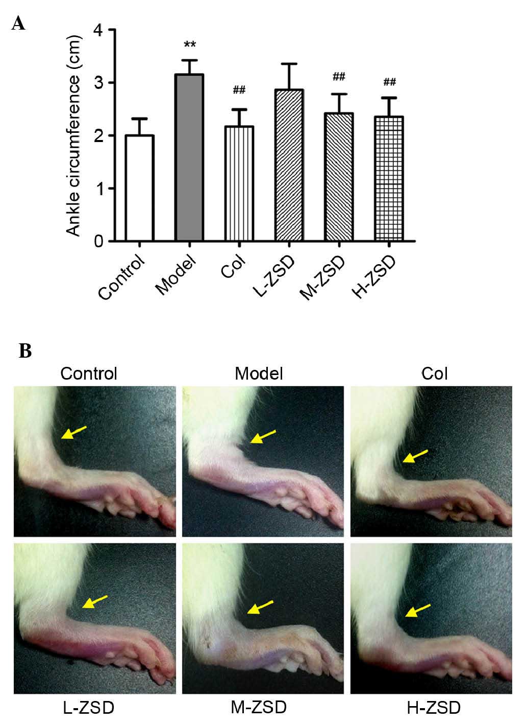
Effects of ZSD on MSU crystal-induced paw
edema in rats
As shown in Fig. 1A and
B, MSU crystals led to a significant increase in the ankle
diameter of the rats, compared with the normal control rats. Of
note, treatment with ZSD (20 and 40 mg/kg) significantly suppressed
the MSU crystal-induced ankle swelling, and this effect was
comparable with that of the positive control drug, colchicine.
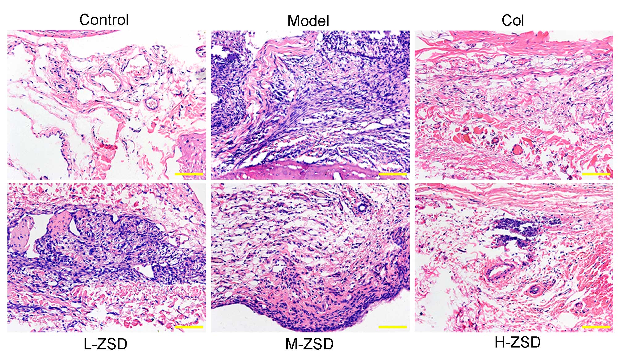
ZSD improves histopathological damage in
rats with gouty arthritis
To observe the effects of ZSD on the histopathology
of the synovium, rat knee joint capsules were removed and analyzed
using H&E staining. As shown in Fig. 2, compared with the control rats,
the sections from the MSU model rats showed marked infiltration of
inflammatory cells and a markedly thickened synovium, whereas this
inflammatory response and synovial hyperplasia was attenuated by
treatment with ZSD, which occurred in a dose-dependent manner. Of
note, the high dose ZSD (40 mg/kg) exerted a similar effect as the
positive control drug, colchicine.
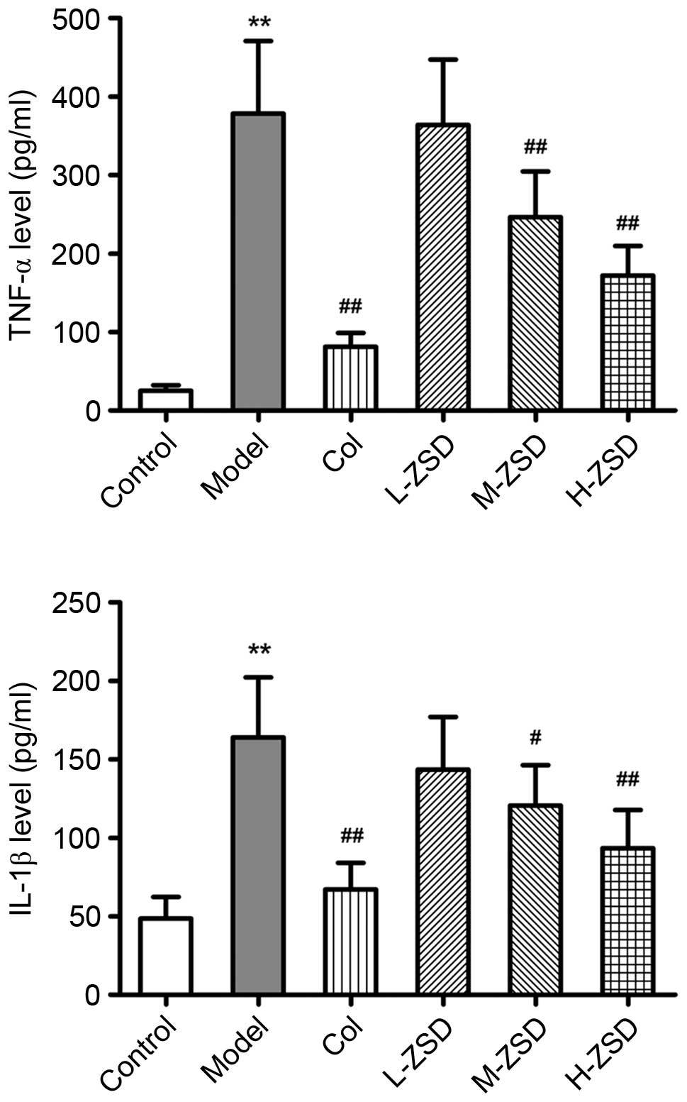
ZSD decreases the levels of IL-1β and
TNF-α in MSU crystal-induced rats
It is well accepted that pro-inflammatory cytokines,
including IL-1β and TNF-α are central to the initiation and
propagation of MSU crystal-induced gouty arthritis (25,26).
Consistent with a previous report (27), the MSU crystals caused a marked
elevation in the levels of IL-1β and TNF-α in the serum of the rats
(Fig. 3). By contrast, these
increased levels of IL-1β and TNF-α were found to be significantly
inhibited by ZSD (20 and 40 mg/kg) administration and colchicine
treatment.
ZSD suppresses the activation of NF-κB in
the synovium of rats with gouty arthritis
The transcription factor, NF-κB, is essential during
inflammatory responses, and the activation of NF-κB has been
identified as a key step in the pathogenesis of gouty arthritis
(28). Therefore, the present
study also investigated the effect of ZSD on the expression levels
of NF-κB p65 and its cytoplasmic inhibitory protein, IκB, in the
synovium of MSU crystal-induced rats using western blot analysis.
As shown in Fig. 4, the expression
levels of nuclear NF-κB p65 in the MSU crystal model rats were
significantly higher, compared with those in the control group,
whereas the protein levels of IκB in the cytoplasm were
significantly reduced by MSU crystal injection, suggesting that MSU
crystals caused NF-κB activation in the synovium. The oral
administration of ZSD (40 mg/kg) inhibited the upregulation of
nuclear p65 protein and downregulation of cytoplasmic IκB protein.
These results indicated that ZSD suppressed the nuclear
translocation and activation of NF-κB p65 induced by MSU
crystals.
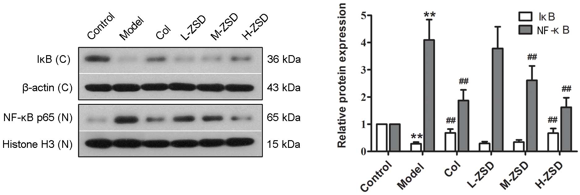 | Figure 4Effect of ZSD on MSU crystal-induced
NF-κB activation in the synovium of rats. The expression levels of
cytoplasmic IκB and nuclear NF-κB p65 in synovial tissues from each
treatment group were measured using western blot analysis.
Representative bands are shown (left), and the relative band
intensity ratio was analyzed (right). Data are presented as the
mean ± standard deviation (n=6). **P<0.01, vs.
control group; ##P<0.01, vs. model group. C,
cytoplasm; N, nuclear. ZSD, Zisheng Shenqi decoction; MSU,
monosodium urate; Col, colchicine; NF-κB, nuclear factor-κB; IκB,
inhibitor of NF-κB. |
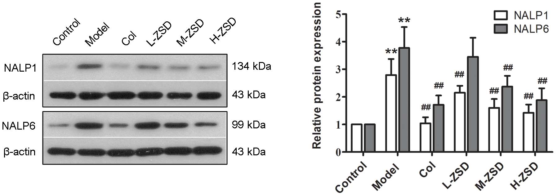
Detection of the protein expression of
NALP1 and NALP6 in the synovium of rats with gouty arthritis rats
and the effects of ZSD
NALP1 and NALP6, two important members of the NALP
family, have been shown to positively regulate the production of
IL-1β and promote the activation of NF-κB in several inflammatory
disorders (29,30). However, whether these two
inflammasomes are also involved in MSU crystal-induced gouty
arthritis remains to be elucidated. The results of the present
study demonstrated that the injection of MSU crystals significantly
upregulated the protein expression levels of NALP1 and NALP6,
compared with the control group (Fig.
5). Of note, treatment with ZSD at 20 and 40 mg/kg inhibited
the upregulation of NALP1 and NALP6 induced by the MSU crystals.
Colchicine also exerted a significant inhibitory effect on the
expression of these two inflammasomes. These findings suggested
that NALP1 and NALP6 inflammasomes were activated in the synovium
of rats with gouty arthritis, and their activation was inhibited by
treatment with ZSD (40 mg/kg) or colchicine.
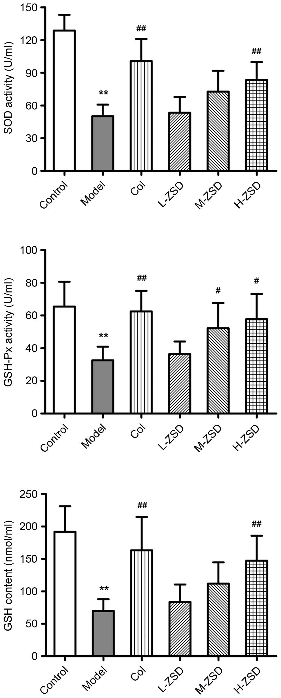
Effects of ZSD on the anti-oxidant status
of the serum of MSU crystal-induced rats
There is substantial evidence to suggest that ROS
overproduction and oxidative stress are also critical for the
pathogenesis of gouty arthritis (8,31).
In order to evaluate whether ZSD also had an anti-oxidative effect
in the experimental model used in the present study, the activities
of SOD and GSH-Px, and the levels of GSH were determined. As shown
in Fig. 6, MSU injection led to
significant decreases in the activities of SOD and GSH-Px, and in
the levels of GSH, compared with the control group. However,
treatment with ZSD (40 mg/kg) markedly enhanced the anti-oxidant
status in the MSU crystal-induced rats, indicating its
anti-peroxidative effect.
Discussion
With continuous improvements in living standards,
the incidence of gouty arthritis has risen. Increasing interest in
treating gouty arthritis has led to a focus on complementary and
alternative medicine (CAM) (32).
Existing evidence suggests that traditional Chinese medicines, as
one of the most popular CAM products, have beneficial efficacy in
the prevention and treatment of gouty arthritis (8,27).
In the present study, the effect of a novel herbal formula, ZSD,
against gouty arthritis were examined in rats with MSU
crystal-induced gouty arthritis rats, and its potential mechanism
was investigated.
A clinically definitive diagnosis of gouty arthritis
is based on the identification of MSU crystals in the synovium or
joint fluid (33). The
experimental model used in the present study represents a
well-established model of gouty arthritis, induced by the injection
of MSU crystals in rats. The most significant symptom of gouty
arthritis is ankle swelling, which was observed in the model group
following 24 h of MSU injection. Of note, ZSD pretreatment (40
mg/kg) markedly prevented the MSU crystal-induced elevation in
ankle swelling. It is well established that the recruitment and
infiltration of neutrophils into the joint fluid and synovium is a
primary hallmark of gouty arthritis (34). Following the activation of
monocytes and neutrophils, these cells actively phagocytoze MSU
crystals, which subsequently trigger the inflammatory caspase
responses. Therefore, inhibiting inflammatory cell infiltration may
be an effective therapeutic strategy against gouty arthritis. The
histopathology results in the present study demonstrated that a ZSD
dose of 40 mg/kg significantly attenuated the inflammatory cell
infiltration into the synovium induced by MSU crystals and improved
synovial hyperplasia. These findings suggested that ZSD protected
the rats from MSU crystal-induced gouty arthritis and synovial
damage.
It is also well known that gouty arthritis is an
inflammatory disease resulting from the deposition of MSU crystals
in the joints. The mechanisms underlying this inflammatory cascade
caused by MSU crystals have been under investigation for several
years, and a number of studies have demonstrated that
pro-inflammatory cytokines, including IL-1β and TNF-α, and the
transcription factor, NF-κB, are important in response to the
injection of MSU crystals into the joint cavity (35,36).
In the pathogenesis of gouty arthritis, NF-κB signals can stimulate
the production of genes encoding pro-inflammatory cytokines.
Conversely, the overexpression of TNF-α and IL-1β can directly
activate the NF-κB pathway, leading to a positive feedback loop,
which further amplifies inflammatory responses and causes joint
injury (35). Normally, NF-κB
binds to an inhibitory protein, IκB, and localizes in the
cytoplasm. Certain stimuli, including MSU crystals, can lead to the
degradation of IκB and translocation of NF-κB into the nucleus
where it regulates the transcription of various target genes
(37). In the present study, the
levels of IL-1β and TNF-α in the serum were significantly increased
in response to MSU crystals, whereas this overproduction was
markedly decreased by ZSD treatment, similar to the positive drug,
colchicine. Additionally, the findings in the present study
demonstrated that ZSD prevented the MSU crystal-induced nuclear
translocation and activation of NF-κB p65 in the synovium. Taken
together, these results suggested that ZSD attenuated MSU
crystal-induced gouty arthritis via anti-inflammatory action, which
may be associated with interrupting the positive feedback loop
between the NF-κB pathway and cytokines, including IL-1β and
TNF-α.
The process involved in the production of IL-1β in
response to MSU crystals is complex, and increasing evidence has
shown that NALP inflammasomes are pivotal in this process. There is
evidence that MSU stimulation triggers the activation of the NALP3
inflammasome, one of the most well-characterized NALP family
members in leukocytes, which in turn converts procaspase-1 to
active caspase-1, which cleaves pro-IL-1β to the active secreted
form, IL-1β (38). In addition,
previous studies have demonstrated that colchicine can reduce NALP3
inflammasome-driven caspase-1 activation by preventing microtubule
assembly and MSU delivery (39).
Similar to NALP3, other members of the NALP family, including NALP1
and NALP6, have also been identified as triggers of caspase-1
activation in the pathogenesis of several inflammatory diseases
(29,30,40).
The studies by Zhu et al (29) and Wang et al (41) demonstrated that the NALP1
inflammasome is involved in the inflammatory reaction process of
rheumatoid arthritis by activating caspase-1. Additionally, NALP6
possesses structural motifs similar to those of molecular sensors,
leading to pro-caspase-1 activation with NALP3 (42). However, whether NALP1 and NALP6
inflammasomes are involved in the pathogenesis of gouty arthritis
has not been reported to date, to the best of our knowledge. In the
present study, it was demonstrated that the injection of MSU
crystals into the joint cavity caused marked activation of the
NALP1 and NALP6 inflammasomes in the synovium. Of note, their
activation by MSU crystals was suppressed by treatment with ZSD (20
and 40 mg/kg) or colchicine. These findings revealed that the NALP1
and NALP6 inflammasomes may be relevant therapeutic targets in the
treatment of gouty arthritis. However, further investigation of the
mechanisms and the detailed regulatory effect of ZSD on these
mechanisms are required.
A previous report revealed that oxidative stress and
ROS are important in the activation of NALP inflammasomes induced
by MSU crystals (36). In
phagocytic cells, the free radicals and ROS can cause damage to the
cells if maintained at higher levels than normal. In order to
circumvent this damage, several defense mechanisms, including SOD,
GSH-Px and catalase, are initiated (31). In agreement with previous reports
(25,31), the results of the present study
showed that the activities of SOD and GSH-Px, and the levels of GSH
were significantly decreased in the rats with MSU crystal-induced
gouty arthritis, compared with the rats in the control group, which
may be due to their increased consumption in response to oxidative
stress. Of note, ZSD treatment (40 mg/kg) markedly enhanced the
anti-oxidant status in the MSU crystal-induced rats. These findings
suggested that ZSD can also prevent MSU crystal-induced gouty
arthritis via anti-oxidative effects.
In conclusion, the present study demonstrated for
the first time, to the best of our knowledge, that ZSD, a novel
Chinese herbal formula with multiple functions in nourishing kidney
and removing dampness, effectively prevented gouty arthritis in the
MSU crystal-induced rat model. The mechanisms involved in this
effect of ZSD on gouty arthritis were associated with the
modulation of multiple anti-oxidant and anti-inflammatory pathways,
as evidenced by increasing activities of SOD and GSH-Px, increased
levels of GSH, downregulation of NALP1 and NALP6 inflammasomes,
reductions in the IL-1β and TNF-α pro-inflammatory cytokines, and
inhibition of the activation of NF-κB. These results suggested that
ZSD may be a promising therapeutic formula for the prevention and
treatment of gouty arthritis in a clinical setting.
Acknowledgments
This study was supported by a grant from the
National Natural Science Foundation of China (grant no.
81173170).
References
|
1
|
Agudelo CA and Wise CM: Gout: Diagnosis,
pathogenesis, and clinical manifestations. Curr Opin Rheumatol.
13:234–239. 2001. View Article : Google Scholar : PubMed/NCBI
|
|
2
|
Woolf AD and Dieppe PA: Mediators of
crystal-induced inflammation in the joint. Br Med Bull. 43:429–444.
1987.PubMed/NCBI
|
|
3
|
Margalit A, Duffin KL, Shaffer AF, Gregory
SA and Isakson PC: Altered arachidonic acid metabolism in urate
crystal induced inflammation. Inflammation. 21:205–222. 1997.
View Article : Google Scholar : PubMed/NCBI
|
|
4
|
Martinon F, Pétrilli V, Mayor A, Tardivel
A and Tschopp J: Gout-associated uric acid crystals activate the
NALP3 inflammasome. Nature. 440:237–241. 2006. View Article : Google Scholar : PubMed/NCBI
|
|
5
|
Ogura Y, Sutterwala FS and Flavell RA: The
inflammasome: First line of the immune response to cell stress.
Cell. 126:659–662. 2006. View Article : Google Scholar : PubMed/NCBI
|
|
6
|
Giamarellos-Bourboulis EJ, Mouktaroudi M,
Bodar E, van der Ven J, Kullberg BJ, Netea MG and van der Meer JW:
Crystals of monosodium urate monohydrate enhance
lipopolysaccharide-induced release of interleukin 1 beta by
mononuclear cells through a caspase 1-mediated process. Ann Rheum
Dis. 68:273–278. 2009. View Article : Google Scholar
|
|
7
|
Schlesinger N and Thiele RG: The
pathogenesis of bone erosions in gouty arthritis. Ann Rheum Dis.
69:1907–1912. 2010. View Article : Google Scholar : PubMed/NCBI
|
|
8
|
Murunikkara V and Rasool M: Trikatu, a
herbal compound that suppresses monosodium urate crystal-induced
inflammation in rats, an experimental model for acute gouty
arthritis. Cell Biochem Funct. 32:106–114. 2014. View Article : Google Scholar
|
|
9
|
Fang ZH and Waizy H: Current concepts in
the treatment of gouty arthritis. Orthop Surg. 5:6–12. 2013.
View Article : Google Scholar : PubMed/NCBI
|
|
10
|
Fam AG: Treating acute gouty arthritis
with selective COX 2 inhibitors. BMJ. 325:980–981. 2002. View Article : Google Scholar : PubMed/NCBI
|
|
11
|
Li XX, Han M, Wang YY and Liu JP: Chinese
herbal medicine for gout: A systematic review of randomized
clinical trials. Clin Rheumatol. 32:943–959. 2013. View Article : Google Scholar : PubMed/NCBI
|
|
12
|
Wilson L and Saseen JJ: Gouty arthritis: A
review of acute management and prevention. Pharmacotherapy. Jun
18–2016.Epub ahead of print. View Article : Google Scholar : PubMed/NCBI
|
|
13
|
Wang W, Bhole VM and Krishnan E: Chronic
kidney disease as a risk factor for incident gout among men and
women: retrospective cohort study using data from the Framingham
Heart Study. BMJ Open. 5:e0068432015. View Article : Google Scholar : PubMed/NCBI
|
|
14
|
Sellin L, Kielstain JT and de Groot K:
Hyperuricemia- more than gout: Impact on cardiovascular risk and
renal insufficiency. Z Rheumatol. 74:322–328. 2015. View Article : Google Scholar : PubMed/NCBI
|
|
15
|
Wang Y, Wang L, Li E, Li Y, Wang Z, Sun X,
Yu X, Ma L and Wang Y and Wang Y: Chuanhu anti-gout mixture versus
colchicine for acute gouty arthritis: A randomized, double-blind,
double-dummy, non-inferiority trial. Int J Med Sci. 11:880–885.
2014. View Article : Google Scholar :
|
|
16
|
Wu CR, Lin LW, Wang WH and Hsieh MT: The
ameliorating effects of LiuWei Dihuang Wang on
cycloheximide-induced impairment of passive avoidance performance
in rats. J Ethnopharmacol. 113:79–84. 2007. View Article : Google Scholar : PubMed/NCBI
|
|
17
|
Lian F, Wu HC, Sun ZG, Guo Y, Shi L and
Xue MY: Effects of Liuwei Dihuang Granule ([symbols; see text]) on
the outcomes of in vitro fertilization pre-embryo transfer in
infertility women with Kidney-yin deficiency syndrome and the
proteome expressions in the follicular fluid. Chin J Integr Med.
20:503–509. 2014. View Article : Google Scholar : PubMed/NCBI
|
|
18
|
Qiu R, Shen R, Lin D, Chen Y and Ye H:
Treatment of 60 cases of gouty arthritis with modified Simiao Tang.
J Tradit Chin Med. 28:94–97. 2008. View Article : Google Scholar : PubMed/NCBI
|
|
19
|
Jiang J, Wu F, Lu J, Lu Z and Xu Q:
Anti-inflammatory activity of the aqueous extract from Rhizoma
smilacis glabrae. Pharmacol Res. 36:309–314. 1997. View Article : Google Scholar
|
|
20
|
Jiang Y, Zhang Y, Chen W, Liu C, Li X, Sun
D, Liu Z, Xu Y, Mao X, Guo Q and Lin N: Achyranthes bidentata
extract exerts osteoprotective effects on steroid-induced
osteonecrosis of the femoral head in rats by regulating
RANKL/RANK/OPG signaling. J Transl Med. 12:3342014. View Article : Google Scholar : PubMed/NCBI
|
|
21
|
Chiang LC, Chiang W, Chang MY and Lin CC:
In vitro cytotoxic, antiviral and immunomodulatory effects of
Plantago major and Plantago asiatica. Am J Chin Med. 31:225–234.
2003. View Article : Google Scholar : PubMed/NCBI
|
|
22
|
Huang Q, Duan Z, Yang J, Ma X, Zhan R, Xu
H and Chen W: SNP typing for germplasm identification of Amomum
villosum Lour. Based on DNA barcoding markers. PloS One.
9:e1149402014. View Article : Google Scholar : PubMed/NCBI
|
|
23
|
Coderre TJ and Wall PD: Ankle joint urate
arthritis (AJUA) in rats: An alternative animal model of arthritis
to that produced by Freund's adjuvant. Pain. 28:379–393. 1987.
View Article : Google Scholar : PubMed/NCBI
|
|
24
|
Li HX, Xiao Y, Cao LL, Yan X, Li C, Shi
HY, Wang JW and Ye YH: Cerebroside C increases tolerance to
chilling injury and alters lipid composition in wheat roots. PloS
One. 8:e733802013. View Article : Google Scholar : PubMed/NCBI
|
|
25
|
Sabina EP, Nagar S and Rasool M: A role of
piperine on monosodium urate crystal-induced inflammation-an
experimental model of gouty arthritis. Inflammation. 34:184–192.
2011. View Article : Google Scholar
|
|
26
|
de Souza MR, de Paula CA, Pereira de
Resende ML, Grabe-Guimarães A, de Souza Filho JD and
Saúde-Guimãraes DA: Pharmacological basis for use of Lychnophora
trichocarpha in gouty arthritis: Anti-hyperuricemic and
anti-inflammatory effects of its extract, fraction and
constituents. J Ethnopharmacol. 142:845–850. 2012. View Article : Google Scholar : PubMed/NCBI
|
|
27
|
Jiang Y, You XY, Fu KL and Yin WL: Effects
of extract from Mangifera indica Leaf on monosodium urate
crystal-induced gouty arthritis in rats. Evid Based Complement
Alternat Med. 2012:9675732012. View Article : Google Scholar
|
|
28
|
Jaramillo M, Godbout M, Naccache PH and
Olivier M: Signaling events involved in macrophage chemokine
expression in response to monosodium urate crystals. J Biol Chem.
279:52797–52805. 2004. View Article : Google Scholar : PubMed/NCBI
|
|
29
|
Zhu L, Li J, Guo L, Yu X, Wu D, Luo L, Zhu
L, Chen W, Chen C, Ye C and Zhang D: Activation of NALP1
inflammasomes in rats with adjuvant arthritis; a novel therapeutic
target of carboxyamidotriazole in a model of rheumatoid arthritis.
Br J Pharmacol. 172:3446–3459. 2015. View Article : Google Scholar : PubMed/NCBI
|
|
30
|
Grenier JM, Wang L, Manji GA, Huang WJ,
Al-Garawi A, Kelly R, Carlson A, Merriam S, Lora JM, Briskin M, et
al: Functional screening of five PYPAF family members identifies
PYPAF5 as a novel regulator of NF-kappaB and caspase-1. FEBS Lett.
530:73–78. 2002. View Article : Google Scholar : PubMed/NCBI
|
|
31
|
Sabina EP, Rasool M, Mathew L, Ezilrani P
and Indu H: 6-Shogaol inhibits monosodium urate crystal-induced
inflammation-an in vivo and in vitro study. Food Chem Toxicol.
48:229–235. 2010. View Article : Google Scholar
|
|
32
|
Venkatesha SH, Rajaiah R, Berman BM and
Moudgil KD: Immunomodulation of autoimmune arthritis by herbal CAM.
Evid Based Complement Alternat Med. 2011:9867972011. View Article : Google Scholar : PubMed/NCBI
|
|
33
|
Schlesinger N: Diagnosing and treating
gout: A review to aid primary care physicians. Postgrad Med.
122:157–161. 2010. View Article : Google Scholar : PubMed/NCBI
|
|
34
|
Torres R, Macdonald L, Croll SD, Reinhardt
J, Dore A, Stevens S, Hylton DM, Rudge JS, Liu-Bryan R, Terkeltaub
RA, et al: Hyperalgesia, synovitis and multiple biomarkers of
inflammation are suppressed by interleukin 1 inhibition in a novel
animal model of gouty arthritis. Ann Rheum Dis. 68:1602–1608. 2009.
View Article : Google Scholar : PubMed/NCBI
|
|
35
|
Ma Y, Zhou LL, Yan HY and Liu M: Effects
of extracts from Paederia scandens (LOUR.) MERRILL (Rubiaceae) on
MSU crystal-induced rats gouty arthritis. Am J Chin Med.
37:669–683. 2009. View Article : Google Scholar : PubMed/NCBI
|
|
36
|
Punzi L, Scanu A, Ramonda R and Oliviero
F: Gout as autoinflammatory disease: New mechanisms for more
appropriated treatment targets. Autoimmun Rev. 12:66–71. 2012.
View Article : Google Scholar : PubMed/NCBI
|
|
37
|
Grall F, Gu X, Tan L, Cho JY, Inan MS,
Pettit AR, Thamrongsak U, Choy BK, Manning C, Akbarali Y, et al:
Responses to the proinflammatory cytokines interleukin-1 and tumor
necrosis factor alpha in cells derived from rheumatoid synovium and
other joint tissues involve nuclear factor kappaB-mediated
induction of the Ets transcription factor ESE-1. Arthritis Rheum.
48:1249–1260. 2003. View Article : Google Scholar : PubMed/NCBI
|
|
38
|
Zheng SC, Zhu XX, Xue Y, Zhang LH, Zou HJ,
Qiu JH and Liu Q: Role of the NLRP3 inflammasome in the transient
release of IL-1β induced by monosodium urate crystals in human
fibroblast-like synoviocytes. J Inflamm (Lond). 12:302015.
View Article : Google Scholar
|
|
39
|
Dalbeth N, Lauterio TJ and Wolfe HR:
Mechanism of action of colchicine in the treatment of gout. Clin
Ther. 36:1465–1479. 2014. View Article : Google Scholar : PubMed/NCBI
|
|
40
|
Bruey JM, Bruey-Sedano N, Luciano F, Zhai
D, Balpai R, Xu C, Kress CL, Bailly-Maitre B, Li X, Osterman A, et
al: Bcl-2 and Bcl-XL regulate proinflammatory caspase-1 activation
by interaction with NALP1. Cell. 129:45–56. 2007. View Article : Google Scholar : PubMed/NCBI
|
|
41
|
Wang T, Zhu CL, Wang S, Mo LW, Yang GD, Hu
J and Zhang F: Role of NLRP3 and NLRP1 inflammasomes signaling
pathways in pathogenesis of rheumatoid arthritis. Asian Pac J Trop
Med. 7:827–831. 2014. View Article : Google Scholar : PubMed/NCBI
|
|
42
|
Anand PK and Kanneganti TD: NLRP6 in
infection and inflammation. Microbes Infect. 15:661–668. 2013.
View Article : Google Scholar : PubMed/NCBI
|