Introduction
Liver fibrosis results from dysregulation of the
normal healing process and scar formation, and is a common and
potentially severe consequence of virtually all chronic liver
diseases (1). Diseases that may
lead to liver fibrosis include: Viral and autoimmune hepatitis,
drug injury, iron deposition, alcohol consumption and biliary
obstruction.
Fibronectin (FN), which is produced by hepatic
stellate cells (HSCs), is a multifunctional glycoprotein and
extracellular matrix (ECM) component that is present in the cell
membrane and cytoplasm (2,3). FN is required in vitro for
collagen matrix assembly, and can be used to support other matrix
proteins (4–6). In addition, FN is associated with
cell cycle progression, participates in cell adhesion and
proliferation, and has an important role in fibrotic progression
(7). Although collagen is the main
ECM component of fibrotic tissue, excessive FN deposition occurs
prior to collagen deposition. Increased expression of the FN
isoforms EIIIA and EIIIB occurs during wound healing and tissue
repair. These tissue-remodeling processes are associated with liver
fibrogenesis, and the expression of these isoforms is often
considered an important indicator of ECM accumulation (8–10).
Kawelke et al (11)
reported that FN protects against excessive liver fibrosis by
modulating stellate cell availability and the responsiveness of
stellate cells to active transforming growth factor-β (TGF-β). In
addition, Kawelke et al (11) demonstrated that loss of FN
expression leads to increased stellate cell activation at baseline
and following TGF-β stimulation. Altrock et al (12) reported that interference in
collagen deposition, via FN matrix formation inhibitors, decreased
fibrosis and improved liver function.
Since FN appears to have an important role in liver
fibrogenesis, FN expression may be considered a critical factor
mediating the long-term consequences of several chronic liver
diseases. Therefore, the present study aimed to determine FN
expression patterns during hepatic fibrogenesis using in
vitro and in vivo models. The former was conducted with
HSCs, which were stimulated with TGF-β, and the process of
fibrogenesis of these cells mimicked acute hepatitis; the latter
model was the CCl4 rat model, used to mimic the process of chronic
liver injury, such as chronic hepatitis.
Materials and methods
Animals
A total of 50 healthy male Wistar rats (age, 6–8
weeks; weight, 180–200 g) were obtained from the Laboratory Animal
Center of the Academy of Military Medical Sciences (Beijing,
China). The rats were housed in a temperature- and
humidity-controlled facility under a 12 h light-dark cycle, and
were given ad libitum access to a standard laboratory diet
and tap water. The experimental procedures and ethical requirements
of the present study conformed to the Laboratory Animal Management
Regulations of the People's Republic of China. All experimental
procedures were approved by the Animal Experimental Committee of
Beijing Friendship Hospital affiliated with Capital Medical
University (Beijing, China).
Carbon tetrachloride
(CCl4)-induced liver fibrosis rat model and sample
collection
The 50 rats were randomly divided into the normal
control group (n=6) and the liver fibrosis model group (n=44). The
liver fibrosis model group received intraperitoneal injections of
0.15 ml/100 g 50% CCl4 in olive oil (5:5, v/v) twice
weekly for eight weeks. Rats in the normal control group received
intraperitoneal injections of the same volume of physiological
saline over the same period. Reversal of liver fibrosis was
investigated for 4 weeks after the final intraperitoneal injection.
Rats were sacrificed at 2, 4, 6, 8, 10 and 12 weeks. Seven rats
were sacrificed at each time point. Two rats succumbed without
investigator intervention during week 12. All rats from the normal
group were sacrificed during week 8.
All rats were euthanized under anesthesia using 1%
sodium pentobarbital (50 mg/kg, i.p). Blood samples were collected
for liver function tests. In addition, liver specimens were
collected, divided into three parts and subjected to the following
analyses: i) mRNA extraction using TRIzol for reverse
transcription-quantitative polymerase chain reaction (RT-qPCR)
analyses; ii) protein expression analysis of fibrotic indices using
western blotting; and iii) histological and immunohistochemical
analyses.
Cell culture
The HSC-T6 immortalized rat cell line (Tumor Cell
Bank of Chinese Academy of Medical Sciences, Beijing, China) was
cultured in Dulbecco's modified Eagle's medium (Gibco; Thermo
Fisher Scientific, Inc., Waltham, MA, USA) supplemented with 10%
(v/v) fetal bovine serum (FBS; Gibco; Thermo Fisher Scientific,
Inc.), 100 IU/ml penicillin and 100 µg/ml streptomycin
(Beijing Suolaibao Biotechnology Co., Ltd., Beijing, China). Cells
were incubated at 37°C in a humidified atmosphere containing 5%
CO2. All experiments were carried out 24 h after the
cells were seeded.
Cells were divided into two groups: The control
group and the transforming growth factor (TGF)-β group. Recombinant
human TGF-β (Sigma -Aldrich; Merck Millipore, Darmstadt, Germany)
was used, since it is a major fibrogenic mediator in the liver
(13,14). TGF-β was administered to the
cultured cells at 0, 1, 2, 5, 10 and 20 ng/ml for 0, 1, 3, 6, 12,
24 and 48 h. Cells were exposed to starvation conditions using
FBS-free medium for 24 h prior to treatment with recombinant human
TGF-β.
FN immunofluorescence assay
Cells were plated at a density of 3×10⁵
cells/well in six-well plates, and were allowed to adhere
overnight. The cells were then cultured in serum-free medium for an
additional 24 h at 37°C. Subsequently, cells were rinsed in
phosphate-buffered saline (3×10 min), and were fixed in 4%
paraformaldehyde (40 min, 37°C) and 3% H2O2
(30 min, 37°C). Following fixation, cells were blocked with 1% FBS
(40 min, 37°C) and were incubated overnight at 4°C with rabbit
anti-mouse elastin primary antibody (FN; 1:100; sc-9068; Santa Cruz
Biotechnology, Inc., Dallas, TX, USA). Goat anti-rabbit fluorescein
isothiocyanate probes (1:200; bs-0295G-FITC; BIOSS, Beijing, China)
were used as secondary antibodies at 37°C for 1 h. The cell nuclei
were stained using 4′,6-diamidino-2-phenylindole (Santa Cruz
Biotechnology, Inc.). Immunofluorescence was analyzed using a
fluorescence microscope. Cells incubated with normal rabbit serum
instead of the primary antibody served as a negative control.
Liver function test
Blood samples were centrifuged at 1,200 × g for 5
min at 4°C to separate serum from the blood. Serum aspartate
transaminase (AST) and alanine transaminase (ALT) (15) activities were determined by the
Clinical Laboratory (Beijing Friendship Hospital, Beijing,
China).
RNA extraction and RT-qPCR
Total RNA was extracted from frozen liver samples or
cultured cells using TRIzol® (Invitrogen; Thermo Fisher
Scientific, Inc.) according to the manufacturer's protocol. RT was
performed using random primers, Moloney Murine Leukemia Virus
Reverse Transcriptase and RNase inhibitor (Fermentas; Thermo Fisher
Scientific, Inc.). The expression levels of all transcripts were
normalized to the expression of the housekeeping gene,
glyceraldehyde 3-phosphate dehydrogenase (GAPDH), in the same
sample. PCR was performed using Power SYBR Green PCR Master Mix
(Applied Biosystems; Thermo Fisher Scientific, Inc.) and an ABI
7500 platform (Applied Biosystems; Thermo Fisher Scientific, Inc.).
RNase-free DNase (Promega Corporation, Madison, WI, USA) was used
to eliminate the contamination of genomic DNA. The first cDNA
strand was synthesized by the RT reaction in a reaction mixture of
20 µl containing 4 µl MgCl2, 3 µl
10X RT buffer, 2 µl dNTP mixture, 0.5 µl recombinant
RNase inhibitor, 0.6 µl AMV reverse transcriptase, 1
µl oligo dT-Adaptor primer and 5 µg RNA sample. The
RT reaction product was then used for the amplification of cDNA
with the SYBR Master Mix reaction system (total, 10 µl)
including 1 µl upstream primers, 1 µl downstream
primers, 1 µl cDNA and 7 µl
diethylpyrocarbonate-treated water. PCR was conducted as follows:
Initial denaturation at 95°C for 10 min, 40 cycles at 95°C for 15
sec and 64°C for 30 sec, followed by 95°C for 15 sec, 60°C for 1
min, 95°C for 15 sec and a final extension step at 60°C for 15 sec.
Data were analyzed according to the 2−ΔΔCq method, as
described by Livak and Schmittgen (16). The following primer pairs were
used: FN, forward 5′-CCAGCCTACGGATGACTC-3′, reverse
5′-AATGACCACTGCCAAAGC-3′; and GAPDH, forward
5′-GGCACAGTCAAGGCTGAGAATG-3′, and reverse
5′-ATGGTGGTGAAGACGCCAGTA-3′ [Sangon Biotech (Shanghai) Co., Ltd.,
Shanghai, China].
Western blot analysis
Liver tissues and cultured HSCs were homogenized in
radioimmunoprecipitation assay buffer and were centrifuged as
previously described (17). The
supernatants were considered whole-cell lysates. Total protein
concentration was determined using a Bicinchoninc Acid Protein
Assay kit (Pierce; Thermo Fisher Scientific, Inc.). Proteins (~80
µg) were separated by 8% sodium dodecyl
sulfate-polyacrylamide gel electrophoresis and were transferred to
polyvinylidene fluoride membranes by electroblotting. Subsequently,
the membranes were incubated for 3 h with 5% non-fat dry milk at
37°C, followed by an overnight incubation at 4°C with mouse anti-FN
polyclonal antibody (1:600; sc-8422; Santa Cruz Biotechnology,
Inc.), rabbit anti-TGF-β antibody (1:800; ab92486; Abcam,
Cambridge, UK), rabbit anti-α-smooth muscle actin (α-SMA) antibody
(1:800; ab5694; Abcam) and mouse anti-β-actin monoclonal antibody
(1:800; ab8226; Abcam). Following the overnight incubation,
membranes were incubated for 1 h at 37°C with peroxidase-conjugated
goat anti-mouse secondary antibodies (1:6,000 in Tris-buffered
saline containing 1% Tween; sc-2005; Santa Cruz Biotechnology,
Inc.). Enhanced chemiluminescence (EMD Millipore, Billerica, MA,
USA) was used to detect immunoreactive bands. Finally,
densitometric analysis of the bands was performed using Image Lab
software, version 4.0 (Bio-Rad Laboratories, Inc., Hercules, CA,
USA).
Histopathology and
immunohistochemistry
Liver samples were fixed in 4% paraformaldehyde,
embedded in paraffin and sectioned at 3 µm. The tissue
sections were then stained with hematoxylin and eosin (H&E) or
Masson's trichrome, and were examined for changes in liver
pathology. The histopathological scores for fibrosis were evaluated
according to previously published criteria (18), as follows: 0, Normal liver; 1,
increased collagen matrix accumulation without septa formation
(small stellate expansions of the portal fields); 2, formation of
incomplete septa from the portal tract to the central vein (septa
that do not interconnect with each other); 3, complete but thin
septa interconnecting with each other to divide the parenchyma into
separate fragments; and 4, similar to grade 3, except for the
presence of thick septa (complete cirrhosis).
Immunohistochemical analysis of paraffin-embedded
sections was performed using a microwave-based antigen retrieval
method, as described previously (19), and antibodies against FN were used
(mouse polyclonal; 1:100; sc-8422; Santa Cruz Biotechnology, Inc.).
In brief, histological sections were heated for 60 min at 60°C,
deparaffinized with xylene and rinsed in a reducing gradient of
alcohol. Subsequently, antigens were retrieved with citrate buffer
in a microwave oven. Subsequent to treatment with hydrogen peroxide
at 37°C for 30 min to block endogenous peroxidase activity, the
sections were incubated with the primary antibody overnight at 4°C
and then incubated with the secondary antibody (PV-9002; OriGene
Technologies, Inc., Beijing, China) for 30 min at room temperature.
Finally stained sections were developed using diaminobenzidine and
were counterstained with hematoxylin and a BX43 optical microscope
(Olympus Corporation, Tokyo, Japan) was used for analysis. FN
expression was analyzed using a quantitative image analysis system
(Image-Pro Plus 6.0; Media Cybernetics Inc., Rockville, MD, USA).
Briefly, 10 fields were randomly selected from each section and
positive signals within the section were highlighted, measured and
expressed as a percentage of the positive areas within the entirety
of the examined liver tissue.
Statistical analysis
Analyses were performed using SPSS software (SPSS
version 17.0; SPSS Inc., Chicago, IL, USA), and the differences
between multiple groups were evaluated using one-way analysis of
variance and the least significant difference test was used for
multiple comparisons. Results are presented as the mean ± standard
deviation and experiments were repeated 6 times. P<0.05 was
considered to indicate a statistically significant difference.
Results
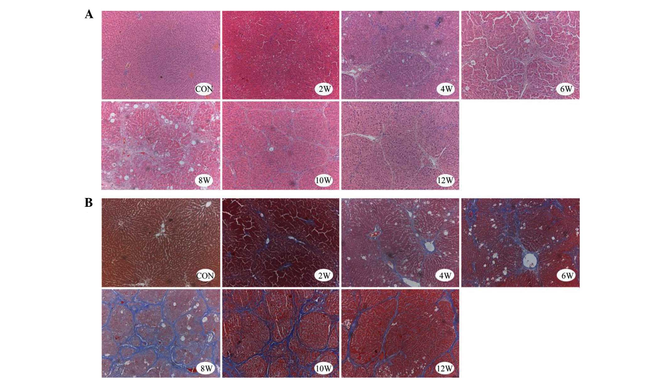
Liver pathology in
CCl4-treated mice
In the liver samples of the control group, normal
lobular architecture was observed with hepatic cords radiating from
the central veins, and the absence of regenerated collagen fibers,
as detected by H&E and Masson's trichrome staining. Conversely,
liver samples from the experimental group (2–12 weeks) exhibited
severe fibrosis. Morphological alterations in the experimental
group included thick fibrotic septa, fatty degeneration, enlarged
hepatocytes with nodular arrangement, focal inflammatory changes,
thickened vessel walls and early capillarization (Fig. 1).
The hepatic lobule structure persisted beyond 4
weeks; however, in the CCl4 treatment group the
appearance of the hepatic lobules became disorganized, and were
characterized by fibrous septa, severe fatty degeneration, hydropic
degeneration and focal necrosis. After 6 and 8 weeks of
CCl4 treatment, pseudolobules, and inflammatory cell
infiltration into the portal areas and fibrous septa were observed.
At 12 weeks (or 4 weeks after the final CCl4 injection),
fibrous septa began to attenuate or exhibit discontinuous shapes,
with some of the fibrous septa becoming barely visible (Fig. 1).
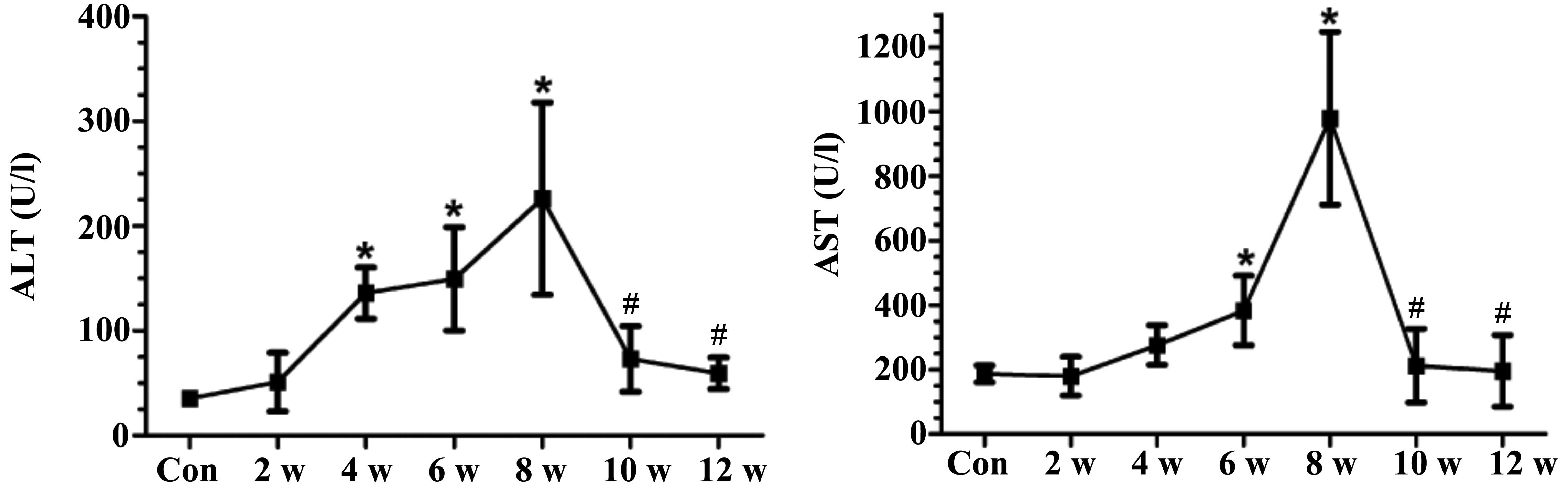
Changes in serum indices of liver
function
Treatment with CCl4 (2–8 weeks) resulted
in a significant elevation in AST and ALT levels. However, at 10
weeks (2 weeks after the final CCl4 injection), AST and
ALT levels were markedly lower compared with those measured at 8
weeks (Fig. 2).
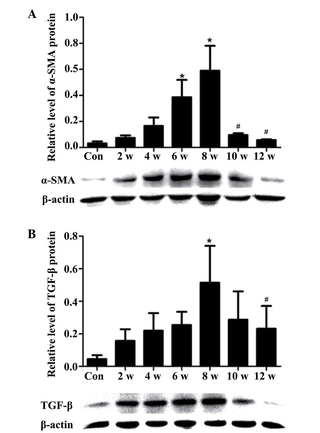
α-SMA and TGF-β protein expression in
vivo
α-SMA expression in HSCs reflects myofibroblast-like
cell activation, has been directly linked to experimental liver
fibrogenesis, and is indirectly associated with fibrosis in chronic
human liver disease (20).
The expression levels of α-SMA were increased in the
CCl4 treatment group compared with in the control group.
However, when CCl4 treatment ceased, protein expression
returned to baseline levels (Fig.
3A). In addition, the expression levels of TGF-β were elevated
in the CCl4 treatment group (2–8 weeks), and this
increase corresponded with the degree of liver fibrosis (Fig. 3B).
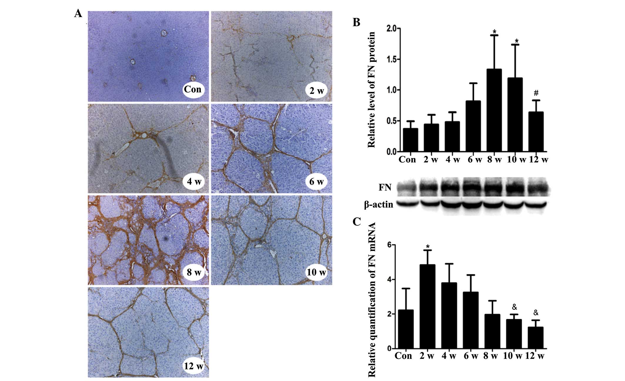
In vivo location, and hepatic mRNA and
protein expression levels of FN during hepatic fibrogenesis
Markedly increased FN staining was detected in
hepatocytes, sinusoidal areas, extracellular spaces, and portal and
central areas in the experimental group tissues compared with in
the control tissues, as determined by immunohistochemical staining
of FN in liver sections. FN expression increased with increasing
fibrosis, and peaked during week 8. However, after week 8 and the
discontinuation of CCl4 treatment, FN staining gradually
decreased (Fig. 4A). The criterion
used to assess FN positivity was yellow or brown staining in the
cell membrane or mesenchyme.
The protein expression levels of FN in
CCl4-injured liver tissues gradually increased during
the course of liver fibrogenesis. As detected by western blotting,
FN expression reached its maximal point during week 8, after which
expression gradually decreased (Fig.
4B). The mRNA expression levels of FN rapidly increased to a
maximum level during the first 2 weeks. After the first 2 weeks,
expression gradually fell (Fig.
4C).
In vitro location, and mRNA and protein
expression levels of FN in HSC-T6 cells
Immunofluorescence assays demonstrated that FN was
primarily located in the cytoplasm of HSC-T6 cells (Fig. 5A). TGF-β stimulation upregulated
the mRNA and protein expression levels of FN in HSC-T6 cells in a
dose- and time-dependent manner (Fig.
5B–E).
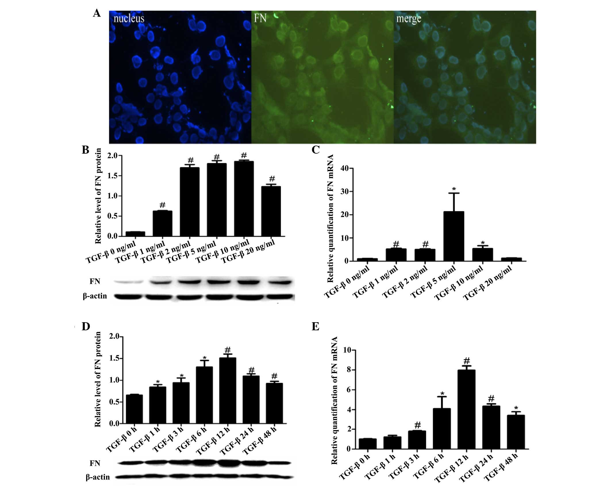 | Figure 5FN location, and mRNA and protein
expression in HSC-T6 cells. (A) FN immunofluorescence localization
in HSC-T6 cells. Blue, nucleus (left); green, target protein
(middle); merged green and blue image (right). Magnification, ×40.
(B) Protein expression levels of FN in the HSC-T6 cells, as
detected by western blotting. (C) mRNA expression levels of FN in
HSC-T6 cells, as detected by RT-qPCR. FN expression was normalized
to GAPDH (internal control). (B and C) Six concentrations of TGF-β
(0, 1, 2, 5, 10 and 20 ng/ml) were assayed. (D) Protein expression
levels of FN in the HSC-T6 cells, as detected by western blotting.
(E) mRNA expression levels of FN in HSC-T6 cells, as detected by
RT-qPCR. FN expression was normalized to GAPDH (internal control).
(D and E) Seven time points (0, 1, 3, 6, 12, 24 and 48 h) were
assayed, and HSC-T6 cells were stimulated with TGF-β (5 ng/ml).
Data are presented as the mean ± standard deviation.
*P<0.05 vs. the Con group; #P<0.01 vs.
the Con group. Con, control; HSC, hepatic stellate cells; FN,
fibronectin; TGF-β, transforming growth factor-β. |
Discussion
Liver fibrosis is a category of interest in liver
disease research due to its importance in the natural history of
chronic liver diseases. Previous studies have revealed that hepatic
fibrogenesis is a dynamic process, and that the capacity for
recovery exists from any degree of fibrosis, including established
cirrhosis (21,22).
The present study induced hepatic fibrosis in male
Wistar rats via intraperitoneal injections of CCl4. This
regimen induced major fibrosis, which regressed following the
discontinuation of CCl4 treatment. Decreased serum ALT
and AST activities, and TGF-β and α-SMA protein levels were
detected following the discontinuation of CCl4
treatment, mimicking the clinically observed expression
patterns.
The progression of liver fibrosis is characterized
by the gradual accumulation of ECM proteins in injured tissue, and
HSCs have a crucial role in this fibrotic response (23). Upon liver injury, quiescent HSCs
are activated and transdifferentiate into myofibroblast-like cells.
These myofibroblast-like cells are characterized by increased
proliferation and excessive deposition of ECM proteins (24). In addition, activated HSCs are able
to upregulate the expression of cytoskeletal proteins, including
α-SMA. Therefore, α-SMA is considered a marker of HSC activation,
which may reflect the degree of liver fibrosis (25,26).
Alterations to the hepatic architecture occur due to the combined
increase in cytoskeletal protein synthesis and the inability to
break down these proteins, resulting in liver fibrosis and,
eventually, cirrhosis (27). The
results of the present study suggested that HSC activation
indirectly indicated the level of ECM deposition and the degree of
liver fibrosis. In the present study, α-SMA expression was detected
at various time points following CCl4 injection, and the
results demonstrated that α-SMA expression increased in response to
CCl4 treatment, in a time-dependent manner. However,
when CCl4 treatment was terminated, α-SMA protein
expression returned to baseline. These results suggested that liver
fibrosis can be reversed.
Increasing evidence has indicated that TGF-β is a
key mediator of liver fibrosis (14,28,29).
Myofibroblast differentiation of HSCs is induced by TGF-β, and
TGF-β induction results in increased ECM protein production
(30). Thus, it is suggested that
TGF-β serves an important role in morphogenesis, proliferation and
differentiation of cells, and is good for cell repair. With
fibrosis of the liver, the expression of TGF-β will increase in
order to repair the damaged liver tissue. Thus, TGF-β expression is
increased in damaged liver compared with the normal liver.
FN is a non-collagenous glycoprotein with several
functions. FN is produced and secreted by fibroblasts, endothelial
cells, macrophages and hepatocytes in vivo (31), and its expression is closely
associated with hepatic fibrosis, cellular damage, differentiation
and repair. In the present study, FN expression increased with
increasing CCl4 levels, and decreased when
CCl4 injections ceased. Furthermore, FN expression
levels were associated with the degree of hepatic fibrosis;
however, it was also confirmed that liver fibrosis could be
reversed. The present study also demonstrated that FN expression
varied during the process of fibrogenesis according to the type of
hepatitis (acute or chronic).
In acute hepatitis, fibroblasts proliferate and FN
levels increase after liver injury (32,33).
Conversely, in chronic hepatitis, FN levels increase due to fibrous
tissue hyperplasia; however, due to significant FN deposition in
the fibrous tissue, the FN content of the liver appears to fall
after the formation of liver fibrosis (34). Nevertheless, the total deposition
of FN consistently reflects the degree of liver fibrosis and the
degree of ECM deposition, and is an important marker of hepatic
fibrosis.
The in vivo experiments conducted in the
present study were designed to simulate chronic hepatitis, and the
results indicated that the mRNA expression levels of FN peaked as
early as at 2 weeks. The time lag between FN mRNA expression and
the expression of the corresponding protein may be related to the
time required for FN synthesis in the liver. The detected
upregulated mRNA levels may represent activation of the promoter or
enhancer, or of upstream transcription factors; however, whereas
protein levels are usually coupled to the biological function of
the proteins themselves, this is not always true for the
corresponding mRNA levels.
The in vitro experiments conducted in the
present study were designed to more closely represent the
conditions present during acute hepatitis. The results indicated
that FN mRNA expression corresponded with FN protein expression,
with peak mRNA and protein expression values observed in response
to the same concentration and duration of TGF-β stimulation. The
above results indicate that the protein expression may not be
consistent with the corresponding RNA expression in the chronic
hepatitis model, however, in the acute hepatitis model, the
expression of these two indexes tended to be consistent.
In conclusion, the present study confirmed the
reversibility of liver fibrosis. This was true even after the
establishment of cirrhosis. The results indicated that FN may have
an early and critical role during the process of liver
fibrogenesis. The in vivo and in vitro models
exhibited marked differences in FN expression, and these
differences may closely resemble those between chronic and acute
human hepatitis.
Acknowledgments
The present study was funded by the National Natural
Science Foundation of China (grant no. 81300334) and the WBE Liver
Fibrosis Foundation (grant no. CFHPC20120145). The authors would
like to thank Medjaden Bioscience Limited for assisting in the
preparation of this manuscript.
References
|
1
|
Cong M, Liu T, Wang P, Fan X, Yang A, Bai
Y, Peng Z, Wu P, Tong X, Chen J, et al: Antifibrotic effects of a
recombinant adeno-associated virus carrying small interfering RNA
targeting TIMP-1 in rat liver fibrosis. Am J Pathol. 182:1607–1616.
2013. View Article : Google Scholar : PubMed/NCBI
|
|
2
|
Xu G, Niki T, Virtanen I, Rogiers V, De
Bleser P and Geerts A: Gene expression and synthesis of fibronectin
isoforms in rat hepatic stellate cells. Comparison with liver
parenchymal cells and skin fibroblasts. J Pathol. 183:90–98. 1997.
View Article : Google Scholar : PubMed/NCBI
|
|
3
|
Matsui S, Takahashi T, Oyanagi Y,
Takahashi S, Boku S, Takahashi K, Furukawa K, Arai F and Asakura H:
Expression, localization and alternative splicing pattern of
fibronectin messenger RNA in fibrotic human liver and
hepatocellular carcinoma. J Hepatol. 27:843–853. 1997. View Article : Google Scholar
|
|
4
|
Sottile J and Hocking DC: Fibronectin
polymerization regulates the composition and stability of
extracellular matrix fibrils and cell-matrix adhesions. MolBiol
Cell. 13:3546–3559. 2002.
|
|
5
|
Velling T, Risteli J, Wennerberg K, Mosher
DF and Johansson S: Polymerization of type I and III collagens is
dependent on fibronectin and enhanced by integrins alpha 11beta 1
and alpha 2beta 1. J Biol Chem. 277:37377–37381. 2002. View Article : Google Scholar : PubMed/NCBI
|
|
6
|
Roeb E and Matern S: Fibronectin-a key
substance in pathogenesis of liver cirrhosis? Leber Magen Darm.
23:239–242. 1993.In German. PubMed/NCBI
|
|
7
|
Manabe R, Oh-e N and Sekiguchi K:
Alternatively spliced EDA segment regulates fibronectin-dependent
cell cycle progression and mitogenic signal transduction. J Biol
Chem. 274:5919–5924. 1999. View Article : Google Scholar : PubMed/NCBI
|
|
8
|
Krzyzanowska-Gołab D, Lemańska-Perek A and
Katnik-Prastowska I: Fibronectin as an active component of the
extracellular matrix. Postepy Hig Med Dosw (Online). 61:655–663.
2007.In Polish.
|
|
9
|
Leite AR, Corrêa-Giannella ML, Dagli ML,
Fortes MA, Vegas VM and Giannella-Neto D: Fibronectin and laminin
induce expression of islet cell markers in hepatic oval cells in
culture. Cell Tissue Res. 327:529–537. 2007. View Article : Google Scholar
|
|
10
|
Mòdol T, Brice N, Ruiz de Galarreta R,
García Garzón A, Iraburu MJ, Martínez-Irujo JJ and López-Zabalza
MJ: Fibronectin peptides as potential regulators of hepatic
fibrosis through apoptosis of hepatic stellate cells. J Cell
Physiol. 230:546–553. 2015. View Article : Google Scholar
|
|
11
|
Kawelke N, Vasel M, Sens C, Av A, Dooley S
and Nakchbandi IA: Fibronectin protects from excessive liver
fibrosis by modulating the availability of and responsiveness of
stellate cells to active TGF-β. PLoS One. 6:e281812011. View Article : Google Scholar
|
|
12
|
Altrock E, Sens C, Wuerfel C, Vasel M,
Kawelke N, Dooley S, Sottile J and Nakchbandi IA: Inhibition of
fibronectin deposition improves experimental liver fibrosis. J
Hepatol. 62:625–633. 2015. View Article : Google Scholar
|
|
13
|
Parsons CJ, Takashima M and Rippe RA:
Molecular mechanisms of hepatic fibrogenesis. J Gastroenterol
Hepatol. 22(Suppl 1): S79–S84. 2007. View Article : Google Scholar : PubMed/NCBI
|
|
14
|
Inagaki Y and Okazaki I: Emerging insights
into Transforming growth factor beta Smad signal in hepatic
fibrogenesis. Gut. 56:284–292. 2007. View Article : Google Scholar : PubMed/NCBI
|
|
15
|
Reitman S and Frankel S: A colorimetric
method for the determination of serum glutamic oxalacetic and
glutamic pyruvic transaminases. Am J Clin Pathol. 28:56–63. 1957.
View Article : Google Scholar : PubMed/NCBI
|
|
16
|
Livak KJ and Schmittgen TD: Analysis of
relative gene expression data using real-time quantitative PCR and
the 2(-Delta DeltaC(T)) method. Methods. 25:402–408. 2001.
View Article : Google Scholar
|
|
17
|
Zhang X, Yang J, Li Y and Liu Y: Both Sp1
and Smad participate in mediating TGF-beta1-induced HGF receptor
expression in renal epithelial cells. Am J Physiol Renal Physiol.
288:F16–F26. 2005. View Article : Google Scholar
|
|
18
|
Ruwart MJ, Wilkinson KF, Rush BD, Vidmar
TJ, Peters KM, Henley KS, Appelman HD, Kim KY, Schuppan D and Hahn
EG: The integrated value of serum procollagen III peptide over time
predicts hepatic hydroxyproline content and stainable collagen in a
model of dietary cirrhosis in the rat. Hepatology. 10:801–806.
1989. View Article : Google Scholar : PubMed/NCBI
|
|
19
|
Lan HY, Mu W, Nikolic-Paterson DJ and
Atkins RC: A novel, simple, reliable, and sensitive method for
multiple immunoenzyme staining: Use of microwave oven heating to
block antibody crossreactivity and retrieve antigens. J Histochem
Cytochem. 43:97–102. 1995. View Article : Google Scholar : PubMed/NCBI
|
|
20
|
Carpino G, Morini S, Ginanni Corradini S,
Franchitto A, Merli M, Siciliano M, Gentili F, Onetti Muda A,
Berloco P, Rossi M, et al: Alpha-SMA expression in hepatic stellate
cells and quantitative analysis of hepatic fibrosis in cirrhosis
and in recurrent chronic hepatitis after liver transplantation. Dig
Liver Dis. 37:349–356. 2005. View Article : Google Scholar : PubMed/NCBI
|
|
21
|
Sohrabpour AA, Mohamadnejad M and
Malekzadeh R: Review article: The reversibility of cirrhosis.
Aliment Pharmacol Ther. 36:824–832. 2012.PubMed/NCBI
|
|
22
|
Brenner DA: Reversibility of liver
fibrosis. Gastroenterol Hepatol (N Y). 9:737–739. 2013.
|
|
23
|
Bataller R and Brenner DA: Liver fibrosis.
J Clin Invest. 115:209–218. 2005. View
Article : Google Scholar : PubMed/NCBI
|
|
24
|
Foo NP, Lin SH, Lee YH, Wu MJ and Wang YJ:
α-Lipoic acid inhibits liver fibrosis through the attenuation of
ROS-triggered signaling in hepatic stellate cells activated by PDGF
and TGF-α. Toxicology. 282:39–46. 2011. View Article : Google Scholar : PubMed/NCBI
|
|
25
|
Friedman SL: Hepatic stellate cells:
Protean, multifunctional, and enigmatic cells of the liver. Physiol
Rev. 88:125–172. 2008. View Article : Google Scholar : PubMed/NCBI
|
|
26
|
Bae MA, Rhee SD, Jung WH, Ahn JH, Song BJ
and Cheon HG: Selective inhibition of activated stellate cells and
protection from carbon tetrachloride-induced liver injury in rats
by a new PPARgamma agonist KR62776. Arch Pharm Res. 33:433–442.
2010. View Article : Google Scholar : PubMed/NCBI
|
|
27
|
Svegliati-Baroni G, De Minicis S and
Marzioni M: Hepatic fibrogenesis in response to chronic liver
injury: Novel insights on the role of cell-to-cell interaction and
transition. Liver Int. 28:1052–1064. 2008. View Article : Google Scholar : PubMed/NCBI
|
|
28
|
Meindl-Beinker NM and Dooley S:
Transforming growth factor-beta and hepatocyte transdifferentiation
in liver fibrogenesis. J Gastroenterol Hepatol. 23(Suppl 1):
S122–S127. 2008. View Article : Google Scholar : PubMed/NCBI
|
|
29
|
Arauz J, Zarco N, Segovia J, Shibayama M,
Tsutsumi V and Muriel P: Caffeine prevents experimental liver
fibrosis by blocking the expression of TGF-β. Eur J Gastroenterol
Hepatol. 26:164–273. 2014. View Article : Google Scholar
|
|
30
|
Gressner AM and Weiskirchen R: Modern
pathogenetic concepts of liver fibrosis suggest stellate cells and
TGF-beta as major players and therapeutic targets. J Cell Mol Med.
10:76–99. 2006. View Article : Google Scholar : PubMed/NCBI
|
|
31
|
Mosesson MW: Fibrinogen and fibrin and
structure and functions. J Thromb Haemost. 3:1894–1904. 2005.
View Article : Google Scholar : PubMed/NCBI
|
|
32
|
Wielockx B, Lannoy K, Shapiro SD, Itoh T,
Itohara S, Vandekerckhove J and Libert C: Inhibition of matrix
metalloproteinases blocks lethal hepatitis and apoptosis induced by
tumor necrosis factor and allows safe antitumor therapy. Nat Med.
7:1202–1208. 2001. View Article : Google Scholar : PubMed/NCBI
|
|
33
|
Leu JI, Crissey MA and Taub R: Massive
hepatic apoptosis associated with TGF-beta1 activation after Fas
ligand treatment of IGF binding protein-1-deficient mice. J Clin
Invest. 111:129–139. 2003. View Article : Google Scholar : PubMed/NCBI
|
|
34
|
Kandemir O, Polat G, Sahin E, Bagdatoglu
O, Camdeviren H and Kaya A: Fibronectin levels in chronic viral
hepatitis and response of this protein to interferon therapy.
Hepatogastroenterology. 51:811–814. 2004.PubMed/NCBI
|