Introduction
Abnormalities in several intestinal endocrine cell
types have been reported in patients with inflammatory bowel
disease (IBD) and in animal models of human IBD (1–20).
The association between the neuroendocrine peptides/amines in the
gut and the immune system has been previously investigated, and it
was suggested that interactions between gut hormones and immune
cells may serve a pivotal role in the pathophysiology of IBD
(8,10,11,21–29).
The etiology of IBD is unknown and the currently
available treatments are not completely satisfactory (2,30).
Treatment with 5-aminosalicylates and corticosteroids are not
effective for the long-term treatment of the majority of patients
with IBD. In addition, thiopurine analogues, mercaptopurine and
azathioprine, as well as methotrexate, have been used. Short and
long-term side effects limit the use of these agents. Biological
agents, such as antibodies against tumor necrosis factor α (TNFα),
have been used for two decades. However, only ~65% of patients with
ulcerative colitis and Crohn's disease respond to treatment with
anti-TNFα, and surgery remains the only option for many IBD
patients (2,30). Understanding the role of the gut
neuroendocrine peptides/amines in the pathophysiology of IBD may
provide an insight into its etiology and lead to the use of
agonists or antagonists to these peptides and amines as a treatment
for IBD (26).
Using a model of human ulcerative colitis (UC) in
dextran sulfate sodium (DSS)-induced rats, a recent study
demonstrated that abnormalities in the large intestine endocrine
cells were strongly correlated with the alterations in immune cells
(31). The present study
investigated the large intestine endocrine cells in an animal model
of Crohn's disease (CD), which involved the induction of colitis in
rats using trinitrobenzene sulfonic acid (TNBS). The aim of the
current study was to determine whether a change in immune cell
number is correlated with abnormalities in the endocrine cells.
Materials and methods
Animal model
A total of 30 male Wistar rats (6 weeks of age;
Wistar Hannover GALAS; Taconic Biosciences, Inc., Lille Skensved,
Denmark), with a mean body weight of 276 g (range, 235–380 g), were
housed in Makrolon III cages with water and food available ad
libitum. They were fed a standard diet (B&K Universal AS,
Nittedal, Norway) and were maintained at a temperature of 20–22°C,
a relative humidity of 50–60% and under 12 h light/dark cycles.
Rats were acclimated to these animal house conditions for a minimum
of 7 days prior to the start of the experiments. They were then
divided equally into the following 2 groups: Control and
TNBS-induced colitis.
Induction of colitis with TNBS
Rats were fasted for 24 h prior to TNBS
administration. A single dose of TNBS (Sigma-Aldrich; Merck
Millipore, Darmstadt, Germany) was administered to the colon of
each rat (25 mg/animal in a 50% ethanol solution; 0.5 ml/rat)
followed by 2 ml air, at 8 cm from the anal margin using an 8.5
cm-long, 2.5-mm-wide round-tipped Teflon feeding tube (AgnTho's AB,
Lidingö, Sweden) under isoflurane (Schering-Plough Pharmaceuticals,
North Wales, USA) anesthesia. The animals were kept in a prone
position with their hind legs raised for at least 2 min following
the administration of the TNBS. They were supervised until recovery
and then monitored several times daily. The control group received
the same treatment as the TNBS group, except that 0.9% saline
instead of TNBS was introduced into the colon. Any rats that
exhibited signs of pain were injected subcutaneously with 1 ml
Temgesic (0.3 mg Temgesic/ml; Merck Sharpe & Dohme, Hoddesdon,
UK).
Colonoscopy
Colonoscopies were performed in the control and TNBS
rats at 3 days following the administration of 0.9% saline and
TNBS, respectively. The bowels were prepared as described
previously (32). Briefly, prior
to the colonoscopy, the rats were deprived of food for 24 h and
received gastric doses of 1 and 2 ml Picoprep (Ferring Holding SA,
Saint Prex, Switzerland) followed by 2 ml water at 24 and 12 h,
respectively. Picoprep was administered using an 8.5 cm-long, 2.5
mm wide round-tipped Teflon feeding tube (AgnTho's AB). Picoprep
(150 ml) contains 10 mg sodium sulfates, 3.5 g magnesium oxide, and
12 g citric acid.
Rats were anesthetized by inhalation of isoflurane
(Merck Sharpe & Dohme) prior to and during the colonoscopy.
They were placed in a supine position and secured to an acrylic
surgical table (World Precision Instruments, Sarasota, FL, USA),
and a warming pad (T/Pad; Gaymar Industries, Inc., Orchard Park,
NY, USA) with a heat therapy pump (Gaymar TP500 T/Pump; Gaymar
Industries, Inc.) was used to maintain normothermia during the
procedure. The top of a video gastroscope (GIF-N180; Olympus
Corporation, Tokyo, Japan) was lubricated with 2% lidocaine
(Xylocaine; AstraZeneca, Södertälje, Sweden) and introduced gently
into the anus.
Endoscopic inflammation was scored according to the
same grading scale as described by Vermeulen et al (33). This scale comprises the following
five subscales (total score, 0–19 points): Degree of inflammation
(0–6 points), extent of disease (0–10 points), stenosis (0 or 1
point), edema (0 or 1 point) and active bleeding (0 or 1
point).
Following the procedure, rats were sacrificed by CO2
inhalation and a postmortem laparotomy was conducted. Tissue
samples obtained from the distal colon were examined
histopathologically, and with immunostaining techniques as
described below.
The local ethical committee for experimental animals
at the University of Bergen (Bergen, Norway), which is responsible
for implementing the European Convention for the Protection of
Vertebrate Animals used for Experimental and Other Scientific
Purposes, approved the protocols employed for the purposes of the
current study.
Histopathological and
immunohistochemical analysis
Rat colon tissue samples were fixed overnight in 4%
buffered paraformaldehyde, embedded in paraffin and sectioned at
into 5 µm-sections. The sections were deparaffinized and then
stained with hematoxylin-eosin, or immunostained using the
ultraView Universal DAB Detection kit (version 1.02.0018; Venata
Medical Systems, Inc., Basel, Switzerland) and the BenchMark Ultra
IHC/ISH staining module (Venata Medical Systems, Inc.). Tissue
sections were incubated with primary antibodies for 32 min at 37°C.
Details of the primary antibodies used are listed in Table I.
 | Table I.Details of primary antibodies
used. |
Table I.
Details of primary antibodies
used.
| Target protein | Species raised
in | Target species | Dilution | Source | Catalogue
number |
|---|
| Chromogranin A | Mousea | N-terminal of
purified chromograninA | 1:1,000 | Dako (Glostrup,
Denmark) | M869 |
| Serotonin | Mousea | Serotonin | 1:1,500 | Dako | 5HT-209 |
| Peptide YY | Rabbitb | Peptide YY | 1:1,600 | Alpha-Diagnostics
International (San Antonio, TX, USA) | PYY 11A |
| Oxyntomodulin | Rabbitb | Porcine
glucagon | 1:200 | Acris Antibodies
GmbH (Herford, Germany) | BP508 |
| Pancreatic | Rabbitb | Synthetic human
polypeptide polypeptide | 1:500 | Diagnostic
Bio-Systems (Pleasanton CA, USA) | 114 |
| Somatostatin | Rabbitb | Synthetic human
somatostatin | 1:800 | Dako | A566 |
| Leukocytes | Mousea | Human CD45 | 1:600 | Dako | M0701 |
| B/T
lymphocytes | Mousea | Human CD5 | 1:500 | Dako | IS082 |
| T lymphocytes | Mousea | Human CD57 | 1:200 | Dako | IS647 |
| B lymphocytes | Mousea | Human CD23 | 1:400 | Dako | IS781 |
| Monocytes and
macrophages | Mousea | Human CD68 | 1:100 | Dako | M0814 |
| Mast cells | Mousea | Human mast cell
tryptase | 1:800 | Dako | M7052 |
Quantification of endocrine and immune
cells
The endocrine and immune cells were quantified by
manually counting each cell type in 10 randomly selected
microscopic fields of view using cellSens imaging software (version
1.7; cellSens; Olympus Corporation). The number of endocrine cells
in the lining epithelium, and the number of immune cells in the
lamina propria were manually counted in each field using a computer
mouse. To achieve this, the epithelial cell area was determined by
manually drawing an enclosed region with the computer mouse. A ×40
objective was used, which, represented a tissue area of 0.035
mm2 for each frame (field) on the monitor. The data are
presented as the number of endocrine cells/mm2 of
epithelium, and the number of immune cells/field of view.
Immunostained sections were coded and mixed, and measurements were
determined by the same person (Professor Magdy El-Salhy), who was
unaware of which group the slides were derived from.
Statistical analysis
Differences between the control and TNBS groups were
analyzed using the nonparametric Mann-Whitney U test. The existence
of a correlation between abnormalities/alterations in the densities
of endocrine cells and immune cells was determined using the
nonparametric Spearman's rank correlation test. The data are
presented as the mean ± standard error of the mean. P<0.05 was
considered to indicate a statistically significant difference.
Results
Histopathological examination
Histopathological examination of the colonic tissues
demonstrated that those derived from control rats displayed a
normal histology, whereas those from the TNBS group exhibited an
abnormal mucosal architecture, the presence of crypt abscesses,
edema, bleeding and infiltration of immune cells into the mucosa
and submucosa (Fig. 1).
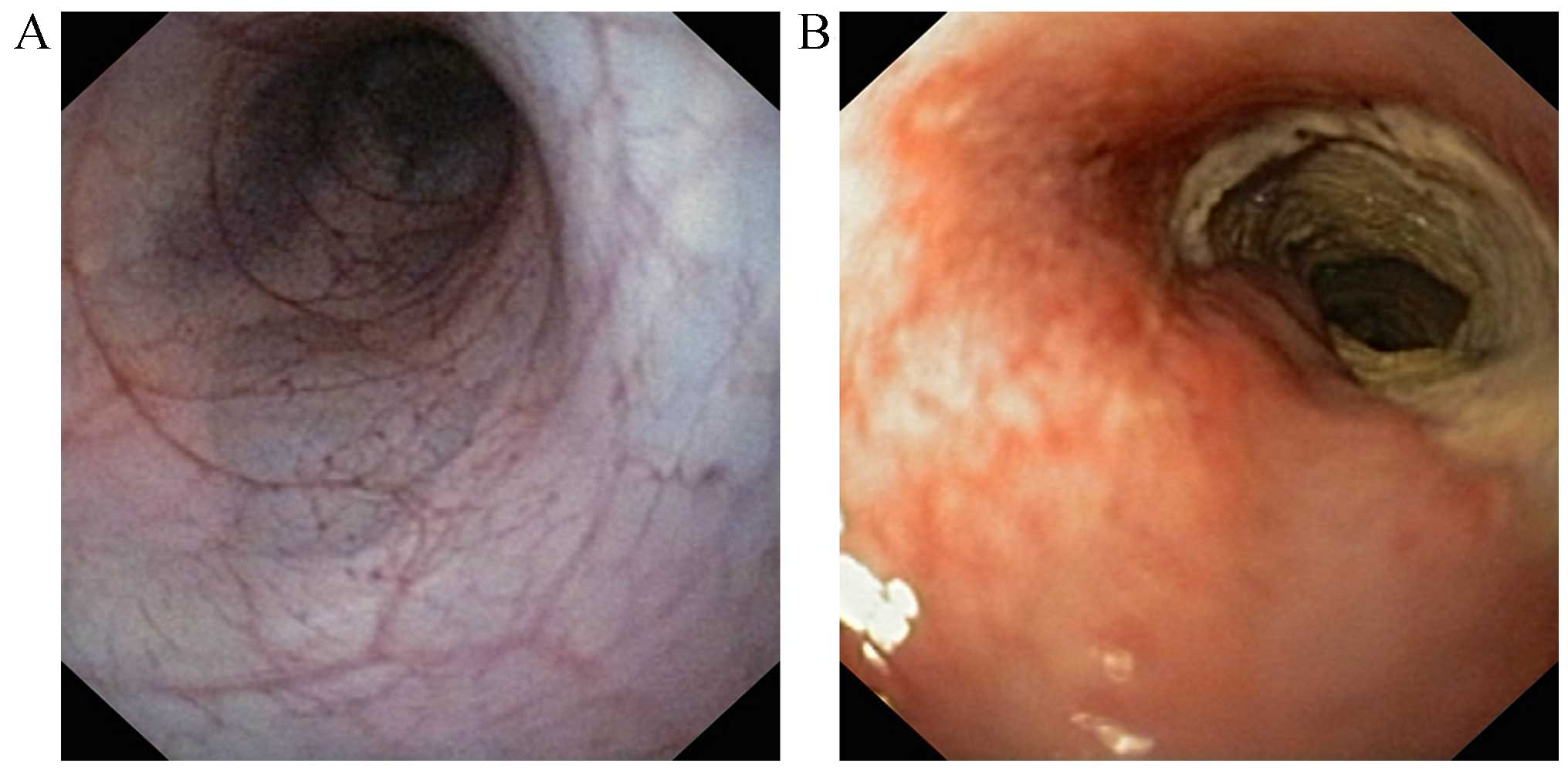
Colonoscopy
Rat colons in the control group displayed a normal
appearance with undamaged mucosa and clear branching of blood
vessels, whereas the colonic mucosa in the TNBS group exhibited
patchy and discontinuous erythema, edema and occasional hemorrhage
(Fig. 2). In addition, aphthoid
ulcers abruptly surrounded by normal mucosa were observed in the
colons of TNBS rats (Fig. 2B). The
deep ulcerations coalesced, which led to mucosal detachment and the
presence of few mucosal islands. Ulcerated stenosis was also
observed in TNBS rat colons (Fig.
2B). The endoscopic inflammation scores were 0 and 6.4±0.8 in
the control and TNBS groups, respectively.
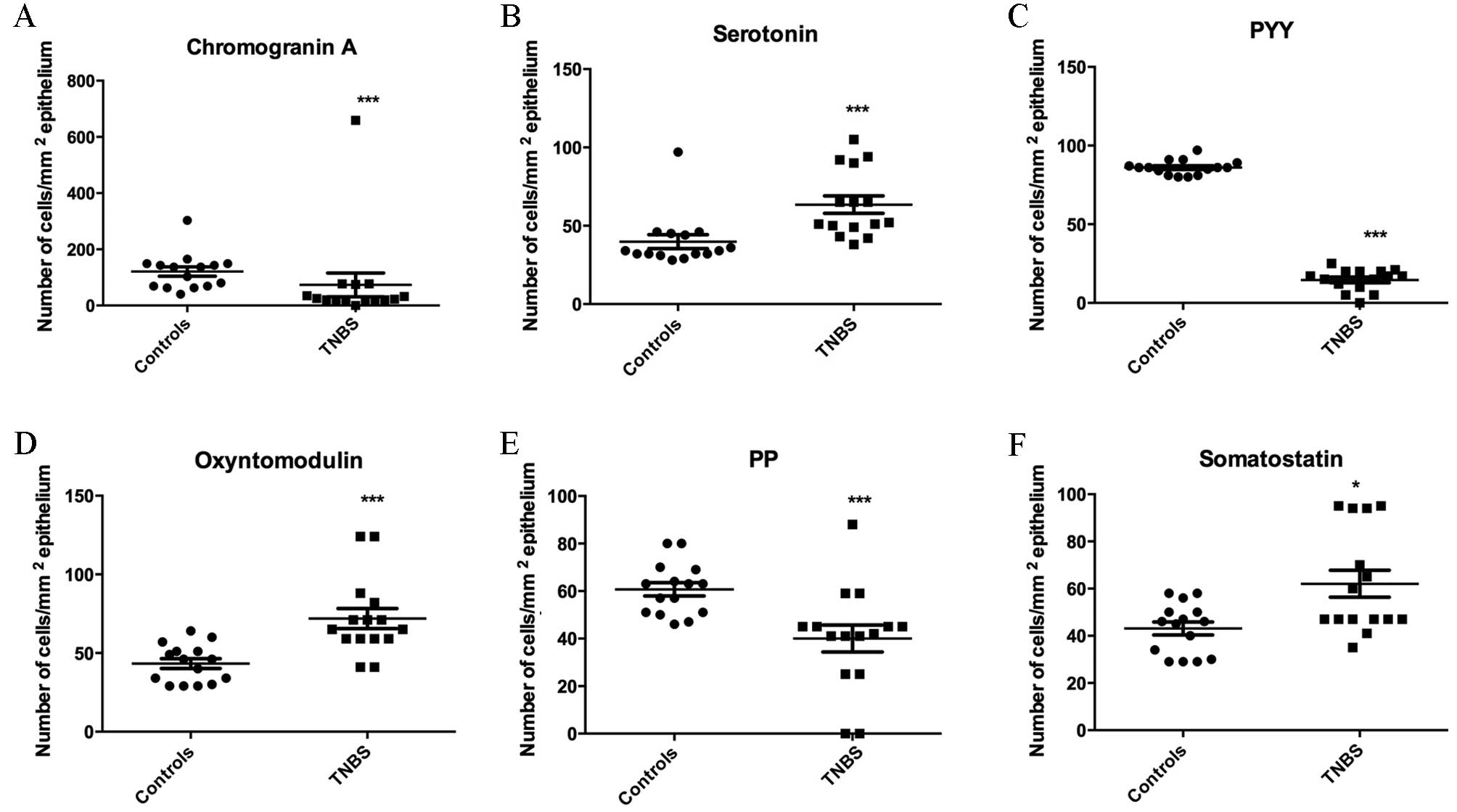
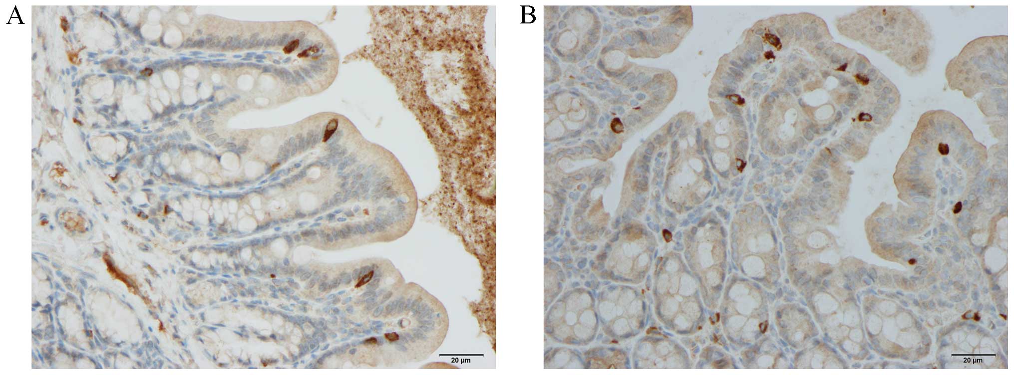
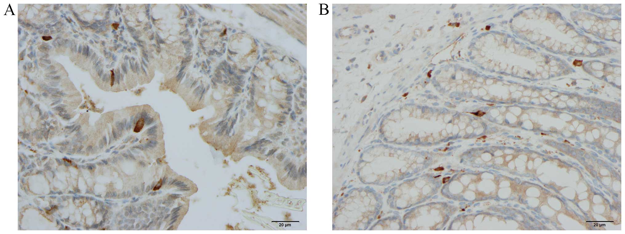
Endocrine cells
The densities of various endocrine cells are
presented in Figs. 3, 4 and 5.
The density of chromogranin A (CgA), peptide YY (PYY) and
pancreatic polypeptide (PP) staining was reduced in the colon
tissues from rats in the TNBS group compared with those of the
control group (P<0.0001, P<0.0001 and P=0.0002, respectively;
Fig. 3). In contrast, serotonin
oxyntomodulin and somatostatin densities were increased in the
colon tissues from the TNBS group compared with those of the
controls (P<0.0001, P<0.0001 and P=0.01, respectively;
Fig. 3).
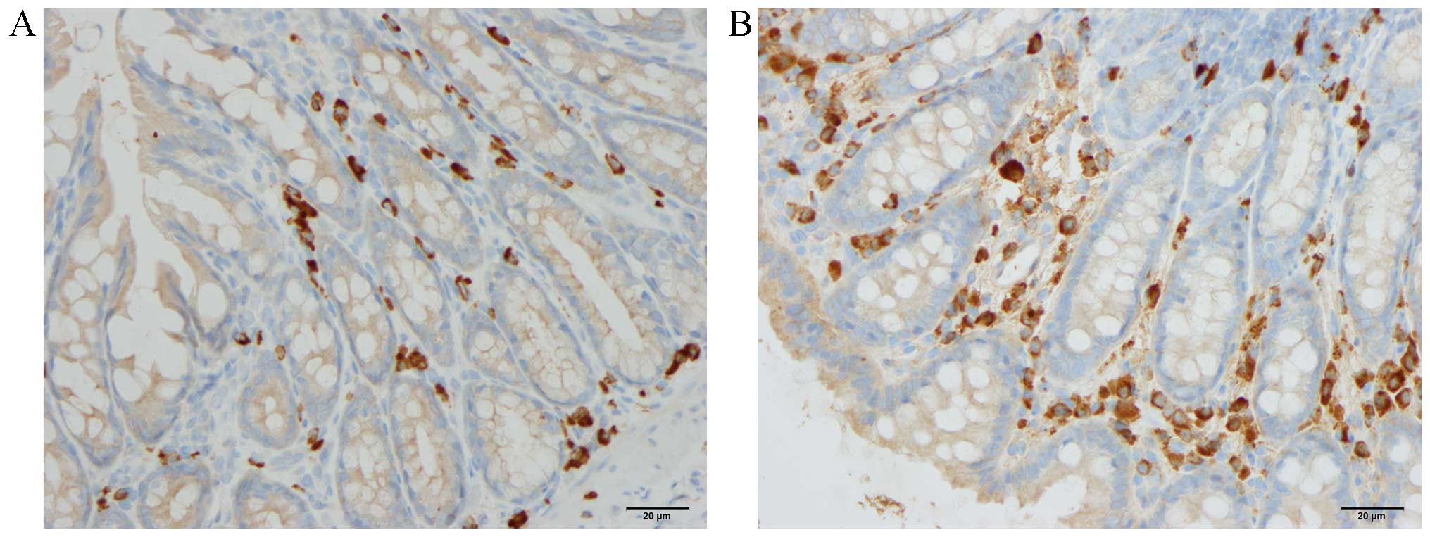
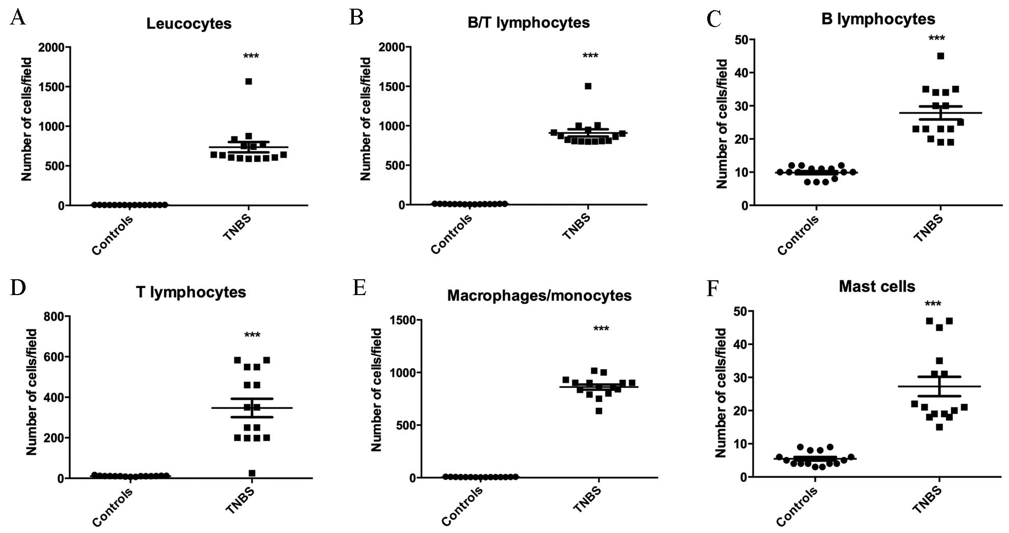
Immune cells
As presented in Figs.
1 and 6, the densities of all
types of immune cells were significantly greater in the TNBS group
compared with the control group [leukocytes, 5.9±0.4 vs. 23.3±2.2
cells/field (P<0.0001); B/T lymphocytes, 9.0±0.7 vs. 35.8±2.3
cells/field (P<0.0001); T lymphocytes, 10.5±0.6 vs. 26.6±2.9
cells/field (P<0.0001); B lymphocytes, 9.7±0.4 vs. 27.7±2.6
cells/field (P<0.0001); macrophages/monocytes, 7.6±0.7 vs.
909.0±46.3 cells/field (P<0.0001); and mast cells 5.5±0.5 vs.
27.3±2.9 cells/field (P<0.0001)].
Correlation between endocrine and
immune cells
The Spearman correlation coefficients and P-values
for the correlations between different endocrine cell types and
various immune cells are presented in Table II. The number of CgA, PYY, and
PP-producing immune cells was observed to be negatively correlated
with the number of specific types of immune cells, whilst positive
correlations were observed for serotonin, oxyntomodulin, and
somatostatin cells.
| Table II.Spearman's rank correlation
coefficients (r) and P-values for the association between endocrine
and immune cells in rats with TNBS-induced colitis. |
Table II.
Spearman's rank correlation
coefficients (r) and P-values for the association between endocrine
and immune cells in rats with TNBS-induced colitis.
|
| Immune cell
type |
|---|
|
|
|
|---|
| Endocrine cell
type | Leukocytes | B/T
lymphocytes | T lymphocytes | B lymphocytes |
Macrophages/monocytes | Mast cells |
|---|
| Chromogranin A | r=−0.7 | r=−0.3 | r=−0.6 | r=−0.6 | r=−0.7 | r=−0.5 |
|
| P=0.04 | P=0.09 | P=0.009 | P=0.02 | P=0.008 | P=0.03 |
| Serotonin | r=0.7 | r=0.7 | r=0.4 | r=0.6 | r=0.2 | r=0.4 |
|
| P=0.005 | P=0.009 | P=0.01 | P=0.02 | P=0.05 | P=0.1 |
| Peptide YY | r=−0.5 | r=−0.7 | r=0.2 | r=−0.7 | r=−0.6 | r=0.7 |
|
| P=0.04 | P=0.002 | P=0.06 | P=0.002 | P=0.02 | P=0.7 |
| Oxyntomdulin | r=0.2 | r=0.5 | r=0.6 | r=0.1 | r=0.1 | r=−0.6 |
|
| P=0.6 | P=0.02 | P=0.01 | P=0.7 | P=0.9 | P=0.02 |
| Pancreatic
polypeptide | r=−0.7 | r=−0.5 | r=−0.2 | r=−0.6 | r=−0.6 | r=−0.7 |
|
| P=0.004 | P=0.8 | P=0.5 | P=0.008 | P=0.01 | P=0.002 |
| Somatostatin | r=0.6 | r=0.7 | r=0.1 | r=0.2 | r=−0.7 | r=−0.6 |
|
| P=0.02 | P=0.8 | P=0.7 | P=0.6 | P=0.0044 | P=0.02 |
Discussion
TNBS-induced colitis in rats closely mimics human CD
(34–39). Although this model exhibits
clinical and morphological features similar to human CD (39–41),
it lacks the chronicity observed in human CD (39). The present study observed that the
frequency of all types of colonic endocrine cells was affected in
rats with TNBS-induced colitis. In addition, abnormalities in the
colonic endocrine cells were strongly correlated with the
alterations in the number of different types of immune cells
following the induction of colitis. These observations support the
previously suggested role of gut hormones in immune activation and
inflammation (21,22,42).
In the present study, the alterations in the number
of colonic endocrine cells observed in rats with TNBS-induced
colitis differ from those observed in rats with DSS-induced colitis
in a previous study (31). In TNBS
and DSS-induced colitis, the densities of serotonin and
oxyntomodulin were increased, while the density of PP was reduced
compared with normal controls. However, the CgA and PYY-producing
immune cell densities were increased in DSS-induced colitis,
whereas they were reduced in TNBS-induced colitis compared with
normal controls. In addition, the density of somatostatin was
reduced in DSS-induced colitis (31), however was increased in
TNBS-induced colitis in the present study. Differences in the
alterations of the number of colonic endocrine cells between CD and
UC have been reported previously by El Salhy et al (1). This study demonstrated that the
densities of CgA and serotonin were increased in CD and UC, while
the densities of PYY and PP were reduced, and oxyntomodulin was
decreased in CD only.
Although the abnormalities in the colonic endocrine
cells in DSS-induced colitis were strongly correlated with
leukocytes, B lymphocytes, T lymphocytes, macrophages/monocytes and
mast cells (31), the results for
TNBS-induced colitis in the present study demonstrated that
specific endocrine cell types were correlated with particular
immune cell types. CgA is a member of the granin family (43,44),
which is localized to gut endocrine cells (45–48),
and is considered to be a common marker for gastrointestinal
endocrine cells (49,50). The reduction in the density of
CgA-producing cells observed in the present study may reflect
reductions in the densities of all colonic endocrine cells
following the induction of colitis by TNBS. The density of
CgA-producing cells was negatively correlated with increases in all
immune cell types except for B/T lymphocytes. CgA suppresses the
release of interleukin (IL)-16 and IL-5, and consequently reduces
the number of lymphocytes at sites of inflammation, and reduces the
pro-inflammatory actions of lymphocytes and monocytes (51–53).
In addition, CgA, inhibits the vascular leakage caused by tumor
necrosis factor-α (54). CgA is
generally considered to exert an anti-inflammatory effect (54). It can therefore be speculated that
the reduced density of CgA results from a direct action exerted by
immune cells.
In the present study, the observed increase in the
density of colonic serotonin-producing cells in TNBS-induced
colitis relative to controls is consistent with previous
observations in patients with UC, CD and microscopic colitis, as
well as animal models of colitis (1,3,55–57).
The increased density of serotonin-producing cells in the present
study was correlated with increases in the number of all types of
immune cells examined, apart from macrophages/monocytes and mast
cells. Lymphocytes, macrophages, and dendritic cells express
serotonin receptors (58), and
IL-13 receptors have been localized on serotonin cells (59). In addition, serotonin inhibits the
apoptosis of immune cells, promotes the recruitment of T cells,
affects the proliferation of lymphocytes and protects natural
killer cells (60–63). Furthermore, a previous study
demonstrated that there are fewer serotonin-producing cells in mice
lacking T-lymphocyte receptors (51). Serotonin stimulates gastric and
intestinal motility, and intestinal secretion (64,65).
Therefore, the increase in serotonin may accelerate
gastrointestinal motility and increase intestinal secretion thus
resulting in diarrhea, which is the primary symptom in TNBS-induced
colitis.
PYY is colocalized with oxyntomodulin in endocrine L
cells (66,67). In the present study, the density of
PYY reduced while oxyntomodulin increased, which indicates that L
cells downregulate the expression of PYY, but upregulate the
expression of oxyntomodulin in TNBS-induced colitis in rats. PYY
stimulates the adhesion of macrophages, chemotaxis, phagocytosis
and the production of superoxide anions (68). PYY mRNA has been detected in mouse
macrophages (69). The precise
interaction between oxyntomodulin and immune cells has not yet been
determined. In the present study, the density of PYY cells was
negatively correlated with increases in the number of B/T
lymphocytes, B lymphocytes and macrophages/monocytes, whereas
oxyntomodulin density was positively correlated with B/T
lymphocytes, T lymphocytes, and mast cells. These results indicate
the presence different interactions of PYY and oxyntomodulin with
immune cells. PYY delays gastric emptying, is a pivotal mediator of
the ileal brake, and stimulates the absorption of water and
electrolytes (70). The reduction
in the number of PYY-producing cells observed in the current study
may have contributed to the acceleration of gastrointestinal
motility and increased intestinal secretion observed in
TNBS-induced colitis.
The reduction in the number of PP-producing cells
observed in the current study is consistent with previous reports
for UC and CD (1). However, the
interaction between PP and immune cells remains to be fully
elucidated. In the present study, PP density was negatively
correlated with the number of B lymphocytes, macrophages/monocytes
and mast cells. PP stimulates gastric acid secretion and the
motility of the stomach and small intestine in addition to relaxing
the gallbladder (64). The
increased density of somatostatin cells in TNBS-induced colitis
observed in the present study contradicts previous observations of
UC, CD and DSS-induced colitis, where somatostatin cell density was
reported to reduce (1,19,20).
Somatostatin inhibits lymphocyte proliferation, immunoglobulin
synthesis and the release of neutrophil elastase (71–75).
In the present study, the density of somatostatin-producing cells
was observed to be positively correlated with the number of
macrophages/monocytes and mast cells. The strong correlation
observed between alterations in PP and somatostatin-producing cell
densities and specific immune cell types, indicate that they may be
involved in the inflammatory process.
The present observations, demonstrating that
alterations in the number of immune cells are strongly correlated
with alterations in large intestinal cells in an animal model of
human of Crohn's disease, support the debated suggestion of an
interaction between intestinal hormones and the gut immune system.
Understanding this interaction may improve our understanding of the
pathophysiological mechanisms involved in IBD, and may provide us
with novel therapeutic approaches to treat this condition.
Acknowledgements
The present study was supported by grants from the
Helse-Vest regional health authority, (Bergen, Norway; grant no.
911978) and Helse-Fonna health organization (Haugesund, Norway;
grant no. 40415).
References
|
1
|
El-Salhy M, Danielsson A, Stenling R and
Grimelius L: Colonic endocrine cells in inflammatory bowel disease.
J Intern Med. 242:413–419. 1997. View Article : Google Scholar : PubMed/NCBI
|
|
2
|
El-Salhy M, Gundersen D, Hatlebakk JG and
Hausken T: Chromogranin a cell density as a diagnostic marker for
lymphocytic colitis. Dig Dis Sci. 57:3154–3159. 2012. View Article : Google Scholar : PubMed/NCBI
|
|
3
|
El-Salhy M, Gundersen D, Hatlebakk JG and
Hausken T: High densities of serotonin and peptide YY cells in the
colon of patients with lymphocytic colitis. World J Gastroenterol.
18:6070–6075. 2012. View Article : Google Scholar : PubMed/NCBI
|
|
4
|
El-Salhy M, Lomholt-Beck B and Gundersen
TD: High chromogranin A cell density in the colon of patients with
lymphocytic colitis. Mol Med Rep. 4:603–605. 2011.PubMed/NCBI
|
|
5
|
Moran GW, Pennock J and McLaughlin JT:
Enteroendocrine cells in terminal ileal Crohn's disease. J Crohns
Colitis. 6:871–880. 2012. View Article : Google Scholar : PubMed/NCBI
|
|
6
|
Moran GW, Leslie FC and McLaughlin JT:
Crohn's disease affecting the small bowel is associated with
reduced appetite and elevated levels of circulating gut peptides.
Clin Nutr. 32:404–411. 2013. View Article : Google Scholar : PubMed/NCBI
|
|
7
|
Besterman HS, Mallinson CN, Modigliani R,
Christofides ND, Pera A, Ponti V, Sarson DL and Bloom SR: Gut
hormones in inflammatory bowel disease. Scand J Gastroenterol.
18:845–852. 1983. View Article : Google Scholar : PubMed/NCBI
|
|
8
|
El-Salhy M, Mazzawi T, Gundersen D,
Hatlebakk JG and Hausken T: The role of peptide YY in
gastrointestinal diseases and disorders (Review). Int J Mol Med.
31:275–282. 2013.PubMed/NCBI
|
|
9
|
Hirotani Y, Mikajiri K, Ikeda K, Myotoku M
and Kurokawa N: Changes of the peptide YY levels in the intestinal
tissue of rats with experimental colitis following oral
administration of mesalazine and prednisolone. Yakugaku Zasshi.
128:1347–1353. 2008. View Article : Google Scholar : PubMed/NCBI
|
|
10
|
Vona-Davis LC and McFadden DW: NPY family
of hormones: Clinical relevance and potential use in
gastrointestinal disease. Curr Top Med Chem. 7:1710–1720. 2007.
View Article : Google Scholar : PubMed/NCBI
|
|
11
|
El-Salhy M, Suhr O and Danielsson A:
Peptide YY in gastrointestinal disorders. Peptides. 23:397–402.
2002. View Article : Google Scholar : PubMed/NCBI
|
|
12
|
Tari A, Teshima H, Sumii K, Haruma K,
Ohgoshi H, Yoshihara M, Kajiyama G and Miyachi Y: Peptide YY
abnormalities in patients with ulcerative colitis. Jpn J Med.
27:49–55. 1988. View Article : Google Scholar : PubMed/NCBI
|
|
13
|
Sciola V, Massironi S, Conte D, Caprioli
F, Ferrero S, Ciafardini C, Peracchi M, Bardella MT and Piodi L:
Plasma chromogranin a in patients with inflammatory bowel disease.
Inflamm Bowel Dis. 15:867–871. 2009. View Article : Google Scholar : PubMed/NCBI
|
|
14
|
Bishop AE, Pietroletti R, Taat CW,
Brummelkamp WH and Polak JM: Increased populations of endocrine
cells in Crohn's ileitis. Virchows Arch A Pathol Anat Histopathol.
410:391–396. 1987. View Article : Google Scholar : PubMed/NCBI
|
|
15
|
Manocha M and Khan WI: Serotonin and GI
disorders: An update on clinical and experimental studies. Clin
Transl Gastroenterol. 3:e132012. View Article : Google Scholar : PubMed/NCBI
|
|
16
|
Stoyanova II and Gulubova MV: Mast cells
and inflammatory mediators in chronic ulcerative colitis. Acta
Histochem. 104:185–192. 2002. View Article : Google Scholar : PubMed/NCBI
|
|
17
|
Yamamoto H, Morise K, Kusugami K, Furusawa
A, Konagaya T, Nishio Y, Kaneko H, Uchida K, Nagai H, Mitsuma T and
Nagura H: Abnormal neuropeptide concentration in rectal mucosa of
patients with inflammatory bowel disease. J Gastroenterol.
31:525–532. 1996. View Article : Google Scholar : PubMed/NCBI
|
|
18
|
Payer J, Huorka M, Duris I, Mikulecky M,
Kratochvílová H, Ondrejka P and Lukác L: Plasma somatostatin levels
in ulcerative colitis. Hepatogastroenterology. 41:552–553.
1994.PubMed/NCBI
|
|
19
|
Watanabe T, Kubota Y, Sawada T and Muto T:
Distribution and quantification of somatostatin in inflammatory
disease. Dis Colon Rectum. 35:488–494. 1992. View Article : Google Scholar : PubMed/NCBI
|
|
20
|
Koch TR, Carney JA, Morris VA and Go VL:
Somatostatin in the idiopathic inflammatory bowel diseases. Dis
Colon Rectum. 31:198–203. 1988. View Article : Google Scholar : PubMed/NCBI
|
|
21
|
Khan WI and Ghia JE: Gut hormones:
Emerging role in immune activation and inflammation. Clin Exp
Immunol. 161:19–27. 2010.PubMed/NCBI
|
|
22
|
Margolis KG and Gershon MD: Neuropeptides
and inflammatory bowel disease. Curr Opin Gastroenterol.
25:503–511. 2009. View Article : Google Scholar : PubMed/NCBI
|
|
23
|
Bampton PA and Dinning PG: High resolution
colonic manometry-what have we learnt?-A review of the literature
2012. Curr Gastroenterol Rep. 15:3282013. View Article : Google Scholar : PubMed/NCBI
|
|
24
|
Ameri P and Ferone D: Diffuse endocrine
system, neuroendocrine tumors and immunity: What's new?
Neuroendocrinology. 95:267–276. 2012. View Article : Google Scholar : PubMed/NCBI
|
|
25
|
Farzi A, Reichmann F and Holzer P: The
homeostatic role of neuropeptide Y in immune function and its
impact on mood and behaviour. Acta Physiol (Oxf). 213:603–627.
2015. View Article : Google Scholar : PubMed/NCBI
|
|
26
|
El-Salhy M and Hausken T: The role of the
neuropeptide Y (NPY) family in he pathophysiology of inflammatory
bowel disease (IBD). Neuropeptides. 55:137–144. 2016. View Article : Google Scholar : PubMed/NCBI
|
|
27
|
Wheway J, Herzog H and Mackay F: NPY and
receptors in immune and inflammatory diseases. Curr Top Med Chem.
7:1743–1752. 2007. View Article : Google Scholar : PubMed/NCBI
|
|
28
|
Wheway J, Herzog H and Mackay F: The Y1
receptor for NPY: A key modulator of the adaptive immune system.
Peptides. 28:453–458. 2007. View Article : Google Scholar : PubMed/NCBI
|
|
29
|
Wheway J, Mackay CR, Newton RA, Sainsbury
A, Boey D, Herzog H and Mackay F: A fundamental bimodal role for
neuropeptide Y1 receptor in the immune system. J Exp Med.
202:1527–1538. 2005. View Article : Google Scholar : PubMed/NCBI
|
|
30
|
El-Salhy M, Gundersen D, Hatlebakk JG and
Hausken T: Clinical presentation, diagnosis, pathogenesis and
treatment options for lymphocytic colitis (Review). Int J Mol Med.
32:263–270. 2013.PubMed/NCBI
|
|
31
|
El-Salhy M, Hatlebakk JG and Gilja OH: The
abnormalities in endocrine and immune cells are correlated in
dextran-sulfate-sodium-induced colitis. Mol Med Rep. in press.
2016.
|
|
32
|
El-Salhy M, Umezawa K, Gilja OH, Hatlebakk
JG, Gundersen D and Hausken T: Amelioration of Severe TNBS Induced
Colitis by Novel AP-1 and NF-κB Inhibitors in Rats. Sci World J.
2014:1–8. 2014. View Article : Google Scholar
|
|
33
|
Vermeulen W, De Man JG, Nullens S,
Pelckmans PA, De Winter BY and Moreels TG: The use of colonoscopy
to follow the inflammatory time course of TNBS colitis in rats.
Acta Gastroenterol Belg. 74:304–311. 2011.PubMed/NCBI
|
|
34
|
Saleh M and Elson CO: Experimental
inflammatory bowel disease: Insights into the host-microbiota
dialog. Immunity. 34:293–302. 2011. View Article : Google Scholar : PubMed/NCBI
|
|
35
|
Carter MJ, Lobo AJ and Travis SP: IBD
Section, British Society of Gastroenterology: Guidelines for the
management of inflammatory bowel disease in adults. Gut. 53:(Suppl
5). V1–V16. 2004. View Article : Google Scholar : PubMed/NCBI
|
|
36
|
Sands BE: New therapies for the treatment
of inflammatory bowel disease. Surg Clin North Am. 86:1045–1064.
2006. View Article : Google Scholar : PubMed/NCBI
|
|
37
|
Lopez A, Billioud V, Peyrin-Biroulet C and
Peyrin-Biroulet L: Adherence to anti-TNF therapy in inflammatory
bowel diseases: A systematic review. Inflamm Bowel Dis.
19:1528–1533. 2013. View Article : Google Scholar : PubMed/NCBI
|
|
38
|
Danese S, Semeraro S, Armuzzi A, Papa A
and Gasbarrini A: Biological therapies for inflammatory bowel
disease: Research drives clinics. Mini Rev Med Chem. 6:771–784.
2006. View Article : Google Scholar : PubMed/NCBI
|
|
39
|
Elson CO, Sartor RB, Tennyson GS and
Riddell RH: Experimental models of inflammatory bowel disease.
Gastroenterology. 109:1344–1367. 1995. View Article : Google Scholar : PubMed/NCBI
|
|
40
|
Dieleman LA, Palmen MJ, Akol H, Bloemena
E, Peña AS, Meuwissen SG and Van Rees EP: Chronic experimental
colitis induced by dextran sulphate sodium (DSS) is characterized
by Th1 and Th2 cytokines. Clin Exp Immunol. 114:385–391. 1998.
View Article : Google Scholar : PubMed/NCBI
|
|
41
|
Low D, Nguyen DD and Mizoguchi E: Animal
models of ulcerative colitis and their application in drug
research. Drug Des Devel Ther. 7:1341–1357. 2013.PubMed/NCBI
|
|
42
|
Öhman L, Törnblom H and Simrén M:
Crosstalk at the mucosal border: Importance of the gut
microenvironment in IBS. Nat Rev Gastroenterol Hepatol. 12:36–49.
2015. View Article : Google Scholar : PubMed/NCBI
|
|
43
|
Buffa R, Mare P, Gini A and Salvadore M:
Chromogranins A and B and secretogranin II in hormonally identified
endocrine cells of the gut and the pancreas. Basic Appl Histochem.
32:471–484. 1988.PubMed/NCBI
|
|
44
|
Eiden LE: Is chromogranin a prohormone?
Nature. 325:3011987. View Article : Google Scholar : PubMed/NCBI
|
|
45
|
Buffa R, Capella C, Fontana P, Usellini L
and Solcia E: Types of endocrine cells in the human colon and
rectum. Cell Tissue Res. 192:227–240. 1978. View Article : Google Scholar : PubMed/NCBI
|
|
46
|
Curry WJ, Johnston CF, Hutton JC, Arden
SD, Rutherford NG, Shaw C and Buchanan KD: The tissue distribution
of rat chromogranin A-derived peptides: Evidence for differential
tissue processing from sequence specific antisera. Histochemistry.
96:531–538. 1991. View Article : Google Scholar : PubMed/NCBI
|
|
47
|
Portela-Gomes GM and Stridsberg M:
Selective processing of chromogranin A in the different islet cells
in human pancreas. J Histochem Cytochem. 49:483–490. 2001.
View Article : Google Scholar : PubMed/NCBI
|
|
48
|
Portela-Gomes GM and Stridsberg M:
Chromogranin A in the human gastrointestinal tract: An
immunocytochemical study with region-specific antibodies. J
Histochem Cytochem. 50:1487–1492. 2002. View Article : Google Scholar : PubMed/NCBI
|
|
49
|
Taupenot L, Harper KL and O'Connor DT: The
chromogranin-secretogranin family. N Engl J Med. 348:1134–1149.
2003. View Article : Google Scholar : PubMed/NCBI
|
|
50
|
Wiedenmann B and Huttner WB: Synaptophysin
and chromogranins/secretogranins-widespread constituents of
distinct types of neuroendocrine vesicles and new tools in tumor
diagnosis. Virchows Arch B Cell Pathol Incl Mol Pathol. 58:95–121.
1989. View Article : Google Scholar : PubMed/NCBI
|
|
51
|
Spiller R: Serotonin and GI clinical
disorders. Neuropharmacology. 55:1072–1080. 2008. View Article : Google Scholar : PubMed/NCBI
|
|
52
|
Egger M, Beer AG, Theurl M, Schgoer W,
Hotter B, Tatarczyk T, Vasiljevic D, Frauscher S, Marksteiner J,
Patsch JR, et al: Monocyte migration: A novel effect and signaling
pathways of catestatin. Eur J Pharmacol. 598:104–111. 2008.
View Article : Google Scholar : PubMed/NCBI
|
|
53
|
Feistritzer C, Mosheimer BA, Colleselli D,
Wiedermann CJ and Kähler CM: Effects of the neuropeptide
secretoneurin on natural killer cell migration and cytokine
release. Regul Pept. 126:195–201. 2005. View Article : Google Scholar : PubMed/NCBI
|
|
54
|
Ferrero E, Magni E, Curnis F, Villa A,
Ferrero ME and Corti A: Regulation of endothelial cell shape and
barrier function by chromogranin A. Ann N Y Acad Sci. 971:355–358.
2002. View Article : Google Scholar : PubMed/NCBI
|
|
55
|
Bertrand PP and Bertrand RL: Serotonin
release and uptake in the gastrointestinal tract. Auton Neurosci.
153:47–57. 2010. View Article : Google Scholar : PubMed/NCBI
|
|
56
|
Qian BF, El-Salhy M, Melgar S, Hammarström
ML and Danielsson A: Neuroendocrine changes in colon of mice with a
disrupted IL-2 gene. Clin Exp Immunol. 120:424–433. 2000.
View Article : Google Scholar : PubMed/NCBI
|
|
57
|
Oshima S, Fujimura M and Fukimiya M:
Changes in number of serotonin-containing cells and serotonin
levels in the intestinal mucosa of rats with colitis induced by
dextran sodium sulfate. Histochem Cell Biol. 112:257–263. 1999.
View Article : Google Scholar : PubMed/NCBI
|
|
58
|
Cloëz-Tayarani I and Changeux JP: Nicotine
and serotonin in immune regulation and inflammatory processes: A
perspective. J Leukoc Biol. 81:599–606. 2007. View Article : Google Scholar : PubMed/NCBI
|
|
59
|
Wang H, Steeds J, Motomura Y, Deng Y,
Verma-Gandhu M, El-Sharkawy RT, McLaughlin JT, Grencis RK and Khan
W: CD4+ T cell-mediated immunological control of enterochromaffin
cell hyperplasia and 5-hydroxytryptamine production in enteric
infection. Gut. 56:949–957. 2007. View Article : Google Scholar : PubMed/NCBI
|
|
60
|
Stefulj J, Cicin-Sain L, Schauenstein K
and Jernej B: Serotonin and immune response: Effect of the amine on
in vitro proliferation of rat lymphocytes. Neuroimmunomodulation.
9:103–108. 2001. View Article : Google Scholar : PubMed/NCBI
|
|
61
|
Betten A, Dahlgren C, Hermodsson S and
Hellstrand K: Serotonin protects NK cells against oxidatively
induced functional inhibition and apoptosis. J Leukoc Biol.
70:65–72. 2001.PubMed/NCBI
|
|
62
|
Laberge S, Cruikshank WW, Beer DJ and
Center DM: Secretion of IL-16 (lymphocyte chemoattractant factor)
from serotonin-stimulated CD8+ T cells in vitro. J Immunol.
156:310–315. 1996.PubMed/NCBI
|
|
63
|
Soga F, Katoh N, Inoue T and Kishimoto S:
Serotonin activates human monocytes and prevents apoptosis. J
Invest Dermatol. 127:1947–1955. 2007. View Article : Google Scholar : PubMed/NCBI
|
|
64
|
El-Salhy M, Seim I, Chopin L, Gundersen D,
Hatlebakk JG and Hausken T: Irritable bowel syndrome: The role of
gut neuroendocrine peptides. Front Biosci (Elite Ed). 4:2783–2800.
2012.PubMed/NCBI
|
|
65
|
El-Salhy M: Irritable bowel syndrome:
Diagnosis, pathogenesis and treatment options. World J
Gastroenterol. 18:5151–5163. 2012.PubMed/NCBI
|
|
66
|
Spångéus A, Forsgren S and el-Salhy M:
Does diabetic state affect co-localization of peptide YY and
enteroglucagon in colonic endocrine cells? Histol Histopathol.
15:37–41. 2000.PubMed/NCBI
|
|
67
|
Pyarokhil AH, Ishihara M, Sasaki M and
Kitamura N: The developmental plasticity of colocalization pattern
of peptide YY and glucagon-like peptide-1 in the endocrine cells of
bovine rectum. Biomed Res. 33:35–38. 2012. View Article : Google Scholar : PubMed/NCBI
|
|
68
|
De la Fuente M, Bernaez I, Del Rio M and
Hernanz A: Stimulation of murine peritoneal macrophage functions by
neuropeptide Y and peptide YY. Involvement of protein kinase C.
Immunology. 80:259–265. 1993.PubMed/NCBI
|
|
69
|
Macia L, Yulyaningsih E, Pangon L, Nguyen
AD, Lin S, Shi YC, Zhang L, Bijker M, Grey S, Mackay F, et al:
Neuropeptide Y1 receptor in immune cells regulates inflammation and
insulin resistance associated with diet-induced obesity. Diabetes.
61:3228–3238. 2012. View Article : Google Scholar : PubMed/NCBI
|
|
70
|
El-Salhy M, Gundersen D, Gilja OH,
Hatlebakk JG and Hausken T: Is irritable bowel syndrome an organic
disorder? World J Gastroenterol. 20:384–400. 2014. View Article : Google Scholar : PubMed/NCBI
|
|
71
|
Payan DG, Hess CA and Goetzl EJ:
Inhibition by somatostatin of the proliferation of T-lymphocytes
and Molt-4 lymphoblasts. Cell Immunol. 84:433–438. 1984. View Article : Google Scholar : PubMed/NCBI
|
|
72
|
Adeyemi EO, Savage AP, Bloom SR and
Hodgson HJ: Somatostatin inhibits neutrophil elastase release in
vitro. Peptides. 11:869–871. 1990. View Article : Google Scholar : PubMed/NCBI
|
|
73
|
Stanisz AM, Befus D and Bienenstock J:
Differential effects of vasoactive intestinal peptide, substance P,
and somatostatin on immunoglobulin synthesis and proliferations by
lymphocytes from Peyer's patches, mesenteric lymph nodes, and
spleen. J Immunol. 136:152–156. 1986.PubMed/NCBI
|
|
74
|
Scicchitano R, Dazin P, Bienenstock J,
Payan DG and Stanisz AM: Distribution of somatostatin receptors on
murine spleen and Peyer's patch T and B lymphocytes. Brain Behav
Immun. 1:173–184. 1987. View Article : Google Scholar : PubMed/NCBI
|
|
75
|
Scicchitano R, Stanisz AM, Payan DG,
Kiyono H, McGhee JR and Bienenstock J: Expression of substance P
and somatostatin receptors on a T helper cell line. Adv Exp Med
Biol 216A. 185–190. 1987. View Article : Google Scholar
|