Introduction
Folium Epimedii, also known as Yin Yang Huo in
China, is derived from the genus Epimedium, which includes
E. sagittatum Maxim., E. pubescens Maxim., E.
koreanum Nakai and E. wushanense T.S. Ying. All these
species have been used for hundreds of years to combat several
diseases, including erectile dysfunction, fatigue, kidney disorders
and joint pain (1). Folium
Epimedii has been confirmed to be effective in the treatment of
cardiovascular diseases (2),
osteoporosis (3) and tumors
(4).
Icariin (ICA), has been suggested to be an
indicative constituent of Folium Epimedii. Previous studies have
indicated that ICA exhibits positive effects in suppressing
inflammation, and promoting cardiovascular functions (5,6) and
antitumor activities (7,8). Furthermore, previous studies have
shown that ICA suppresses cartilage and bone degradation in mice
with collagen-induced arthritis (9), and inhibits cell growth and induces
apoptosis in Burkett lymphoma cell lines (10).
Previously, the quantitative and qualitative
analyses of ICA had been performed primarily using high-performance
liquid chromatography (HPLC) and thin-layer chromatography (TLC)
(11,12). However, these methods have various
limitations, including high cost, component degradation, prolonged
duration, low recovery rates and complicated pretreatment,
particularly for in vivo investigations of metabolism.
Therefore, it is necessary to establish a novel, simple method for
ICA analysis.
The enzyme-linked immunosorbent assay (ELISA)
method, based on specific monoclonal antibodies (MAbs) has become
an important methodology for the qualitative or quantitative
analysis of food or natural products (13,14).
This method is rapid and requires only minimal sample
pre-treatment. In addition, the method can be used simultaneously
for a large number of samples. Previously, MAbs against certain
compounds in traditional Chinese medicines (TCMs) have been
reported (15–21); however, no MAbs specific for ICA
have been described.
In our previous studies, preparations of MAbs have
been developed against baicalin (15), puerarin (16), geniposide (17), glycyrrhizic acid (18), paeoniflorin (19), ginsensoide Re (20) and ginsenoside Rh1 (21), and their ELISA methods have been
established. Consequently, these assays were applied to examine the
pharmacokinetics and pharmacokinetic interactions between these
bioactive compounds. The comparably low quantities of sample
required, for example 5 µl of serum, for the determination in mice
is particularly beneficial for pharmacokinetic investigations.
Furthermore, the development of immunoaffinity chromatography based
on anti-gisenoside Rh1 MAb has been indicated as a potential method
for the separation of epimers (21).
In the present study, the formation and
characterization of an anti-ICA MAb were investigated, and an
indirect competitive (ic)ELISA method was established. The MAb and
icELISA were used to detect ICA in the complex chemical
constituents of TCM. This icELISA method may be of use for further
investigations of ICA.
Materials and methods
Chemicals and reagents
ICA was purchased from Welch Materials, Inc.
(Shanghai, China; purity of 95%). Sodium periodate was obtained
from Sinopharm Chemical Reagent Co., Ltd. (Beijing, China). Bovine
serum albumin (BSA), ovalbumin (OVA) and Freund's complete and
incomplete reagents were obtained from Sigma-Aldrich; Merck
Millipore (Darmstadt, Germany). All other chemicals and reagents
were of analytical grade and were purchased from Sinopharm Chemical
Reagent Co., Ltd.
The composition of the four TCM detected were as
follows: i)≈Zhuanggu guanjie wan (bolus) (China Resources Sanjiu
Medical and Pharmaceutical, Shenzhen, China): Cibotii Rhizoma,
Epimedii Folium, Dipsaci Radix, Psoraleae Fructus, Spatholobi
Caulis, Olibanum, Myrrha, Aucklandiae Radix, Drynariae Rhizoma,
Taxilli Herba, Angelicae Pubescentis Radix, Rehmanniae Radix
Praeparata; ii) Pishen Shuangbu decoction: Taxilli Herba (30 g),
Corn Stigma (30 g), Polygoni Multiflori Radix (24 g), Chuanxiong
Rhizoma (9 g), Eucommiae Cortex (9 g), Magnetitum (3 g), Fossil
Fragments (30 g) and Epimedii Folium (9 g); iii) Erxian decoction:
Curculiginis Rhizoma (9 g), Epimedii Folium (9 g), Morindae
officinalis Radix (9 g), Phellodendri Chinensis Cortex (4.5 g),
Angelicae Sinensis Radix (9 g) and Anemarrhenae Rhizoma (4.5 g);
iv) Tongbi Prescription: Aconiti Kusnezoffii Radix (15 g), Aconiti
Radix (10 g), Asari Radix Et Rhizoma (6 g), Ephedrae Herba (10 g),
Taxilli Herba (15 g), Zingiberis Rhizoma (10 g), Glycyrrhizae Radix
Et Rhizoma (6 g), Epimedii Folium (15 g), Eucommiae Cortex (10 g)
and Dipsaci Radix (15 g). All aforementioned Chinese herbal
medicine was purchased from Tongrentang (Beijing, China).
The compositions of the buffers and solutions used
in the present study were as follows: Phosphate-buffered saline
(PBS; pH 7.4): NaCl (137 mmol/l), Na2HPO4•12
H2O (10 mmol/l), KCl (2.68 mmol/l) and
KH2PO4 (1.47 mmol/l); carbonate buffer
solution (CBS; pH 9.6): Na2CO3 (15 mmol/l)
and NaHCO3 (35 mmol/l); washing buffer: PBS with 0.05%
Tween-20 (PBST); blocking buffer: 10 mg/ml gelatine in PBS (GPBS);
tetramethylbenzidine (TMB) substrate solution: Combination of part
A (0.5 ml of stock comprising 24.3 ml of 0.1 mol/ml citric acid,
25.7 ml of 0.2 mol/ml Na2HPO4 and 50 ml
deionized water), part B (10 ml of stock comprising 2 mg of TMB
dissolved in 1 ml of methanol) and part C (32 µl of 0.75%
H2O2); stopping solution: 2 mol/ml
H2SO4 and 1 mM HCl (pH 4.0; 20 ml/g);
hypoxanthine-aminopterin-thymidine (HAT); polyethylene glycol
(PEG); and hypoxanthine-thymidine (HT).
Synthesis of ICA antigen
conjugate
The conjugates were synthesized using a periodate
oxidation procedure according to a previously reported protocol
with modifications (22,23). Briefly, the ICA was dissolved in
CBS at 1 mg/ml, following which 1 ml of freshly prepared sodium
periodate solution (8 mg) was added drop-wise to 5 ml of the ICA
solution. The mixture was stirred at 25°C for 1 h, following which
5 mg of BSA dissolved in 1 ml of CBS was added, and the final pH
was adjusted to 9.0 using 0.05 M carbonate buffer (pH 9.6).
Following stirring at 25°C for 6 h, the mixture was dialyzed six
times against PBS. The dialysate of the ICA-BSA conjugate was
stored at 4°C for detection and immunization. The ICA-OVA conjugate
was synthesized using the same method described above.
Animal treatment
A total of 5 female BALB/c mice (6 weeks old) were
purchased from Vital River Laboratories (Beijing, China). The mice
were fed a standard rodent diet (Keaoxieli Animal Feed Co., Ltd.,
Beijing, China) ad libitum and housed in an environmentally
controlled (23±2°C; 12 h light/dark cycle) animal facility. Mice
were sacrificed by cervical dislocation. The present study was
performed according to the Guidelines for the Care and Use of
Laboratory Animals and was approved by the Joint Ethical Review
Committee of the Beijing University of Chinese Medicine (Beijing,
China; approval no. 2013 BZHYLL00106).
Immunization
The immunizations were performed at 2-week
intervals. The mice were subcutaneously injected with a 50 µg
volume of the ICA-BSA conjugate in PBS, emulsified with an equal
volume of Freund's complete adjuvant in the initial immunization.
The second and third immunizations, which contained 50 µg of the
ICA-BSA conjugate in Freund's incomplete adjuvant, were injected
subcutaneously 2 and 4 weeks following the initial injection. Blood
was obtained from the tail vein of the mice and centrifuged at 4°C
and 2,227 × g for 10 min following the third immunization,
and a titre of sera was examined by indirect ELISA, using ICA-OVA
as the solid-phase antigen. After 2 weeks the fourth immunization
involved injection with a solution of ICA-BSA (100 µg) in PBS
without adjuvant.
Cell fusion and preparation of the
anti-ICA MAb
At 3 days following the final immunization the
spleen from immunized mice was removed using a cell strainer. Using
the head of a clean syringe, the spleen was ground to dissociate
the splenocytes. The cells were suspended following filtering and
splenocytes were isolated and fused with the
hypoxanthine-aminopterin-thymidine (HAT)-sensitive SP2/0 mouse
myeloma cell line (ScienCell Research Laboratory; Carlsbad, CA,
USA), according to the PEG method (24,25).
Briefly, following centrifugation at room temperature at 180 ×
g for 10 min of the blended splenocytes and myeloma cells
(at a ratio of 5:1), 1 ml of PEG was added drop-wise to the cell
pellet and then incubated for 1 min at 37°C. The HAT medium
(Sigma-Aldrich; Merck Millipore) was then added. The hybridoma was
transferred to 96-well plates for cell culture. The cells producing
MAbs reactive to ICA, as identified using indirect ELISA, were
cloned according to the limiting dilution method (26). The established hybridoma was then
cultured in the HT medium with 5% CO2 at 37°C for 10
days.
A total of 20 mice (10-week-old, male) were
purchased from Vital River Laboratories (Beijing, China). The mice
were fed a standard rodent diet (Keaoxieli Animal Feed Co., Ltd.,
Beijing, China) ad libitum and housed in an environmentally
controlled (23±2°C; 12 h light/dark cycle) animal facility, which
had been injected with 300 µl Freund's incomplete adjuvant on the
previous day. The hybridomas were transplanted into the abdominal
cavity of 10-week-old male BALB/c mice, which had been injected
with Freund's incomplete adjuvant on the previous day. The mice
were sacrificed prior to the injection by cervical dislocation.
After 5–7 days, fluid was drained from the resulting ascites. This
fluid was then purified by centrifugation at 4°C and 5,702 ×
g for 10 min, followed by caprylic acid precipitation and
protein quantification (27).
Establishment of the icELISA
method
The reactivity of the anti-ICA MAbs with ICA-OVA was
determined using indirect ELISA (iELISA). The ICA-OVA was dissolved
in CBS, following which 100 µl was added to each well of a 96-well
maxisorp immunoplate and incubated for 1 h. Each well was then
treated with 200 µl of GPBS for 1 h to inhibit non-specific
absorption. The plate was washed three times with PBST prior to the
addition of 100 µl of anti-ICA MAbs (1:10,0000). Following
incubation for 1 h, the plate was washed three times and incubated
with 100 µl of peroxidase-labelled goat anti-mouse IgG solution
(1:10,000; cat. no. C1308; Applygen Technologies Inc., Beijing,
China) for 30 min. The plate was then washed three times, and 100
µl of TMB substrate solution was added to each well, followed by
incubation for 15 min. The reaction was terminated by adding 50 µl
of 2 M H2SO4, and the absorbance was measured
at 450 nm using a BioTek ELx 800 microplate reader (BioTek China,
Beijing Chin). All reactions were performed at 37°C.
The reactivity of the anti-ICA MAbs was determined
using an icELISA. The protocol for this assay was identical to that
used for the iELISA, with the exception that the primary antibodies
used were 50 µl of ICA and 50 µl of anti-ICA MAb, and the
subsequent incubation duration was 1 h.
Assay sensitivity and specificity
The icELISA was established with ICA-OVA (1:2,000)
as the solid-phase antigen and ascites induced by the transplant of
the hybridoma (1:200,000). Various quantities of ICA were added to
compete with the coated antigen, by which the standard curve of
inhibition and measuring range were established.
Cross-reactivity (CR) is the most important factor
in phytochemical investigations, as there are several
structurally-related compounds. In the present study, the assay
specificities were examined using icELISA with various associated
compounds; the CR% of ICA and the associated compounds were
determined according to Weiler's equation (28).
Assay variation
Intra-assay variation was assessed by evaluating the
relative standard deviation (RSD%) of the ICA samples of varying
concentrations (20, 40, 160 and 640 ng/ml) plated in six replicates
across a microtitre plate. Inter-assay variation was determined by
evaluating the ICA samples on three different microtitre plates for
3 days consecutively.
Recovery
The ICA stock solution was spiked into PBS solutions
at different volumes (0, 100, 200, 400 and 800 ng/ml), and recovery
was determined using icELISA, as described above. The ratio of the
value obtained for the known concentration was used to evaluate the
matrix effect.
Quantitative analysis of ICA using
HPLC
HPLC analysis was performed according to a
previously reported protocol (29)
with modifications. The HPLC system used in the present study was
an Agilent 1260 Infinity with an Agilent ZORBAX SB-C18 column
(Agilent Technologies, Inc., Santa Clara, CA, USA; 5 µm; 0.46×150
mm), maintained at room temperature. The components were separated
by gradient elution using water (solvent A) and acetonitrile
(solvent B) at a constant flow rate of 1.0 ml min−1. The
isocratic profile was as follows: 30% solvent B for 0–20 min. Each
sample (10 ml) was injected and monitored at 270 nm. The column was
held at 30°C.
Sample preparation
Dried samples were pulverized and extracted with 50
ml of 70% aqueous ethanol solution in an ultrasonic bath at 50°C
for 30 min prior to filtering. The processed samples were analyzed
using HPLC. The samples were diluted in the effective measuring
range of 10 to 1,000 ng/ml analyzed using icELISA.
Correlation between HPLC and ELISA for
the analyses of total ICA in TCM prescriptions using MAb
In the present study, four types of TCM prescription
were prepared in accordance with the traditional method (15). The Folium Epimedii extract and
other herb extracts were prepared, as follows: Powdered herbs (30
g) were extracted with boiling water (60 ml) for 30 min and
filtered through gauze. Following the addition of ethanol to 70%,
the solution was stored at 4°C overnight and filtered.
Subsequently, the solvents were vaporized to obtain dry powders,
which were diluted with distilled water (10 ml). The samples were
diluted in the effective measuring range of 10 to 1,000 ng/ml
analyzed using icELISA.
Results and Discussion
Production of MAb against ICA
Six-week-old female BALB/c mice were immunized with
the ICA-BSA conjugate, and the desired hybridoma-secreting MAb
against ICA was cloned using the limiting dilution method following
screening using icELISA. The anti-ICA MAb was successfully obtained
for further experiments.
Assay sensitivity and specificity
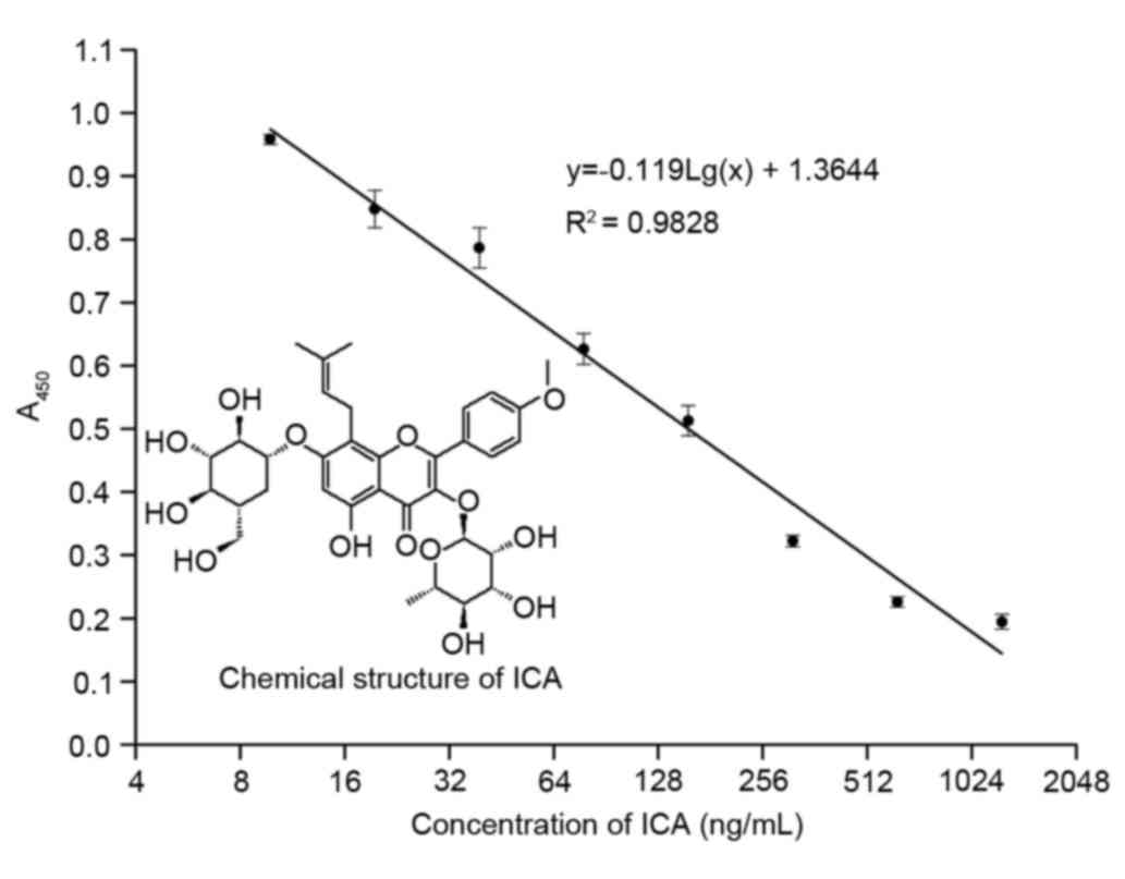
As shown in Fig. 1,
competitive inhibition occurred between the MAb and ICA-OVA with
various concentrations of ICA, resulting in a calibration curve for
ICA, analyzed using icELISA. The concentrations of the standard
solutions were 9.77, 19.53, 39.06, 78.12, 156.25, 312.50, 625.00
and 1,250.00 ng/ml. Under these conditions, a linear regression
coefficient of 0.9828 and linear regression equation, y =
−0.119Lg(x) + 1.3644 (9.77 ng/ml - 1.25 µg/ml), were achieved with
a half maximal inhibitory concentration of 156 ng/ml. The full
measuring range of the assay extended between 10 and 1,000
ng/ml.
As shown in Table
I, several compounds associated with ICA were found to have
cross-reactivity with the anti-ICA MAb, including polygala acid
(0.20%) and quercetin (0.26%). The MAb had no cross-reactivity with
other compounds (<0.09%).
 | Table I.Cross-reactivities of anti-icariin
monoclonal antibodies against natural occurring compounds. |
Table I.
Cross-reactivities of anti-icariin
monoclonal antibodies against natural occurring compounds.
| Compound | Cross-reactivity
(%) |
|---|
| Polygala acid | 0.20 |
| Quercetin | 0.26 |
| Puerarin | <0.09 |
| Baicalin | <0.09 |
| Daidzin | <0.09 |
| Scutellarein | <0.09 |
| Hesperidin | <0.09 |
| Naringin | <0.09 |
| Quercetin | <0.09 |
| Hyperoside | <0.09 |
| Geniposide | <0.09 |
| Vitexin | <0.09 |
| Curculigoside | <0.09 |
| Rutin | <0.09 |
| Paeoniflorin | <0.09 |
| Ginsenoside
Rg1 | <0.09 |
| Saikosaponin A | <0.09 |
Assay variation
Reproducibility and precision are important criteria
for an immunoassay. Standard curves for the ICA analyzed using
icELISA on 3 days (consecutive) were compared and the variations
were calculated. The variations between the well-to-well
(intra-assay) and plate-to-plate (inter-assay) replicates were
measured. As shown in Table II,
the intra-assay RSDs were <4% and the inter-assay RSDs were
<10%, which indicated that this assay was accurate and
stable.
| Table II.Intra- and inter-assay precisions of
ICA analysis via an enzyme-linked immunosorbent assay using
anti-ICA monoclonal antibody. |
Table II.
Intra- and inter-assay precisions of
ICA analysis via an enzyme-linked immunosorbent assay using
anti-ICA monoclonal antibody.
|
| Relative standard
deviation (%) |
|---|
|
|
|
|---|
| ICA (ng/ml) | Intra-assay | Inter-assay |
|---|
| 20 | 3.76 | 6.50 |
| 40 | 2.51 | 7.13 |
| 160 | 3.44 | 9.84 |
| 640 | 2.33 | 5.56 |
Recovery of ICA by icELISA with
spiked-in samples
Various quantities of ICA were added to the Folium
Epimedii extract, following which the spiked-in samples were
dissolved and mixed evenly, and ICA content was calculated using
icELISA. For each level, six samples were analyzed. The levels of
ICA in the Folium Epimedii extract were determined using icELISA.
ICA recovery ranged between 93.80 and 118.80%, with an average of
105.00% (Table III).
| Table III.Recoveries of ICA from samples with
various concentrations of ICA added, determined by enzyme-linked
immunosorbent assay using anti-icariin monoclonal antibody. |
Table III.
Recoveries of ICA from samples with
various concentrations of ICA added, determined by enzyme-linked
immunosorbent assay using anti-icariin monoclonal antibody.
| ICA added
(ng/ml) | Quantity measured
(ng/ml) | Recovery (%) |
|---|
|
0 | 42.24±4.54 |
|
| 100 | 133.42±13.44 | 93.80 |
| 200 | 256.88±88.69 | 106.15 |
| 400 | 525.10±49.70 | 118.80 |
| 800 | 852.66±27.16 | 101.27 |
Quantitative analysis of ICA in
various TCM prescriptions using icELISA
In the present study, the four TCM prescriptions,
which varied in their composition ratios, were determined using
icELISA without pre-treatment. The concentrations of ICA in the
four TCM prescriptions are shown in Table IV.
| Table IV.Contents of ICA in four traditional
Chinese medicines using ELISA with anti-ICA monoclonal antibodies
and HPLC. |
Table IV.
Contents of ICA in four traditional
Chinese medicines using ELISA with anti-ICA monoclonal antibodies
and HPLC.
|
| ELISA | HPLC |
|---|
|
|
|
|
|---|
| Sample | ICA (µg/ml) | CV (%) | ICA (µg/ml) | CV(%) |
|---|
| 1 | 27.61±6.46 | 7.17 | 30.62±0.37 | 1.20 |
| 2 | 253.94±9.78 | 3.85 | 230.69±1.60 | 0.70 |
| 3 | 105.68±8.07 | 7.64 | 103.62±1.19 | 1.15 |
| 4 | 312.79±18.57 | 5.94 | 398.57±0.37 | 0.22 |
Correlation between HPLC and ELISA
analyses of total content of ICA using MAb
Powdered samples were extracted as described above.
As shown in Table IV, the
contents determined using ELISA were consistent with those
determined using HPLC.
The results of the experiments in the present study
showed that the anti-ICA MAb had high sensitivity and specificity,
and that the icELISA method had its own unique advantages, compared
with HPLC. It is typical that intra-assay variations are generally
lower, compared with inter-assay variations. The factors
contributing to these variations are considered to include the
hapten quality, coating, plate wells and multichannel pipettor,
edge effects due to evaporation, uneven temperature during
incubation, and day-to-day variation in the preparation of the
reagents. Thus, the creation of a novel standard curve is required
every time to reduce the variation.
To the best of our knowledge, the present study
developed the first MAb against ICA and established the subsequent
ELISA method, which provided a simpler, more efficient and
sensitive approach for determining the ICA content in drug
materials and biological samples. This method can also serve as a
useful tool for investigating the pharmacokinetics and targets of
ICA.
Acknowledgements
The present study was supported by the National
Natural Science Foundation of China (grant nos. 81473338 and
81373542), the National Key Basic Research Development Program (973
program; grant no. 2011CB505101) and the Classical Prescription
Basic Research Team of Beijing University of Chinese Medicine. The
authors would like to thank Elsevier Language Editing Services for
providing language assistance.
Glossary
Abbreviations
Abbreviations:
|
BSA
|
bovine serum albumin
|
|
CBS
|
carbonate buffer solution
|
|
GPBS
|
10 mg/ml gelatine in PBS
|
|
HAT
|
hypoxanthine-aminopterin-thymidine
|
|
HT
|
hypoxanthine-thymidine
|
|
ICA
|
icariin
|
|
MAb
|
monoclonal antibody
|
|
OVA
|
ovalbumin
|
|
PBS
|
phosphate-buffered saline
|
|
PBST
|
PBS with 0.05% Tween-20
|
|
PEG
|
polyethylene glycol
|
|
TCM
|
traditional Chinese medicine
|
References
|
1
|
Zhang D, Yuan C, Zhu Z, Jin X and Li L:
Influence of the mixture of Epimedii Herba and ginkgo folium
extracts on the coronary flow of isolated hearts in rats.
Pharmacogn Mag. 9:290–293. 2013. View Article : Google Scholar : PubMed/NCBI
|
|
2
|
Zhou H, Yuan Y, Liu Y, Deng W, Zong J,
Bian ZY, Dai J and Tang QZ: Icariin attenuates angiotensin
II-induced hypertrophy and apoptosis in H9c2 cardiomyocytes by
inhibiting reactive oxygen species-dependent JNK and p38 pathways.
Exp Ther Med. 7:1116–1122. 2014.PubMed/NCBI
|
|
3
|
Zhang DW, Cheng Y, Wang NL, Zhang JC, Yang
MS and Yao XS: Effects of total flavonoids and flavonol glycosides
from Epimedium koreanum Nakai on the proliferation and
differentiation of primary osteoblasts. Phytomedicine. 15:55–61.
2008. View Article : Google Scholar : PubMed/NCBI
|
|
4
|
Wang Q, Hao J, Pu J, Zhao L, Lü Z, Hu J,
Yu Q, Wang Y, Xie Y and Li G: Icariin induces apoptosis in mouse
MLTC-10 Leydig tumor cells through activation of the mitochondrial
pathway and down-regulation of the expression of piwil4. Int J
Oncol. 39:973–980. 2011.PubMed/NCBI
|
|
5
|
Xu CQ, Liu BJ, Wu JF, Xu YC, Duan XH, Cao
YX and Dong JC: Icariin attenuates LPS-induced acute inflammatory
responses: Involvement of PI3K/Akt and NF-kappaB signaling pathway.
Eur J Pharmacol. 642:146–153. 2010. View Article : Google Scholar : PubMed/NCBI
|
|
6
|
Song YH, Cai H, Gu N, Qian CF, Cao SP and
Zhao ZM: Icariin attenuates cardiac remodelling through
down-regulating myocardial apoptosis and matrix metalloproteinase
activity in rats with congestive heart failure. J Pharm Pharmacol.
63:541–549. 2011. View Article : Google Scholar : PubMed/NCBI
|
|
7
|
Tong JS, Zhang QH, Huang X, Fu XQ, Qi ST,
Wang YP, Hou Y, Sheng J and Sun QY: Icaritin causes sustained
ERK1/2 activation and induces apoptosis in human endometrial cancer
cells. PLoS One. 6:e167812011. View Article : Google Scholar : PubMed/NCBI
|
|
8
|
Guo Y, Zhang X, Meng J and Wang ZY: An
anticancer agent icaritin induces sustained activation of the
extracellular signal-regulated kinase (ERK) pathway and inhibits
growth of breast cancer cells. Eur J Pharmacol. 658:114–122. 2011.
View Article : Google Scholar : PubMed/NCBI
|
|
9
|
Sun P, Liu Y, Deng X, Yu C, Dai N, Yuan X,
Chen L, Yu S, Si W, Wang X, et al: An inhibitor of cathepsin K,
icariin suppresses cartilage and bone degradation in mice of
collagen-induced arthritis. Phytomedicine. 20:975–979. 2013.
View Article : Google Scholar : PubMed/NCBI
|
|
10
|
Li ZJ, Yao C, Liu SF, Chen L, Xi YM, Zhang
W and Zhang GS: Cytotoxic effect of icaritin and its mechanisms in
inducing apoptosis in human burkitt lymphoma cell line. Biomed Res
Int. 2014:3915122014.PubMed/NCBI
|
|
11
|
Jin J, Li Y, Tanui E Kipletting, Han L,
Jia Y, Zhang L, Wang Y, Zhang X and Zhang Y: Fishing and knockout
of bioactive compounds using a combination of high-speed
counter-current chromatography (HSCCC) and preparative HPLC for
evaluating the holistic efficacy and interaction of the components
of Herba Epimedii. J Ethnopharmacol. 147:357–365. 2013. View Article : Google Scholar : PubMed/NCBI
|
|
12
|
Pozharitskaya ON, Kosman VM, Shikov AN,
Demchenko DV, Eschenko AY and Makarov VG: Comparison between HPLC
and HPTLC densitometry for the determination of icariin from
Epimedium koreanum extracts. J Sep Sci. 30:708–712. 2007.
View Article : Google Scholar : PubMed/NCBI
|
|
13
|
Xu T, Wei KY, Wang J, Eremin SA, Liu SZ,
Li QX and Li J: Development of an enzyme-linked immunosorbent assay
specific to Sudan red I. Anal Biochem. 405:41–49. 2010. View Article : Google Scholar : PubMed/NCBI
|
|
14
|
Daduang S, Sattayasai N, Sattayasai J,
Tophrom P, Thammathaworn A, Chaveerach A and Konkchaiyaphum M:
Screening of plants containing Naja naja siamensis cobra venom
inhibitory activity using modified ELISA technique. Anal Biochem.
341:316–325. 2005. View Article : Google Scholar : PubMed/NCBI
|
|
15
|
Zhao Y, Kong H, Sun Y, Feng H, Zhang Y, Su
X, Qu H and Wang Q: Assessment of baicalin in mouse blood by
monoclonal antibody-based icELISA. Biomed Chromatogr. 28:1864–1868.
2014. View
Article : Google Scholar : PubMed/NCBI
|
|
16
|
Qu H, Zhang G, Li Y, Sun H, Sun Y, Zhao Y
and Wang Q: Development of an enzyme-linked immunosorbent assay
based on anti-puerarin monoclonal antibody and its applications. J
Chromatogr B Analyt Technol Biomed Life Sci 953–954. 120–125.
2014.
|
|
17
|
Qu HH, Sun Y, Wu TT, Zhang GL, Cheng JJ,
Wang XQ, Feng HB, Zhao Y and Wang QG: Pharmacokinetics of
geniposide by monoclonal antibody-based icELISA in mice after oral
administration of Huanglian-Jiedu-Tang. Biol Pharm Bull.
37:1525–1533. 2014. View Article : Google Scholar : PubMed/NCBI
|
|
18
|
Zhang Y, Qu H, Zeng W and Zhao Y, Shan W,
Wang X, Wang Q and Zhao Y: Development of an enzyme-linked
immunosorbent assay and immunoaffinity chromatography for
glycyrrhizic acid using an anti-glycyrrhizic acid monoclonal
antibody. J Sep Sci. 38:2363–2370. 2015. View Article : Google Scholar : PubMed/NCBI
|
|
19
|
Zhao Y, Qu H, Wang X, Zhang Y, Shan W,
Zhao Y and Wang Q: A sensitive and specific indirect competitive
Enzyme-linked immunosorbent assay for detection of paeoniflorin and
its application in pharmacokinetic interactions between
paeoniflorin and glycyrrhizinic acid. Planta Med. 81:765–770. 2015.
View Article : Google Scholar : PubMed/NCBI
|
|
20
|
Qu H, Sai J, Wang Y, Sun Y, Zhang Y, Li Y,
Zhao Y and Wang Q: Establishment of an enzyme-linked immunosorbent
assay and application on determination of ginsenoside Re in human
saliva. Planta Med. 80:1143–1150. 2014. View Article : Google Scholar : PubMed/NCBI
|
|
21
|
Qu H, Wang Y, Shan W, Zhang Y, Feng H, Sai
J, Wang Q and Zhao Y: Development of ELISA for detection of Rh1 and
Rg2 and potential method of immunoaffinity chromatography for
separation of epimers. J Chromatogr B Analyt Technol Biomed Life
Sci. 985:197–205. 2015. View Article : Google Scholar : PubMed/NCBI
|
|
22
|
Phrompittayarat W, Putalun W, Tanaka H,
Jetiyanon K, Wittaya-Areekul S and Ingkaninan K: Determination of
pseudojujubogenin glycosides from Brahmi based on immunoassay using
a monoclonal antibody against bacopaside I. Phytochem Anal.
18:411–418. 2007. View
Article : Google Scholar : PubMed/NCBI
|
|
23
|
Zhu S, Shimokawa S, Shoyama Y and Tanaka
H: A novel analytical ELISA-based methodology for pharmacologically
active saikosaponins. Fitoterapia. 77:100–108. 2006. View Article : Google Scholar : PubMed/NCBI
|
|
24
|
Galfrè G and Milstein C: Preparation of
monoclonal antibodies: Strategies and procedures. Methods Enzymol.
73:3–46. 1981. View Article : Google Scholar : PubMed/NCBI
|
|
25
|
North SM, Styles JM, Hobbs SM and Dean CJ:
Monoclonal antibodies to rat sarcomata. I. Immunization procedures
and source of lymphoid cells for hybridoma production. Immunology.
47:397–405. 1982.PubMed/NCBI
|
|
26
|
Goding JW: Antibody production by
hybridomas. J Immunol Methods. 39:285–308. 1980. View Article : Google Scholar : PubMed/NCBI
|
|
27
|
Liu HF, Ma J, Winter C and Bayer R:
Recovery and purification process development for monoclonal
antibody production. MAbs. 2:480–499. 2010. View Article : Google Scholar : PubMed/NCBI
|
|
28
|
Auten AA, Beauchamp LN, Taylor J and
Hardinger KL: Hidden sources of grapefruit in beverages: Potential
interactions with immunosuppressant medications. Hosp Pharm.
48:489–493. 2013. View Article : Google Scholar : PubMed/NCBI
|
|
29
|
Jian Z, Xu G, Chen H, Wang H and Hu X:
Study on the differences of major pharmaceutical ingredients in
different parts and processed medicinal material of Epimedium
brevicornu maxim in Taihang mountain. Nutr Hosp. 32:913–917.
2015.PubMed/NCBI
|