Introduction
Bladder cancer is the fourth most commonly occurring
cancer worldwide and the most common genitourinary malignant cancer
in China. Previous studies have demonstrated that of 5,647
newly-diagnosed bladder cancer cases in 2009, 2,223 were expected
to be fatal (1,2). Environmental and genetic factors are
important in the development and progression of bladder cancer;
however, the mechanisms underlying carcinogenesis remain to be
fully elucidated. Thus, identifying potential carcinogenic genes is
important to develop novel therapeutic strategies and specify novel
biomarkers for the diagnosis and prognosis of bladder cancer.
B23 (also known as nucleophosmin, numatrin or NO38)
is a nucleolar phosphoprotein that shuttles continuously between
the nucleus and the cytoplasm (3).
Previous studies have indicated that B23is important in various
cellular processes, including ribosome biogenesis,
nucleocytoplasmic transport, centrosome duplication, apoptosis,
cell proliferation, and in pathological conditions including cancer
development and progression (4–6). B23
expression is increased in cancer and proliferating cells compared
with healthy resting cells. The overexpression of B23 at the mRNA
and protein levels contributes to tumorigenesis and is associated
with poor prognosis in numerous cancers, including astrocytomas,
colorectal cancer, hepatocellular carcinomas, breast cancer,
ovarian carcinomas and prostate carcinomas (7–10).
However, the association between the expression of B23 and survival
and prognosis in bladder urothelial carcinoma remains to be
elucidated.
The present study analyzed the mRNA and protein
expression levels of B23 in bladder urothelial carcinoma and
matched adjacent tissues. It was observed that the protein
expression levels of B23 were increased in bladder urothelial
carcinoma and that augmented B23 levels were associated with poor
prognosis. Subsequently, the present study investigated the effect
of B23 on cell growth and tumorigenesis in bladder cancer cells and
observed that increased levels of B23 promoted cancer cell growth
and tumorigenesis via regulation of extracellular signal-regulated
kinase (ERK) phosphorylation.
Materials and methods
Clinical samples
The Second Affiliated Hospital of Harbin Medical
University (Harbin, China) provided 95 well-documented surgically
matching pairs of bladder urothelial carcinoma tissue samples, and
the corresponding adjacent tissue samples, obtained from 2006 to
2009. The characteristics of the patients and their tumors were
collected via a review of medical records and pathological reports.
The patients were followed postoperatively for a mean of 81.5
months (range, 60–105 months). Informed consent was obtained from
patients between 2006 and 2009. The present study was approved by
the ethics committee of the Second Affiliated Hospital of Harbin
Medical University. None of the patients underwent chemotherapy or
radiotherapy prior to surgery, and there was no co-occurrence of
other diagnosed cancers.
Sections of the dissected tumor samples were fixed
in formalin and embedded in paraffin. Sections of paraffin-embedded
tissue were used for immunohistochemical analysis (IHC). Further
tumor samples and their corresponding adjacent tissue samples from
resected bladders were frozen in liquid nitrogen and stored at
−80°C for protein and nucleic acid extraction.
IHC
Tissue samples were processed according to routine
procedures. In brief, the paraffin-embedded bladder urothelial
carcinoma tissue samples and the corresponding adjacent tissue
samples were sectioned at 4 µm and mounted on glass slides. The
slides were subsequently deparaffinized, hydrated, incubated with
3% H2O2 and microwaved for 20 min at room
temperature to block endogenous peroxidase activity and expose
antigens concealed by formalin fixation. Non-specific
antigen-antibody reactions were inhibited using an
immunohistochemistry Protein Blocker-serum and Azide Free
(MB-071-0100, Rockland Immunochemicals, Inc., Pottstown, PA, USA)
for 5 min, following which the slides were washed thoroughly with
PBS. The slides were subsequently incubated overnight with a rabbit
polyclonal primary antibody against B23 (1:200; cat. no.
10306-1-AP; Proteintech Group, Inc., Rosemont, USA) at 4°C. A
biotinylated goat anti-rabbit secondary antibody (1:200; cat. no.
ab6720; Abcam, Cambridge, UK) was applied for 20 min at room
temperature, followed by further washing with buffer to remove any
unbound antibody. A complex of avidin conjugated to horseradish
peroxidase was then applied for 20 min at room temperature. For
color development, the slides were incubated with
3,3′diaminobenzidine (Sigma-Aldrich; Merck Millipore, Darmstadt,
Germany) and counterstained with hematoxylin. Staining results were
evaluated using Aperio VERSA Brightfiled, Fluorescence, FISH
Digital pathology scanner (Leica Microsystems, Ltd., Milton Keynes,
UK) by two independent observers blinded to clinicopathological
data. Regarding the cases with discordant evaluation, two
pathologists performed a consensus adjudication review using a
multi-headed light microscope.
Cell culture and reagents
The RT-4, BI-87,253 J, SV-HUC-1, T24 and J82 human
bladder tumor cell lines were purchased from China Academia Sinica
Cell Repository (Shanghai, China; www.cellbank.org.cn). T24, RT-4, and 253 J cells were
cultured in RPMI 1640 (cat. no. 22400-089; Gibco; Thermo Fisher
Scientific, Inc., Waltham, MA, USA) supplemented with 10% fetal
calf serum (Gibco; Thermo Fisher Scientific, Inc.). J82 cells were
maintained in Opti-Minimal Essential Medium® I (cat. no.
51985-042; Gibco; Thermo Fisher Scientific, Inc.) containing 10%
fetal calf serum (Gibco; Thermo Fisher Scientific, Inc.). SV-HUC-1
cells were cultured in F12K medium (cat. no. N3520; Sigma-Aldrich;
Merck Millipore) supplemented with 10% fetal calf serum (Gibco;
Thermo Fisher Scientific, Inc.) All cells were cultured in a
humidified incubator at 37°C and 5% CO2. The following
antibodies were used: Anti-B23 (cat. no. 10306-1-AP; Proteintech
Group, Inc., Rosemont, USA), mouse anti-GAPDH (cat. no. sc-32233;
Santa Cruz Biotechnology, Inc., Dallas, TX, USA), rabbit anti-ERK
(cat. no. 4372S; Cell Signaling Technology, Inc., Danvers, MA, USA)
and rabbit anti-phosphorylated (p)-ERK1/2 (cat. no. 4370S; Cell
Signaling Technology, Inc.). U0126 (cat. no. 9903; Cell Signaling
Technology, Inc.) was selected as the mitogen activated protein
kinase (MAPK)/ERK inhibitor and the cells were treated with 2 µM
U0126 for 1, 2, 3 days at 37°C.
Lentivirus transfection
B23 short hairpin RNA(shRNA) was cloned using the
PLKO.1 vector (cat. no. SHCLNG-NM002520; Sigma-Aldrich; Merck
Millipore, USA); the targeting sequence was 5-GCC AAG AAT GTG TTG
TCC AAA-3. The empty vector PLKO.1 was used as the control. Stable
knockdown T24 and J82 cells were established as previously
described (11,12). To generate lentivirus expressing
B23, HEK 293T cells cultured on a 6 cm dish and were transfected
with 2 µg pCDH-Flag-B23 or empty vector (cat. no. CD510B-1, System
Biosciences, Palo Alto, CA, USA), 1.5 µg psPax2 and 0.5 µg pMD2 G.
Cells were cultured with DMEM containing 10% FBS 24 h after the
transfection for an additional 24 h. The culture medium containing
lentiviral particles was centrifuged at 1,000 × g for 5 min. Viral
particles collected in the supernatant were used for infection. In
order to establish the stable cell line, puromycin (cat. no. P7130;
Sigma-Aldrich; Merck Millipore) was used as a selection marker for
the infected cells. The expression efficiency was evaluated by
western blot analysis.
RNA extraction and reverse
transcription-quantitative polymerase chain reaction (RT-qPCR)
TRIzol® (Ambion; Thermo Fisher
Scientific, Inc.) was used to isolate total RNA, of which 1 µg was
used to synthesize cDNA with the PrimeScript™ RT reagent kit (cat.
no. DRR037A; Takara Biotechnology Co., Ltd., Dalian, China)
according to the manufacturer's protocol. SYBR®Premix Ex
Taq (RR430A; Takara Biotechnology Co., Ltd.) and ROX (RR430A;
Takara Biotechnology Co., Ltd.) were used to perform qPCR (95°30
sec; 60°C 60 sec; 40 cycles), and results were analyzed with
Statagene Mx3000p (Agilent Technologies, Inc., Santa Clara, CA,
USA) (13). The primer sequences
used for PCR were as follows: Forward, 5-CTC CAT CCT GGC CTC
GCTGT-3 and reverse, 5-GCT GTC ACC TTC ACC GTT CC-3 for actin; and
forward, 5-TTC AGG GCC AGT GCA TAT TAG-3 and reverse, 5-TTC TGT GGA
ACC TTG CTA CC-3 for B23.
Western blot analysis
The antibodies used for western blot analysis were
diluted as followed: anti-B23 (1:1,000), mouse anti-GAPDH
(1:5,000), rabbit anti-ERK (1:1,000) and rabbit anti-phosphorylated
(p)-ERK1/2 (1:1,000). Total protein was extracted from bladder
urothelial carcinoma and non-cancerous adjacent tissues or T24 and
J82 cell lysates, for use in immunoblotting. The protocol used was
as previously described (11).
Cell viability and colony formation
assays
Cell viability was detected using a Cell Counting
kit-8 assay (cat. no. CK04-13; Dojindo Molecular Technologies,
Inc., Kumamoto, Japan). Cells were seeded in 96-well plates at a
density of 5,000 cells in 100 µl medium per well, 24 h prior to the
experiment. For the colony formation assay, T24 and J82 cells were
trypsinized and 1,000 viable cells were subcultured in 6-well
plates in triplicate. Cells were allowed to adhere and colonize for
10 days. To visualize colonies, media was removed and cells were
fixed in 96% ethanol for 10 min and stained with crystal violet
staining solution.
Statistical analysis
Pearson's chi-square test was used to determine
differences in B23 expression between bladder urothelial carcinoma
tissues and the corresponding adjacent tissues, and to determine
the association between B23 expression and the clinical parameters
of gender, age, tumor size, initial clinical stage, pathological
grade and recurrence. Overall and disease-free survival following
tumor removal was calculated by the Kaplan-Meier method, and
difference in survival curves was analyzed by the log-rank test.
The Cox proportional hazards model was used for multivariate
analysis of prognostic factors. P<0.05 was considered to
indicate a statistically significant difference. All statistical
analysis was performed using SPSS software version 16.0 (SPSS,
Inc., Chicago, IL, USA).
Results
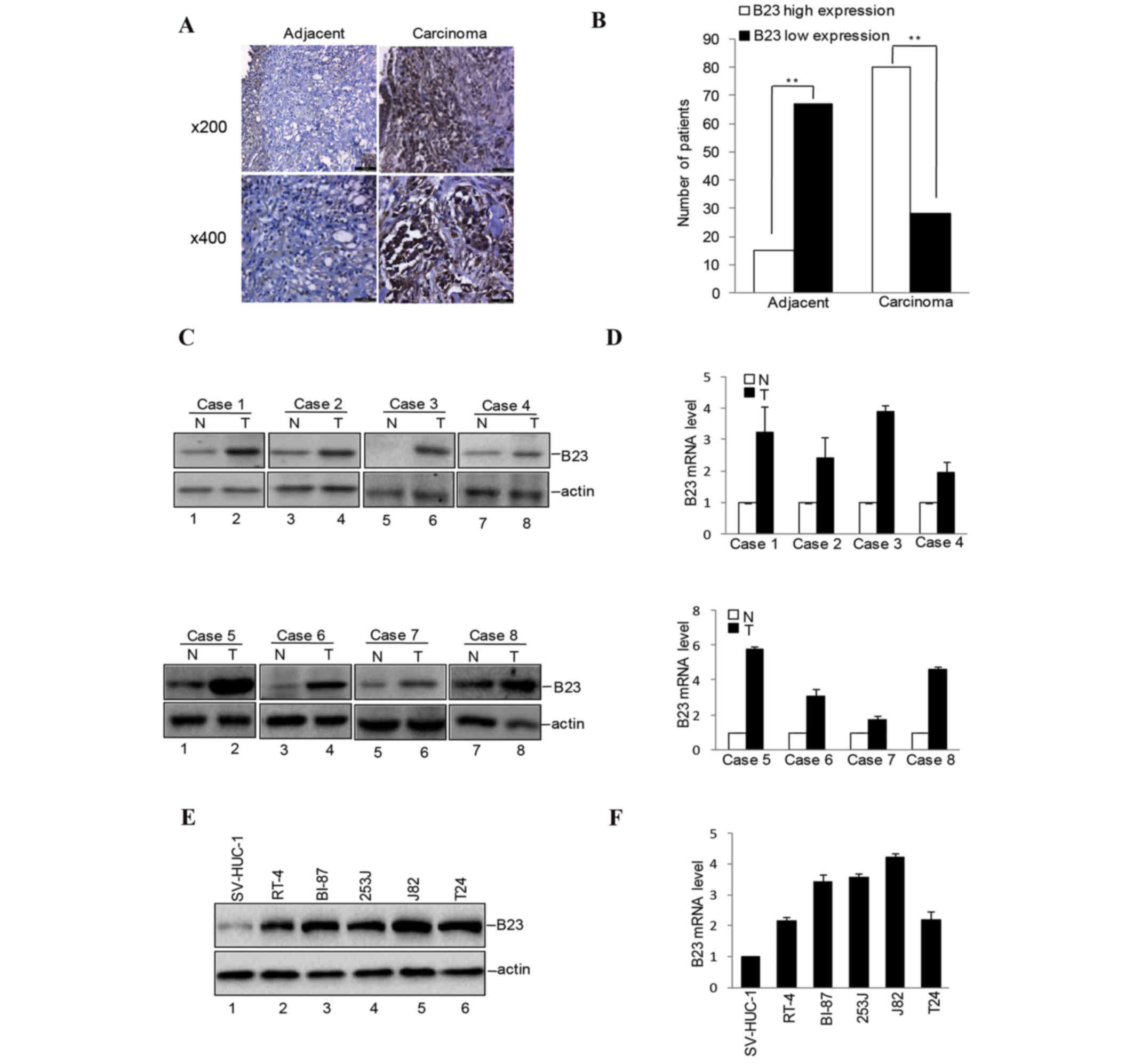
Overexpression of B23 in bladder
urothelial carcinoma tissues and cells
B23 expression was detected in 70.5% (67/95) of
bladder urothelial carcinoma tissues, and in 16% (15/95) of
adjacent tissues, as assessed by IHC. The difference in B23
expression between bladder urothelial carcinoma tissues and
adjacent tissues was statistically significant (P<0.01 vs.
adjacent tissues; Student's t-test) Fig. 1A and B). The present study detected
the expression of B23 in bladder urothelial carcinoma tissues and
their corresponding adjacent non-cancer tissues by western blot
analysis and RT-qPCR, and it was observed that the protein
(Fig. 1C) and mRNA (Fig. 1D) expression levels of B23 were
increased in carcinoma tissues compared with adjacent tissues. The
expression of B23 in bladder cancer cells and normal bladder cells
was then investigated. High protein (Fig. 1E) and mRNA (Fig. 1F) expression levels of B23 were
observed in the bladder cancer cells. This data suggested that the
expression levels of B23 were increased in bladder urothelial
carcinoma tissues and cells.
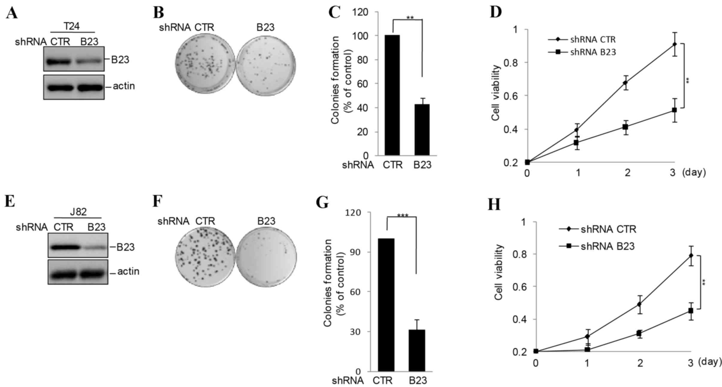
B23 promotes bladder cancer cell
proliferation
Based on the data described above, the present study
hypothesized that B23 may be important for bladder cancer
tumorigenesis. Stable B23 knockdown cells were constructed in the
T24 and J82 bladder cancer cell lines. B23 knockdown T24 and J82
cells demonstrated a dramatic decrease in cell viability and colony
formation, compared with cells with the empty vector (Fig. 2).
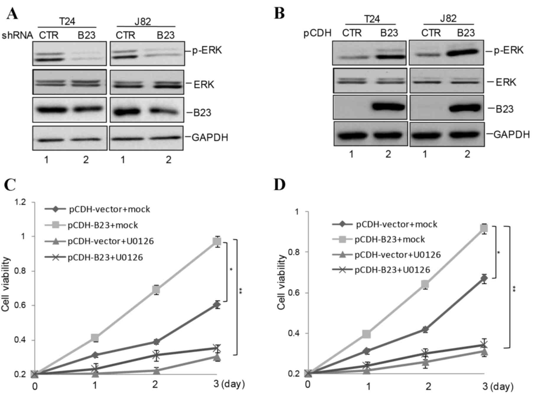
B23 regulates bladder cancer cell
growth via the ERK signaling pathway
Previous studies have reported that B23 is a
positive regulator of the ERK signaling pathway and may promote
cell proliferation via ERK activity in prostate cancer (14–16).
Therefore, to investigate whether B23 affected bladder cancer cell
growth via the ERK signaling pathway, the present study detected
the protein expression levels of p-ERK, and it was observed that
the phosphorylation of ERK was inhibited by B23 knockdown; however,
the total protein expression levels of ERK remained unaltered
(Fig. 3A). The phosphorylation of
ERK was increased in B23 overexpressing bladder cancer cells
(Fig. 3B). To confirm that B23
promoted bladder cancer growth via upregulation of ERK
phosphorylation, T24 and J82 bladder cancer cells were treated with
the pharmacological ERK inhibitor U0126. The effect of B23 on cell
growth was suppressed by ERK inhibition via U0126 (Fig. 3C and D).
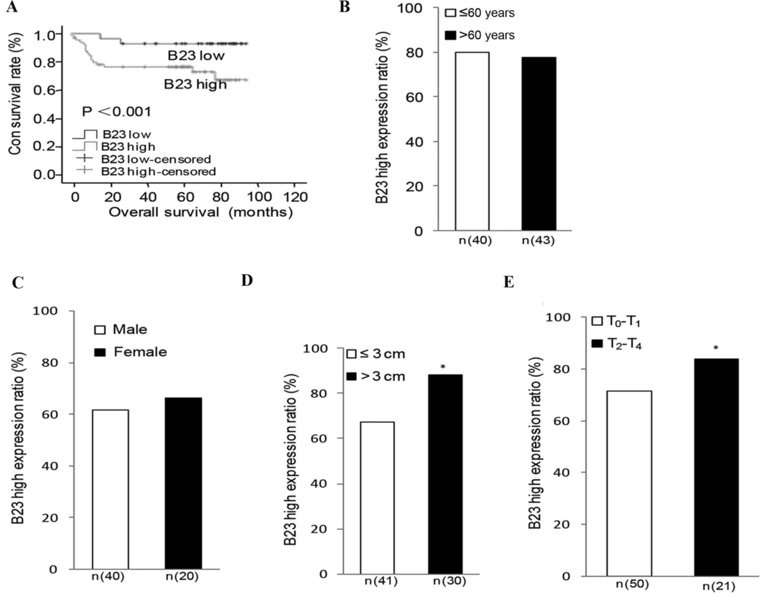
Association between B23 expression and
overall survival rate in bladder urothelial carcinoma patients
The association between B23 expression and patient
survival rate was determined using Kaplan-Meier analysis and the
log-rank test. As presented in Fig.
4A, a statistical correlation was observed between B23 protein
expression levels and overall survival in bladder urothelial
carcinoma patients was determined by IHC. Patients with high B23
expression had a markedly lower survival rate compared with
patients with low B23 expression (P<0.001). There was no
statistical significance observed between B23 expression and other
clinical parameters including age and gender (Fig. 4B and C). Subsequently, the
association between B23 expression and different clinical
parameters, including tumor size, initial clinical stage and tumor
stage were investigated. Significant positive correlations between
B23 expression and tumor size (P=0.041; Fig. 4D), tumor stage (P=0.030; Fig. 4E), initial clinical stage (P=0.021)
and recurrence (P=0.049) were observed.
Discussion
Bladder urothelial carcinoma is a common malignant
cancer that results in ~150,000 fatalities per year worldwide
(17). It has been suggested that
environmental and genetic factors are important in the development
and progression of bladder cancer; however, the underlying
mechanisms of bladder carcinogenesis remain to be fully elucidated.
It is important to understand these mechanisms to predict tumor
behavior and identify novel therapeutic targets.
The present study reported that the mRNA and protein
expression levels of B23 were increased in human bladder urothelial
carcinoma tissues compared with adjacent non-cancerous tissues, and
that the increased B23 expression promoted bladder cancer cell
growth via the ERK signaling pathway. Following this, the
association between B23 expression and prognosis in 95 bladder
urothelial carcinoma patients was investigated. It was observed
that augmented B23 expression was correlated with poor
prognosis.
B23 is a multifunctional protein involved in a
complex network of interactions. Previous studies have demonstrated
the overexpression of B23 in numerous solid tumors, including
gastric, prostate and liver cancer (8–10,18).
However, the function of B23 in bladder urothelial carcinoma
remains to be elucidated. The present study, to the best of our
knowledge, was the first to analyze the expression of B23 in
bladder urothelial carcinoma tissues and matched adjacent tissues.
It was observed that the mRNA and protein expression levels of B23
were increased in bladder urothelial carcinoma tissues compared
with adjacent non-cancerous tissues. Following this, the expression
of B23 in bladder cancer and normal bladder cells was investigated,
and it was demonstrated that B23 was upregulated in cancer compared
with normal cells. The data from the present study indicated that
B23 was overexpressed in bladder urothelial carcinoma tissues and
cells; however, the underlying molecular mechanism remains to be
elucidated, and will be investigated in future studies.
An increase in B23 expression levels has been
associated with an increase in proliferating cells, compared with
normal resting cells (19).
Therefore, the present study investigated whether B23 was important
in bladder cancer cells. B23 knockdown T24 and J82 cells
demonstrated a marked decrease in cell viability and colony
formation, compared with control cells. A previous study indicated
the abnormal activation of the MAPK signaling pathway was important
for bladder cancer (20). To
determine whether B23 was involved in the activation of the MAPK
signaling pathway in bladder cancer, the present study detected the
expression of phosphorylation of ERK and revealed that knockdown of
B23 inhibited the activation of ERK. Following this, it was
demonstrated B23 promoted bladder cancer cell growth via regulation
of the MAPK pathway.
The correlation between B23 expression and prognosis
in the 95 bladder urothelial carcinoma patients was subsequently
investigated. A significant positive correlation was observed
between B23 expression and tumor size, initial clinical stage,
tumor stage and recurrence. No statistical significance was
observed between B23 expression and age or gender. In addition, it
was observed that patients with high B23 expression had a markedly
reduced survival rate compared with patients with low B23
expression.
In conclusion, the results of the present study
revealed that B23 expression levels were increased in bladder
urothelial carcinoma tissues, and B23 was a reliable and
independent prognostic factor for bladder urothelial carcinoma
patients. In addition, it was observed that elevated B23 expression
accelerated bladder cancer cell growth and tumorigenesis via the
MAPK signaling pathway. Therefore, the evidence suggested that B23
may be a potential therapeutic target for the treatment of bladder
urothelial carcinoma.
Acknowledgements
The present study was supported by the Health and
Family Planning Commission of Heilongjiang Province (grant nos.
2007315 and 2005251).
References
|
1
|
Jemal A, Bray F, Center MM, Ferlay J, Ward
E and Forman D: Global cancer statistics. CA Cancer J Clin.
61:69–90. 2011. View Article : Google Scholar : PubMed/NCBI
|
|
2
|
Gu F: Changing constituents of
genitourinary cancer in recent 50 years in Beijing. Chin Med J
(Engl). 116:1391–1393. 2003.PubMed/NCBI
|
|
3
|
Grisendi S, Mecucci C, Falini B and
Pandolfi PP: Nucleophosmin and cancer. Nat Rev Cancer. 6:493–505.
2006. View
Article : Google Scholar : PubMed/NCBI
|
|
4
|
Chan WY, Liu QR, Borjigin J, Busch H,
Rennert OM, Tease LA and Chan PK: Characterization of the cDNA
encoding human nucleophosmin and studies of its role in normal and
abnormal growth. Biochemistry. 28:1033–1039. 1989. View Article : Google Scholar : PubMed/NCBI
|
|
5
|
Feuerstein N, Chan PK and Mond JJ:
Identification of numatrin, the nuclear matrix protein associated
with induction of mitogenesis, as the nucleolar protein B23.
Implication for the role of the nucleolus in early transduction of
mitogenic signals. J Biol Chem. 263:10608–10612. 1988.PubMed/NCBI
|
|
6
|
Hingorani K, Szebeni A and Olson MO:
Mapping the functional domains of nucleolar protein B23. J Biol
Chem. 275:24451–24457. 2000. View Article : Google Scholar : PubMed/NCBI
|
|
7
|
Gimenez M, Souza VC, Izumi C, Barbieri MR,
Chammas R, Oba-Shinjo SM, Uno M, Marie SK and Rosa JC: Proteomic
analysis of low- to high-grade astrocytomas reveals an alteration
of the expression level of raf kinase inhibitor protein and
nucleophosmin. Proteomics. 10:2812–2821. 2010. View Article : Google Scholar : PubMed/NCBI
|
|
8
|
Nozawa Y, Van Belzen N, Van der Made AC,
Dinjens WN and Bosman FT: Expression of nucleophosmin/B23 in normal
and neoplastic colorectal mucosa. J Pathol. 178:48–52. 1996.
View Article : Google Scholar : PubMed/NCBI
|
|
9
|
Skaar TC, Prasad SC, Sharareh S, Lippman
ME, Brünner N and Clarke R: Two-dimensional gel electrophoresis
analyses identify nucleophosmin as an estrogen regulated protein
associated with acquired estrogen-independence in human breast
cancer cells. J Steroid Biochem Mol Biol. 67:391–402. 1998.
View Article : Google Scholar : PubMed/NCBI
|
|
10
|
Yun JP, Miao J, Chen GG, Tian QH, Zhang
CQ, Xiang J, Fu J and Lai PB: Increased expression of
nucleophosmin/B23 in hepatocellular carcinoma and correlation with
clinicopathological parameters. Br J Cancer. 96:477–484. 2007.
View Article : Google Scholar : PubMed/NCBI
|
|
11
|
Han C, Gu H, Wang J, Lu W, Mei Y and Wu M:
Regulation of L-threonine dehydrogenase in somatic cell
reprogramming. Stem Cells. 31:953–965. 2013. View Article : Google Scholar : PubMed/NCBI
|
|
12
|
Han C, Jin L, Mei Y and Wu M: Endoplasmic
reticulum stress inhibits cell cycle progression via induction of
p27 in melanoma cells. Cell Signal. 25:144–149. 2013. View Article : Google Scholar : PubMed/NCBI
|
|
13
|
Livak KJ and Schmittgen TD: Analysis of
relative gene expression data using real-time quantitative PCR and
the 2(−Delata C(T)) Method. Methods. 11:402–408. 2001. View Article : Google Scholar
|
|
14
|
Loubeau G, Boudra R, Maquaire S,
Lours-Calet C, Beaudoin C, Verrelle P and Morel L: NPM1 silencing
reduces tumour growth and MAPK signalling in prostate cancer cells.
PLoS One. 9:e962932014. View Article : Google Scholar : PubMed/NCBI
|
|
15
|
Yang X, Du T, Wang X, Zhang Y, Hu W, Du X,
Miao L and Han C: IDH1, a CHOP and C/EBPβ-responsive gene under ER
stress, sensitizes human melanoma cells to hypoxia-induced
apoptosis. Cancer Lett. 365:201–210. 2015. View Article : Google Scholar : PubMed/NCBI
|
|
16
|
Zhang D, Zhu L, Li C, Mu J, Fu Y, Zhu Q,
Zhou Z, Liu P and Han C: Sialyltransferase7A, a Klf4-responsive
gene, promotes cardiomyocyte apoptosis during myocardial
infarction. Basic Res Cardiol. 110:282015. View Article : Google Scholar : PubMed/NCBI
|
|
17
|
Mitra AP and Cote RJ: Molecular
pathogenesis and diagnostics of bladder cancer. Annu Rev Pathol.
4:251–285. 2009. View Article : Google Scholar : PubMed/NCBI
|
|
18
|
Mina N, Soubani AO, Cote ML, Suwan T,
Wenzlaff AS, Jhajhria S, Samarah H and Schwartz AG: The
relationship between chronic obstructive pulmonary disease and lung
cancer in African American patients. Clin Lung Cancer. 13:149–156.
2012. View Article : Google Scholar : PubMed/NCBI
|
|
19
|
Chen J, Sun J, Yang L, Yan Y, Shi W, Shi
J, Huang Q, Chen J and Lan Q: Upregulation of of B23 promotes tumor
cell proliferation and predicts poor prognosis in glioma. Biochemn
Biophys Res Commum. 10:124–130. 2015. View Article : Google Scholar
|
|
20
|
Koul HK, Pal M and Koul S: Role of p38 MAP
kinase signal transduction in solid tumors. 9:2013.342–359
|