Introduction
Candida albicans (C. albicans), a
commensal yeast in the oral cavity, can incorporate into biofilms
that form on denture surfaces (1,2),
leading in some cases to mucosal infections referred to as denture
stomatitis (3–5).
Lysozyme, an antimicrobial protein present in
exocrine secretions (6) and in
phagocytes (7), contributes along
with other innate non-immune factors in saliva to the control of
the oral microflora, thereby preserving health of the mucosa and
the dental surfaces (3,8,9). The
antimicrobial action of lysozyme is mediated through its muramidase
activity (10) which catalyzes the
hydrolysis of N-acetyl-muramic/N-acetyl-glucosamine bonds in
peptidoglycans composing the cell wall in Gram-positive bacteria
(11). Lysozyme in the oral cavity
comes from the salivary glands, particularly the submandibular and
sublingual glands (12,13), and from neutrophils arriving in the
oral environment through gingival fluid in the healthy mouth
(14) or crevicular fluid in
periodontitis (15,16). Physiological lysozyme
concentrations in saliva range from 1 to 57 µg/ml (17).
Previous studies have investigated the effect of
lysozyme on C. albicans blastoconidia (3,18),
but the mechanisms of action remain unclear. The three modes of
action of lysozyme on C. albicans are its muramidase-like
activity (19,20), its cationic nature capable of
destabilizing the cell membrane (21), and its agglutination property
(22). Although no peptidoglycan
substrate for muramidase activity exists in C. albicans,
lysozyme-induced wall-like material deposits between its chitin
wall and its cell membrane have been observed by electron
microscopy (19). Synergistic
action of lysozyme with other salivary proteins has been suggested
by several authors as a mechanism of action towards C.
albicans, and some oral care products already combine lysozyme
with lactoferrin and lactoperoxidase (23–25).
Indeed, in vitro investigations have shown that C.
albicans susceptibility to lysozyme increases when combined
with peroxidase (26,27) but not with lactoferrin (28), while hyaluronic acid inhibits the
lysozyme effect on C. albicans (27,29).
Lysozyme also enhances the activity of antimycotic drugs such as
polyenes and azoles (30–33).
A lower excretion of antimicrobial proteins in
saliva has been demonstrated to promote C. albicans growth
in the oral cavity (34). The
in vitro effect of lysozyme on C. albicans biofilms
remains controversial: certain studies demonstrated no beneficial
effect of artificial saliva-containing lysozyme and other salivary
proteins on yeast adhesion to acrylic resin (35), while others demonstrated a
preventive role for lysozyme with respect to C. albicans
biofilm formation on dentures (33). To the best of our knowledge, no
studies have explored the link between C. albicans biofilm
formation and lysozyme agglutination properties, previously
described at 1 mg/ml concentration (22). The aim of the present in
vitro study was to investigate whether lysozyme modulates C.
albicans biofilm production when present in physiological
concentrations, by triggering distinct mechanisms, namely
inhibition and aggregation.
Materials and methods
Microorganisms
Yeasts were grown aerobically at 37°C on Sabouraud
agar with 0.4 g/l chloramphenicol and 0.04 g/l gentamycin (BD
Diagnostics, Franklin Lakes, NJ, USA). All in vitro
investigations were conducted on a third subculture of C.
albicans ATCC 10231 (Oxoid; Thermo Fisher Scientific, Inc.,
Waltham, MA, USA) suspended in Sabouraud broth (cat. no. CM147;
Oxoid™; Thermo Fisher Scientific, Inc.) or in distilled water. The
suspension was adjusted to 1–20×106 blastoconidia per ml by
dilution, following a blastoconidia count using a Thoma cell
counting chamber (Marienfeld™, Lauda-Königshofen, Germany). Wild
strains were isolated by swabbing from dentures and identified on
the basis of their colony aspect on CHROMagar™ medium (BD
Diagnostics), by chlamydoconidia formation on BT™ Rice Extract agar
(BD Diagnostics) and by the API yeast identification system
(bioMérieux, Marcy-l'Etoile, France).
Lysozyme
Lysozyme from chicken egg white was purchased from
Sigma-Aldrich; Merck Millipore (Darmstadt, Germany) with a
molecular mass of 14,307 kDa and an isoelectric point of 11.35.
Final concentrations in reaction media ranged from 3–1,000 µg/ml
(0.2-70 µM).
Biofilm production
Yeast biofilms were prepared in polystyrene flat
bottom 96-well plates (Greiner Bio-One, Frickenhausen, Germany) by
seeding 2×10⁶ yeast cells per well and incubating at 37°C for 24 h
in liquid Sabouraud medium with increasing concentrations of
lysozyme (3–1,000 µg/ml in 250 µl total volume per well). Yeast
growth was evaluated by spectrophotometry at 600 nm on a Packard
SpectraCount microplate reader (Thermo Fisher Scientific, Inc.) and
the attached biomass was quantified by crystal violet staining
following washing in 0.9% NaCl and fixation in 100% methanol. For a
set of 4 experiments, 125 µl from each well was transferred
directly after incubation into a new 96-well plate, washed in 0.9%
NaCl and evaluated by spectrophotometry at 600 nm. Data were
compared with untreated control. Negative controls, namely wells
without any yeast seeded, attested to the absence of accidental
cross-well contamination during handling. Controls with 25 µg/ml
amphotericin B (Gibco™, Thermo Fisher Scientific, Inc.) assessed
the efficiency of a reference antifungal drug.
Crystal violet staining
Biofilm biomass was evaluated by crystal violet
staining [procedure adapted from (36)]. Following aspiration of the well
contents and three washes with sterile 0.9% NaCl (250 µl per well),
the attached biomass was fixed with 250 µl 100% methanol
(Sigma-Aldrich; Merck Millipore) for 15 min. Biofilms were then
stained by the addition of 2% Hucker crystal violet solution for 5
min and rinsed under running tap water and dried. Crystal violet
dye in the biofilm was solubilized in 2 M acetic acid (250 µl per
well) for 30 min. The absorbance of each well was measured at 600
nm on a Packard SpectraCount microplate reader (Thermo Fisher
Scientific, Inc.). Absorbance readings >2 were determined by
diluting samples 10-fold in 2 M acetic acid, then measuring the
absorbance and multiplying by 10.
C. albicans adhesion onto resin
pieces
Acrylic resin (Palapress, Heraeus Kulzer, Hanau,
Germany) pieces (2.56×18.30±0.10×4.60±0.10 mm) were produced by a
conventional muffle formatting method similar to that used for
removable denture manufacturing; one side was polished (smooth
surface) and the other was not (rough surface). Acrylic pieces were
stored at 4°C in 0.1% (w/v) sodium azide. For biofilm production,
each resin piece was transferred to a Falcon polypropylene 15 ml
conical bottom tube (BD Biosciences), washed three times with 4 ml
sterile distilled water for 5 min at room temperature with gentle
stirring at 3 rpm (SB3 Stuart Rotator Holder, Bibby Scientific,
Stone, Staffordshire, UK), rinsed in Sabouraud liquid medium for 5
min, and finally immersed in 4 ml fresh Sabouraud medium containing
a C. albicans ATCC 10231 suspension (105 blastoconidia per
ml) and lysozyme (10 or 1,000 µg/ml). Controls were performed
without lysozyme. Following incubation for 4 h at 37°C with
continuous rotary agitation at 3 rpm to prevent cells from
sedimentation, the liquid phase was aspirated and each resin piece
was transferred into a new clean Falcon tube by sliding. Resin
pieces were then washed three times in sterile 0.9% NaCl. Swabs
from each surface (rough or smooth) were serially seeded onto four
different Petri dishes containing Sabouraud solid medium with 0.4
g/l chloramphenicol and 0.04 g/l gentamycin, in order to recover
the adherent yeast cells. All plates processed from resin pieces
were incubated for 48 h at 37°C before a colony forming unit (CFU)
count was performed.
Fluorescence staining assay
Fresh solutions of fluorescein diacetate (FDA; 5
mg/ml in acetone) and ethidium bromide (EB; 5 mg/ml in PBS) were
separately diluted 100-fold in PBS and then mixed at a 1:1 ratio.
Fluorescent reagent and C. albicans biofilm suspended in PBS
were mixed at a 1:1 ratio and incubated for 15 min at 37°C prior to
microscopic examination (Leica DM2000; Leica Microsystems, GmbH,
Wetzlar, Germany). Green-fluorescence was considered as a marker of
living cells and orange as dead cells. As a control, the assay was
conducted with C. albicans ATCC 10231 blastoconidia before
and after a 30 min incubation at 80°C, which resulted in 100% live
and dead cells, respectively.
Statistical analysis
Data were analyzed by the Kolmogorov-Smirnov test,
one-sample t-test, unpaired Student's t-test, analysis of variance
(ANOVA) with Dunnett's post hoc test, two-way ANOVA, the
Mann-Whitney test, the Wilcoxon signed rank test, and the
Kruskal-Wallis test with Dunn's multiple comparison post hoc test
using GraphPad Prism version 7.01 (GraphPad Software, Inc., La
Jolla, CA, USA). Data are expressed as the mean ± standard error of
the mean, unless indicated otherwise. P<0.05 was considered to
indicate a statistically significant difference.
Results
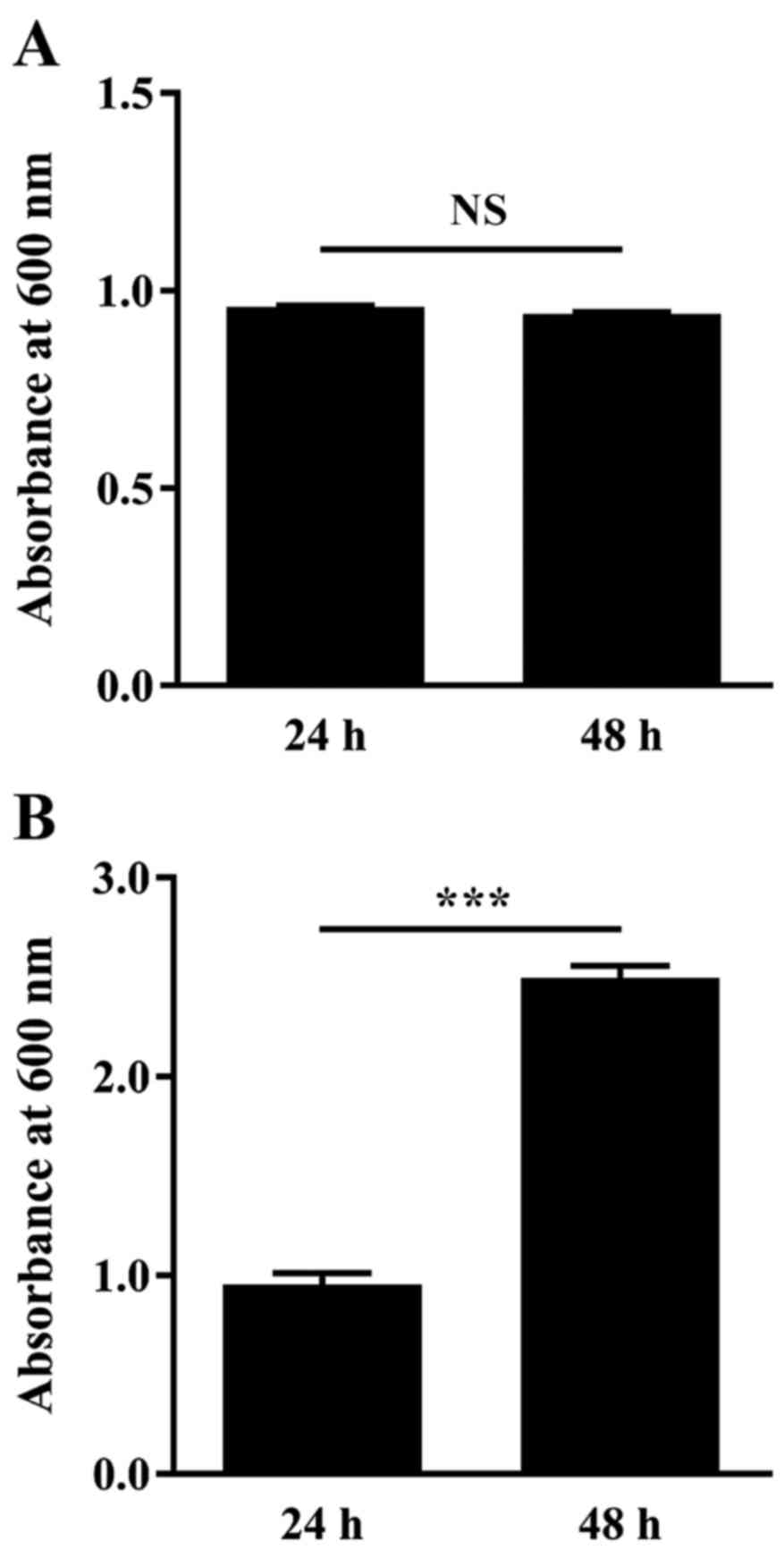
C. albicans ATCC 10231 biofilm
formation on polystyrene
In a preliminary experiment, C. albicans ATCC
10231 was cultured at 37°C for 24 and 48 h, then Sabouraud broth
turbidity and attached biomass were measured by absorbance at 600
nm and crystal violet staining, respectively. Turbidity absorbance
measurements were 0.958±0.003 following incubation for 24 h and
remained similar following 48 h incubation (0.941±0.005; Fig. 1A). However, measurements of the
attached biomass were significantly increased, by 2.6-fold, at 48 h
compared with 24 h (P<0.0001; Fig.
1B).
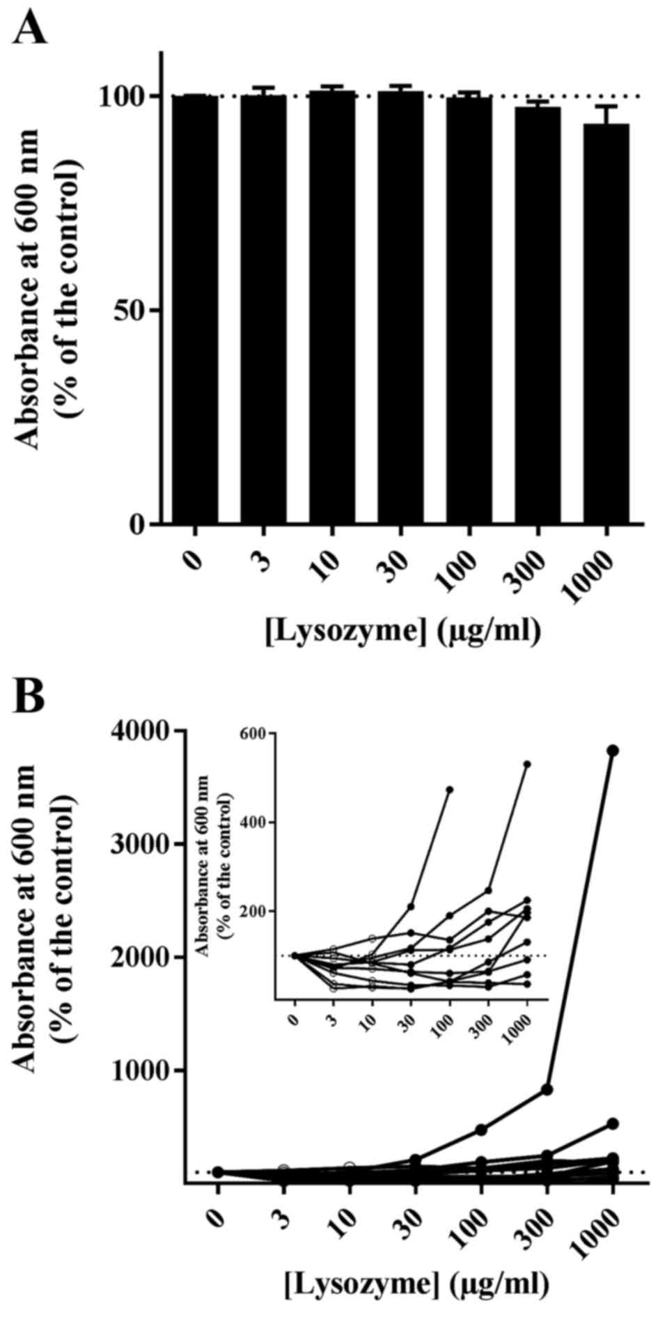
The effect of various concentrations of lysozyme
(0–1,000 µg/ml) on C. albicans ATCC 10231 growth in
Sabouraud liquid medium was measured next (Fig. 2). Lysozyme concentrations ≥300
µg/ml significantly reduced C. albicans growth compared with
the untreated control (P<0.0001); a reduction of 5.3±0.9 and
9.0±1.6% relative to the untreated control was measured at 300 and
1,000 µg/ml of lysozyme, respectively (Fig. 2A). Lysozyme concentrations <100
µg/ml had no effect on C. albicans growth compared with the
untreated control (Fig. 2A).
Amphotericin B (25 µg/ml) completely prevented growth and,
consequently, the formation of biofilms (Fig. 2A). Fig. 2B illustrates the effect of lysozyme
treatment on C. albicans ATCC 10231 biofilm formation in
96-well plates. Lysozyme produced a significant biphasic effect on
biofilm formation (Fig. 2B),
despite its relatively limited effect on yeast growth (Fig. 2A). Lysozyme acted as a biofilm
promotor at the highest concentration tested (1,000 µg/ml), but as
a biofilm limiting factor at the lowest concentrations (10–30
µg/ml). At 1,000 µg/ml, the attached biomass averaged 286.8±49.0%
relative to the control, however, at 10 µg/ml lysozyme, the
attached biomass was 60.9±8.3% relative to the control (Fig. 2B). An ANOVA analysis with Dunnett's
multiple comparison test and a one-sample t-test confirmed the
biofilm promoting effect of 1,000 µg/ml lysozyme (P<0.001 and
P=0.004, respectively; Fig. 2B).
The attached biomass in the presence of 10–30 µg/ml lysozyme was
significantly different from the control by one-sample
t-test (P=0.0059 and P=0.0371, respectively; Fig. 2B), but analysis by ANOVA did not
reveal significant differences (Fig.
2B).
 | Figure 2.Effect of lysozyme on Candida
albicans ATCC 10231 growth. Yeast cells were grown at 37°C for
24 h in the presence of 0, 10, 30, 100, 300 or 1,000 µg/ml
lysozyme. Amph B treatment (25 µg/ml) was used as a negative growth
control. (A) Growth was measured by turbidimetry. (B) In
vitro biofilm formation was evaluated by crystal violet
staining. Data are expressed as the mean ± standard error (n=7-10
independent experiments, 8 technical replicates each). NS, not
significant, *P<0.05, **P< 0.01, ***P<0.001. Dunnett's
post-test compares each column with control without lysozyme. Amph
B, amphotericin B. |
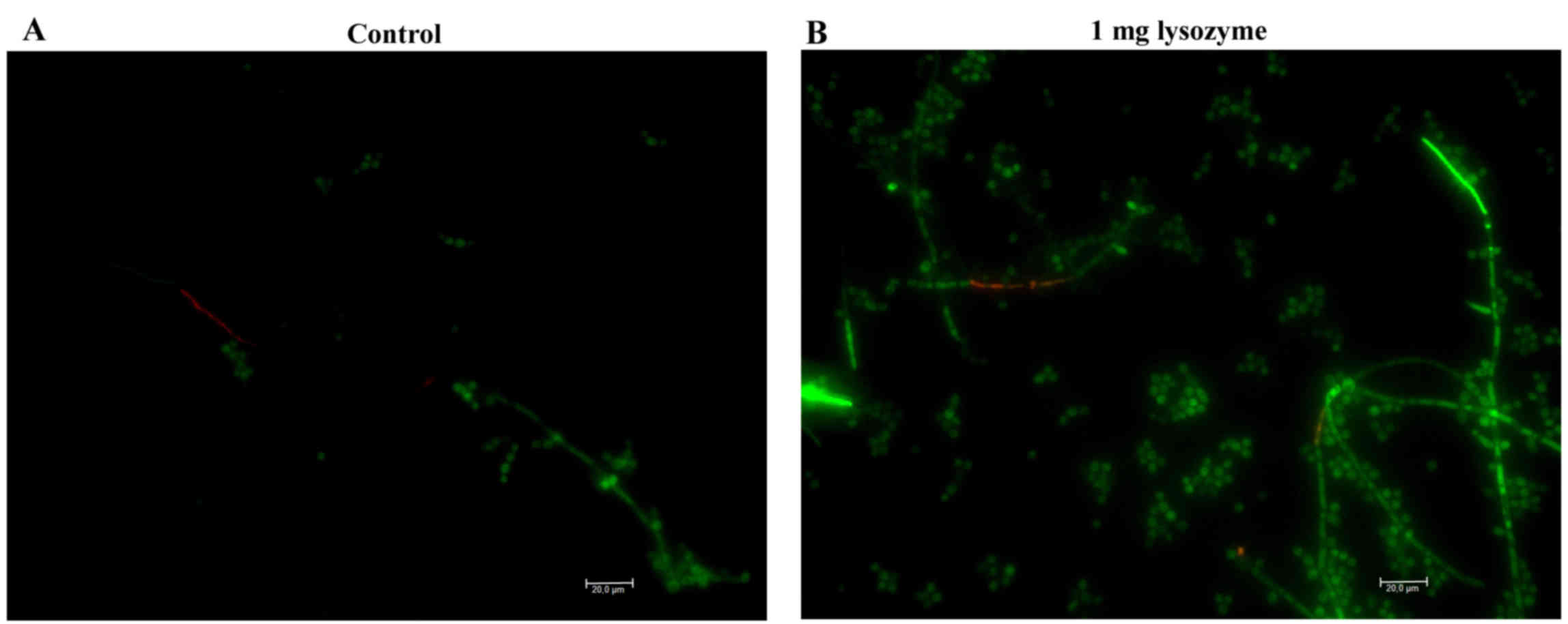
Fig. 3 illustrates
the characteristics of C. albicans biofilms harvested from
the bottom of the wells following incubation with 1,000 µg/ml
lysozyme. Microscopic examination revealed that the majority
(>95%) of C. albicans cells (blastoconidia with some
hyphal structures) were live as in the control without lysozyme
(Fig. 3). C. albicans ATCC
10231 attached biomass was evaluated by 2 different experimental
designs following 48 h incubation at 37°C in 96-well plates: Direct
addition of lysozyme at time zero (Table IA), as previously performed for the
24 h observations displayed in Figs.
2 and 3; and culture medium
renewal with the addition of lysozyme following a 2 h preincubation
of C. albicans cells to allow time for adherence (Table IB). Independent of the incubation
time (24 vs. 48 h), addition of lysozyme to planktonic cells
resulted in an increase in attached biomass compared with the
untreated control, with the increase being higher after 48 h
(Table I). Treatment with 1,000
µg/ml lysozyme resulted in a 9.6-fold increase in attached biomass
compared with control (P<0.0001; Table IA). When biofilm production was
examined following C. albicans pre-adherence, a less
significant 1.4-fold increase in attached biomass was observed in
the presence of 1,000 µg/ml lysozyme compared with control
(P=0.0008; Table IB), while
lysozyme at physiological concentrations (<30 µg/ml) always
reduced biomass (Table I).
 | Table I.Effect of lysozyme on Candida
albicans ATCC 10231 attached biomass. |
Table I.
Effect of lysozyme on Candida
albicans ATCC 10231 attached biomass.
| A, All reagents
added at time zero |
|---|
|
|---|
| Lysozyme
(µg/ml) | 0 | 3 | 10 | 30 | 100 | 300 | 1,000 |
|---|
| % (mean ± SEM) | 100.0±2.5 | 81.1±4.1 | 98.4±4.1 | 124.6±10.3 | 482.4±25.4 | 923.6±33.8 | 958.2±21.1 |
| N | 16 | 8 | 8 | 8 | 8 | 8 | 8 |
| P-value (vs. 0
µg/ml) | – | 0.8786 | 0.9999 | 0.7064 | <0.0001 | <0.0001 | <0.0001 |
|
| B, Culture medium
renewal and lysozyme addition after 2 h pre-incubation |
|
| Lysozyme
(µg/ml) | 0 | 3 | 10 | 30 | 100 | 300 | 1,000 |
|
| % (mean ± SEM) | 100.0±6.0 | 60.1±1.6 | 81.8±4.1 | 73.5±11.1 | 106.8±10.0 | 118.0±6.8 | 141.3±10.7 |
| N | 16 | 8 | 8 | 8 | 8 | 8 | 8 |
| P-value (vs. 0
µg/ml) | – | 0.0013 | 0.2950 | 0.0533 | 0.9643 | 0.3081 | 0.0008 |
Biofilm formation on polystyrene by C.
albicans wild strains
The effect of lysozyme on 10 wild C. albicans
strains isolated from different dentures was then examined by
turbidimetric growth and biofilm evaluation. No significant effect
of lysozyme (0–1,000 µg/ml) on the growth of wild strains in
Sabouraud liquid medium was observed, apart from a slight but
non-significant decrease at 1,000 µl/ml, which was the highest
concentration tested (Fig. 4A).
Amphotericin B (25 µg/ml) completely prevented the growth of yeast
and biofilm formation (data not shown), similar to the results for
the reference strain (Fig. 2A).
The effect of lysozyme (0–1,000 µg/ml) on the ability of the wild
strains to form biofilms on 96-well plates was subsequently
assessed (Fig. 2B). The results
demonstrated variable patterns of biofilm formation (Fig. 4B). Some strains demonstrated large
quantities of biofilm production in the presence of a high lysozyme
concentration (1,000 µg/ml) but poor biofilm production at low
concentrations (Fig. 4B). Other
strains that exhibited poor biofilm production with a high lysozyme
concentration were further inhibited by weak concentrations (3–30
µg/ml; Fig. 4B). Only one wild
strain out of ten was inhibited by lysozyme in a dose-dependent
manner (Fig. 4B). Overall, in the
10 clinical strains isolated from dentures, the attached biomass
following incubation with 1,000 µg/ml lysozyme ranged from 36.1 to
3825.0% compared with untreated controls (mean, 548.4%; median,
191.4%; non-Gaussian distribution; Fig. 4B). However, at 3 µg/ml lysozyme,
the attached biomass ranged from 26.3 to 114.8% compared with
untreated controls (mean, 73.8%; median, 73.8%; Gaussian
distribution; Fig. 4B). Thus, the
inhibitory effect of lysozyme was observed when this was added in
concentrations consistent with its physiological range. The
non-physiological concentration of 1,000 µg/ml significantly
enhanced biofilm production in six strains out of 10 compared with
control (Fig. 4B).
The difference in the attached biomass between the
two tested lysozyme concentrations (10 and 1,000 µg/ml) was
significant for both the reference (Fig. 2B) and the clinical (Fig. 4B) strains (Mann Whitney test,
P<0.0001 and P=0.0185, respectively). The increase in the amount
of biofilm in the presence of 1,000 µg/ml lysozyme was not
significant in clinical strains (Mann Whitney test, P=0.1425) but
was significant in the reference strain (Mann Whitney test,
P=0.0013) when compared with control without lysozyme. In contrast,
the decrease in attached yeast in the presence of 3 µg/ml lysozyme
compared with untreated controls was significant in 8 out of 10
clinical strains (Mann Whitney test, P=0.0230). Again, the
inhibitory effect of lysozyme was observed in concentrations lower
than 30 µg/ml, which is considered as the physiological
concentration in saliva, while non-physiological concentrations
above 100 µg/ml enhanced biofilm production in 6 strains out of
10.
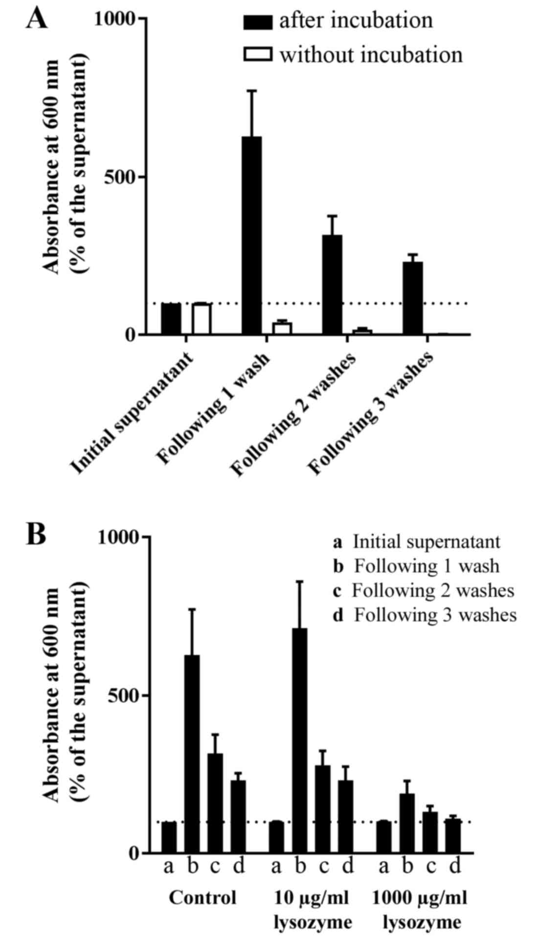
Lysozyme effect on C. albicans biofilm
cohesion
Biofilm cohesion was examined by turbidity variation
in wells where C. albicans ATCC 10231 cells were cultured
for 24 h at 37°C and subsequently washed three times with sterile
0.9% NaCl (Fig. 5). As a control,
wells were inoculated with cultures in Sabouraud broth and
immediately processed without incubation. In the absence of
incubation (and thus without biofilm formation), the turbidity
underwent a gradual decrease to a residual turbidity of 1.7±0.7%
relative to the initial supernatant (Fig. 5A). The turbidity of the incubated
samples was 627.4±144.4% relative to the control after the first
washing; turbidity after the second and third washes was 316.3±59.7
and 230.4±23.6% of that of the culture medium, respectively
(Fig. 5A). The effect of lysozyme
on cell detachment during the washing process was further examined
(Fig. 5B). In the presence of 10
µg/ml lysozyme, the progress of the washing liquid turbidity was
similar to the control without lysozyme (Fig. 5B). However, in the presence of
1,000 µg/ml lysozyme, the turbidity following washes was not much
greater than the supernatant (Fig.
5B). A two-way ANOVA analysis showed that the washing steps and
the lysozyme concentration significantly affected the turbidity of
the liquid (P<0.0001; Fig. 5B).
However, the washing steps did not have the same effect at the two
concentrations of lysozyme (10 and 1,000 µg/ml, respectively) as
shown by the two-way ANOVA analysis for interaction between factors
(P=0.0101; Fig. 5B).
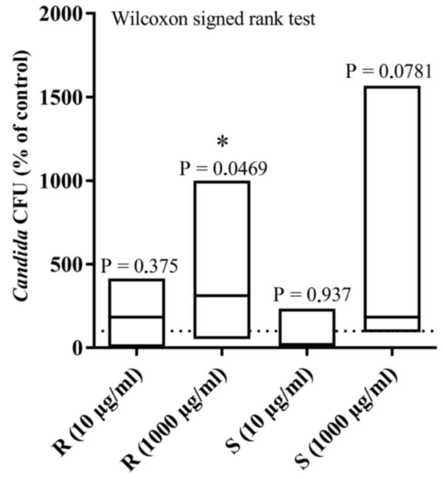
C. albicans adhesion on resin
The effect of 10 and 1,000 µg/ml lysozyme on the
ability of C. albicans to adhere to resin pieces (before and
after surface polishing) was examined following 4 h of incubation
at 37°C (Fig. 6). A Kolmogorov
Smirnov test revealed that the data did not conform to a Gaussian
distribution. Lysozyme at 1,000 µg/ml resulted in a mean increase
of 10- (P=0.0469) and 15.7-fold (P=0.0781) relative to untreated
controls for the rough side (median 312.3% of the control) and the
smooth side (median 184.0% of the control) of the resin foil,
respectively (Fig. 6). Lysozyme at
10 µg/ml did not lead to significant changes in the attached yeast
cell count: 4.1-fold for the rough side (median 184.0% of the
control) and 2.3-fold for the smooth side (median 19.8% of the
control). Analysis of the results by the Wilcoxon signed rank test
demonstrated that the high lysozyme concentration (1,000 µg/ml)
resulted in significant changes in adherence to the rough resin
(P=0.0469), while the effect on the smooth resin was close to
significant (P=0.0781). Finally, in the absence of lysozyme,
polishing negatively affected C. albicans adherence to the
acrylic surface, as the CFU count on the smooth resin corresponded
to 40.8±7.9% of that observed on the paired rough resin
(P=0.0247).
Discussion
Previous investigations have demonstrated that the
effect of lysozyme can differ from one bacterial species to another
and even within the same bacterial species (37); susceptibility to lysozyme is also
dependent on the monitored parameter (growth, viability, cell
lysis). In the present study, investigations of C. albicans
wild strains isolated from dentures revealed several,
non-consistent patterns of susceptibility to lysozyme. To the best
of our knowledge, no previous studies have focused on biofilm
formation and its inhibition by lysozyme from exocrine secretions.
In the present study, egg white lysozyme produced a biphasic effect
on biofilm formation, while it slightly affected yeast growth
tested by turbidimetry. In the C. albicans ATCC 10231
reference strain, lysozyme acted as a biofilm promotor at the
highest concentration tested (1,000 µg/ml), but as a biofilm
limiting factor at the lowest concentration (10 µg/ml). In the
clinical strains isolated from contaminated dentures, a variety of
responses to lysozyme were observed, with no apparent uniform
pattern. The inhibitory effect of lysozyme was observed at
concentrations <30 µg/ml, which are physiological concentrations
in saliva (17), while
non-physiological doses >100 µg/ml enhanced biofilm growth. The
different results on polystyrene vs. resin are not necessarily
attributable to the material itself, since a different method of
attached biomass evaluation was used for each material (crystal
violet staining vs. CFU counts following seeding, respectively).
However, because of crystal violet adsorption on resin pieces in
the absence of C. albicans cells (data not shown), a
different method had to be employed. Although less sensitive than a
colorimetric assay, CFU counts on Petri dishes presented the
advantage of confirming the viability of attached biomass without
additional manipulation.
Pro-biofilm effect of lysozyme on C.
albicans
In the present study, a pro-biofilm property for
lysozyme was suggested in different strains, on two different
support materials (polystyrene, widely used in the laboratory, and
acrylic resin, widely used in the fabrication of dentures), by two
different experimental designs (C. albicans adhesion to the
material surface and biofilm production), and by two different
biofilm production assessments (crystal violet staining and CFU
counts). Lysozyme capacity to increase attached biomass was
observed at high but non-physiological concentrations. The FDA/EB
staining assay revealed that the majority of cells attached in the
presence of 1 mg/ml lysozyme were alive. The pro-biofilm property
of lysozyme on C. albicans ATCC 10231 is more pronounced
when added simultaneously with the C. albicans cells,
suggesting a crucial role in the adherence phase of the biofilm
formation. The pro-biofilm effect was also confirmed by the
assessment of biofilm cohesion during three successive washes with
saline. In the presence of 1,000 µg/ml lysozyme, the turbidity of
the washing media did not increase, which suggests low yeast cell
loss by the biofilm. Previous studies have focused on the
agglutination or flocculation power of lysozyme (at a cited
concentration of 1 mg/ml) in non-ionic solutions upon air
saprophytes (22), bacterial
suspensions (38) or C.
albicans yeasts (22). The
presence of lysozyme in the in vivo acquired exogenous
pellicle on teeth is well documented but no data concerning its
effect on biofilm formation exist to date (39). The present study demonstrated an
increase in C. albicans adherence in six out of ten wild
C. albicans strains when higher concentrations of lysozyme
were present, suggesting that the use of high concentrations on
lysozyme in oral care products may be unfitting. Intra-species
variability in lysozyme susceptibility has also previously been
demonstrated in bacteria (37).
Anti-biofilm effect of lysozyme on C.
albicans
In the present study, low concentrations of lysozyme
(<30 µg/ml) resulted in a reduction in the attached biomass. At
a concentration of 10 µg/ml, biofilm reduction on polystyrene was
observed in C. albicans ATCC 10231 and in 3 out of 10
clinical strains investigated. The anti-biofilm effect was not
observed for the reference strain on either a rough (P=0.375) or
smooth (P=0.937) resin support. However, a previous study has
demonstrated that a similar concentration range of lysozyme (6–100
µg/ml) significantly inhibited C. albicans biofilm formation
on an acrylic resin surface (33).
In contrast, a separate study demonstrated no significant reduction
in C. albicans attachment on acrylic resin disks immersed in
a solution containing lysozyme, lactoferrin and glucose oxidase
complex/lactoperoxidase (35).
These conflicting observations are consistent with the present
study, where the susceptibility of C. albicans wild strains
to lysozyme differed from one another.
Inhibitory effect of lysozyme upon C.
albicans growth
The present study demonstrated a significant effect
of egg white lysozyme on yeast growth at lysozyme concentrations
>300 µg/ml for the reference strain, but not for the wild
strains. The growth reduction effect on C. albicans ATCC
10231 in Sabouraud liquid medium upon exposure to 1,000 and 300
µg/ml lysozyme was 9.0±1.6 and 5.3±0.9%, respectively, compared
with control. In the clinical isolates, a similar mild reduction
was observed but was not statistically significant. It is possible
that this small effect may be due to the incorporation of
planktonic cells into the attached biomass on the lateral sides of
the microplate wells, thus escaping the photometric evaluation. The
effect on the turbidimetric measurement can then be explained by
the pro-biofilm property of the lysozyme. Previously, C.
albicans cells have been observed to be lysed by lysozyme (in a
concentration range from 40 to 5,000 µg/ml) independently of the
amount of glucose present, but this effect was prevented by the
addition of NaCl to the culture medium (40).
The present study demonstrated differing effects of
lysozyme upon in vitro C. albicans biofilm formation
dependent on its concentration: anti-biofilm at physiological
concentrations and pro-biofilm with concentrations >300 µg/ml.
The present study thus indicates that the concentration of active
compounds should be stated in oral care products. In addition, the
present study illustrates the challenges of comprehensively
understanding the complex relationships between oral microflora and
salivary antimicrobial systems. As well as careful and detailed
reporting, new tools should be developed to study the relationship
between the microbiome and its exocrine environment, taking into
account the diversity of antimicrobial factors and the
concentration of each. The present study emphasizes the necessity
of developing strategies for biofilm control based on in
vitro experiments, and to implement them in clinical trials
prior to the incorporation of exocrine proteins, such as lysozyme,
into hygiene products. Further studies should extend these
investigations to other Candida species, and to fungi and
bacteria present in oral biofilms, using a more global
approach.
Acknowledgements
The present study was supported by a grant (grant
no. BRIC-12/143) from the Xenophilia Funds (Université Libre
de Bruxelles, Brussels, Belgium). The authors thank P. Keyzer for
manufacturing the acrylic pieces and Professor M. Stas for her
review of the manuscript.
References
|
1
|
Radford DR, Challacombe SJ and Walter JD:
Denture plaque and adherence of Candida albicans to
denture-base materials in vivo and in vitro. Crit Rev Oral Biol
Med. 10:99–116. 1999. View Article : Google Scholar : PubMed/NCBI
|
|
2
|
Courtois Ph: Candida biofilms on
oral biomaterials. Biomaterials-Physics and chemistry. Pignatello
R: Intech; Rjieka: pp. 475–490. 2011
|
|
3
|
Epstein JB, Truelove EL and Izutzu KT:
Oral candidiasis: Pathogenesis and host defense. Rev Infect Dis.
6:96–106. 1984. View Article : Google Scholar : PubMed/NCBI
|
|
4
|
Webb BC, Thomas CJ, Willcox MD, Harty DW
and Knox KW: Candida-associated denture stomatitis.
Aetiology and management: A review. Part 1. Factors influencing
distribution of Candida species in the oral cavity. Aust
Dent J. 43:45–50. 1998. View Article : Google Scholar : PubMed/NCBI
|
|
5
|
Ramage G, Tomsett K, Wickes BL,
López-Ribot JL and Redding SW: Denture stomatitis: A role for
Candida biofilms. Oral Surg Oral Med Oral Pathol Oral Radiol
Endod. 98:53–59. 2004. View Article : Google Scholar : PubMed/NCBI
|
|
6
|
Fleming A: On a remarkable bacteriolytic
element found in tissues and secretions. Proc R Soc Lond B Biol
Sci. 93:306–317. 1922. View Article : Google Scholar
|
|
7
|
Gordon S, Todd J and Cohn ZA: In vitro
synthesis and secretion of lysozyme by mononuclear phagocytes. J
Exp Med. 139:1228–1248. 1974. View Article : Google Scholar : PubMed/NCBI
|
|
8
|
Vukosavljevic D, Custodio W and Siqueira
WL: Salivary proteins as predictors and controls for oral health. J
Cell Commun Signal. 5:271–275. 2011. View Article : Google Scholar : PubMed/NCBI
|
|
9
|
Fábián TK, Hermann P, Beck A, Fejérdy P
and Fábián G: Salivary defense proteins: Their network and role in
innate and acquired oral immunity. Int J Mol Sci. 13:4295–4320.
2012. View Article : Google Scholar : PubMed/NCBI
|
|
10
|
Phillips DC: The three-dimensional
structure of an enzyme molecule. Sci Am. 215:78–90. 1966.
View Article : Google Scholar : PubMed/NCBI
|
|
11
|
Masschalck B and Michiels CW:
Antimicrobial properties of lysozyme in relation to foodborne
vegetative bacteria. Crit Rev Microbiol. 29:191–214. 2003.
View Article : Google Scholar : PubMed/NCBI
|
|
12
|
MacKay BJ, Goodman H, Cox D, Grossbard BL,
Iacono VJ and Pollock JJ: Development of an enzyme-linked
immunosorbent assay for determination of lysozyme in human parotid
and submandibular-sublingual salivas. J Clin Microbiol. 19:844–848.
1984.PubMed/NCBI
|
|
13
|
Noble RE: Salivary alpha-amylase and
lysozyme levels: A non-invasive technique for measuring parotid vs
submandibular/sublingual gland activity. J Oral Sci. 42:83–86.
2000. View Article : Google Scholar : PubMed/NCBI
|
|
14
|
Brandtzaeg P and Mann WV Jr: A comparative
study of the lysozyme activity of human gingival pocket fluid,
serum and saliva. Acta Odontol Scand. 22:441–455. 1964. View Article : Google Scholar : PubMed/NCBI
|
|
15
|
Eisenberg RJ, Bowers GM and Bergquist JJ:
Lysozyme activity in gingival crevicular fluid. J Baltimore Coll
Dent Surg. 32:83–85. 1977.PubMed/NCBI
|
|
16
|
Sakalauskiene J, Surna A, Ivanauskiene E,
Zekonis G and Gleiznys A: Secretory function of neutrophilic
leucocytes of the patients with periodontal diseases.
Stomatologija. 7:90–94. 2005.PubMed/NCBI
|
|
17
|
Stuchell RN and Mandel ID: A comparative
study of salivary lysozyme in caries-resistant and
caries-susceptible adults. J Dent Res. 62:552–554. 1983. View Article : Google Scholar : PubMed/NCBI
|
|
18
|
Woods CM, Hooper DN, Ooi EH, Tan LW and
Carney AS: Human lysozyme has fungicidal activity against nasal
fungi. Am J Rhinol Allergy. 25:236–240. 2011. View Article : Google Scholar : PubMed/NCBI
|
|
19
|
Marquis G, Montplaisir S, Garzon S,
Strykowski H and Auger P: Fungitoxicity of muramidase.
Ultrastructural damage to Candida albicans. Lab Invest.
46:627–636. 1982.PubMed/NCBI
|
|
20
|
Marquis G, Garzon S, Strykowsky H and
Auger P: Cell walls of normal and lysozyme-damaged blastoconidia of
Candida albicans: Localization of surface factor 4 antigen
and vicinal-glycol staining. Infect Immun. 59:1312–1318.
1991.PubMed/NCBI
|
|
21
|
Edgerton M and Koshlukova SE: Salivary
histatin 5 and its similarities to the other antimicrobial proteins
in human saliva. Adv Dent Res. 14:16–21. 2000. View Article : Google Scholar : PubMed/NCBI
|
|
22
|
Kamaya T: Flocculation phenomenon of
Candida albicans by lysozyme. Mycopathol Mycol Appl.
37:320–330. 1969. View Article : Google Scholar : PubMed/NCBI
|
|
23
|
Tenovuo J: Clinical applications of
antimicrobial host proteins lactoperoxidase, lysozyme and
lactoferrin in xerostomia: Efficacy and safety. Oral Dis. 8:23–29.
2002. View Article : Google Scholar : PubMed/NCBI
|
|
24
|
Gil-Montoya JA, Guardia-López I and
González-Moles MA: Evaluation of the clinical efficacy of a
mouthwash and oral gel containing the antimicrobial proteins
lactoperoxidase, lysozyme and lactoferrin in elderly patients with
dry mouth: A pilot study. Gerodontology. 25:3–9. 2008. View Article : Google Scholar : PubMed/NCBI
|
|
25
|
Güneri P, Alpöz E, Epstein JB, Çankaya H
and Ates M: In vitro antimicrobial effects of commercially
available mouth-wetting agents. Spec Care Dentist. 31:123–128.
2011. View Article : Google Scholar : PubMed/NCBI
|
|
26
|
Lee JY, Kim YY, Chang JY, Park MS and Kho
HS: The effects of peroxidase on the enzymatic and candidacidal
activities of lysozyme. Arch Oral Biol. 55:607–612. 2010.
View Article : Google Scholar : PubMed/NCBI
|
|
27
|
Cho MA, Kim YY, Chang JY and Kho HS:
Interactions between hyaluronic acid, lysozyme, and the glucose
oxidase-mediated lactoperoxidase system in enzymatic and
candidacidal activities. Arch Oral Biol. 58:1349–1356. 2013.
View Article : Google Scholar : PubMed/NCBI
|
|
28
|
Samaranayake YH, Samaranayake LP, Wu PC
and So M: The antifungal effect of lactoferrine and lysozyme on
Candida krusei and Candida albicans. APMIS.
105:875–883. 1997. View Article : Google Scholar : PubMed/NCBI
|
|
29
|
Kang JH, Kim YY, Chang JY and Kho HS:
Influences of hyaluronic acid on the anticandidal activities of
lysozyme and the peroxidase system. Oral Dis. 17:577–583. 2011.
View Article : Google Scholar : PubMed/NCBI
|
|
30
|
Collins MS and Pappagianis D:
Lysozyme-enhanced killing of Candida albicans and
Coccidioides immitis by amphoteracin B. Sabouraudia.
12:329–340. 1974. View Article : Google Scholar : PubMed/NCBI
|
|
31
|
Nishiyama Y, Nakaoka C, Hiratani T, Abe S,
Uchida K and Yamaguchi H: Synergy of lysozyme and lanoconazole on
the morphology of Candida albicans. J Electron Microsc
(Tokyo). 50:41–49. 2001. View Article : Google Scholar : PubMed/NCBI
|
|
32
|
Anil S and Samaranayake LP: Impact of
lysozyme and lactoferrin on oral Candida isolates exposed to
polyene antimycotics and fluconazole. Oral Dis. 8:199–206. 2002.
View Article : Google Scholar : PubMed/NCBI
|
|
33
|
Samaranayake YH, Cheung BP, Parahitiyawa
N, Seneviratne CJ, Yau JY, Yeung KW and Samaranayake LP:
Synergistic activity of lysozyme and antifungal agents against
Candida albicans biofilms on denture acrylic surfaces. Arch
Oral Biol. 54:115–126. 2009. View Article : Google Scholar : PubMed/NCBI
|
|
34
|
Tanida T, Okamoto T, Okamoto A, Wang H,
Hamada T, Ueta E and Ozaki T: Decreased excretion of antimicrobial
proteins and peptides in saliva of patients with oral candidiasis.
J Oral Pathol Med. 32:586–594. 2003. View Article : Google Scholar : PubMed/NCBI
|
|
35
|
Silva MP, Junior Chibebe J, Jorjão AL,
Machado AK, Oliveira LD, Junqueira JC and Jorge AO: Influence of
artificial saliva in biofilm formation of Candida albicans
in vitro. Braz Oral Res. 26:24–28. 2012. View Article : Google Scholar : PubMed/NCBI
|
|
36
|
Stepanovic S, Vukovic D, Dakic I, Savic B
and Svabic-Vlahovic M: A modified microtiter-plate test for
quantification of staphylococcal biofilm formation. J Microbiol
Methods. 40:175–179. 2000. View Article : Google Scholar : PubMed/NCBI
|
|
37
|
Iacono VJ, MacKay BJ, DiRienzo S and
Pollock JJ: Selective antibacterial properties of lysozyme for oral
microorganisms. Infect Immun. 29:623–632. 1980.PubMed/NCBI
|
|
38
|
Salton MR: Cell structure and the the
enzymic lysis of bacteria. J Gen Microbiol. 9:512–523. 1953.
View Article : Google Scholar : PubMed/NCBI
|
|
39
|
Edgerton M and Levine MJ: Characterization
of acquired denture pellicle from healthy and stomatitis patients.
J Prosthet Dent. 68:683–691. 1992. View Article : Google Scholar : PubMed/NCBI
|
|
40
|
Kamaya T: Lytic action of lysozyme on
Candida albicans. Mycopathol Mycol Appl. 42:197–207. 1970.
View Article : Google Scholar : PubMed/NCBI
|