Introduction
Bone quantity and quality is maintained by bone
remodeling, a biphasic process that consists of bone resorption and
formation (1). The process is
dependent on two antagonistic functional cell types: Mesenchymal
stem cell-derived osteoblasts and hematopoietic stem cell-derived
osteoclasts, which are responsible for bone formation and
resorption, respectively (1).
Dysregulation of bone remodeling leads to metabolic bone disorders,
including osteoporosis and perturbation of fracture healing
(1). Numerous humoral factors,
including cytokines, growth factors and prostaglandins (PGs), have
been demonstrated to affect bone remodeling (2). Osteoprotegerin (OPG), which belongs
to the tumor necrosis factor receptor superfamily, is an essential
osteoblast-secretory protein and decoy receptor for the receptor
activator of nuclear factor-κB ligand (RANKL) (3). OPG prevents osteoclastogenesis from
blocking RANK-RANKL binding, which is an essential step of bone
resorption (3). It has previously
been reported that OPG-deficient mice display severe osteoporosis
(4). The competitive antagonism
between RANKL and OPG for RANK binding is a crucial regulatory
system in bone remodeling (5).
PGs, which are lipid signaling molecules, are
involved in various physiological processes (6). PGs act as autacoids in bone
metabolism, and modulate bone cell function (6). Although PGs have conventionally been
recognized as bone resorptive agents (7), there is accumulating evidence
supporting the involvement of PGs in the process of bone formation
(7,8). It has previously been reported that
PGE1 stimulates the synthesis of OPG via activation of
p38 mitogen-activated protein (MAP) kinase and stress-activated
protein kinase/c-Jun N-terminal kinase (SAPK/JNK) in
osteoblast-like MC3T3-E1 cells (9). PGE1 has also been
demonstrated to stimulate the secretion of interleukin-6 (IL-6), a
multifunctional cytokine that modulates bone metabolism (10,11),
via the cAMP protein kinase A pathway in osteoblast-like MC3T3-E1
cells (12).
Mimosine, which is a natural plant amino acid
present in the Leucaena genus, is an iron chelator that
suppresses DNA replication in mammals (13). In addition, mimosine is a normoxic
hypoxia-inducible factor (HIF) inducer (14). HIFs act as DNA-binding
transcription factors with specific nuclear cofactors under low
oxygen concentrations, and activate a series of hypoxia-associated
genes in response to hypoxic environments (15). Numerous genes, including glucose
transporter protein 1, erythropoietin and vascular endothelial
growth factor (VEGF), have been identified as HIF target genes
(14). Therefore, HIFs are
recognized to be implicated in the regulation of glucose
metabolism, erythropoiesis, and angiogenesis (14). It has previously been demonstrated
that hypoxia enhances osteoclast-mediated bone resorption (15), suggesting that HIFs are also
involved in bone metabolism. In addition, the complex HIF-1α
reportedly promotes bone formation by the direct stimulation of
osteoblast proliferation and induces angiogenesis, resulting in the
stimulation of bone regeneration (16,17).
It has previously been reported that mimosine upregulates HIF-1α
protein levels and inhibits PGF2α-induced OPG synthesis
without affecting IL-6 release in osteoblast-like MC3T3-E1 cells
(18). However, the precise
mechanism underlying the effects of HIFs on osteoblasts is yet to
be elucidated.
In the present study, the effects of two normoxic
HIF inducers, mimosine and deferoxamine (19) on PGE1-stimulated
synthesis of OPG and IL-6 in osteoblast-like MC3T3-E1 cells were
investigated. The results strongly suggest that normoxic HIF
inducers suppress PGE1-stimulated OPG synthesis without
affecting IL-6 production in osteoblasts.
Materials and methods
Materials
Mimosine, deferoxamine and PGE1 were
obtained from Sigma-Aldrich; Merck Millipore. Mouse OPG
enzyme-linked immunosorbent assay (ELISA, cat. no. MOP00) and mouse
IL-6 ELISA (cat. no. SM6000B) kits were obtained from R&D
Systems, Inc. (Minneapolis, MN, USA). Phospho-specific p38 MAP
kinase antibodies (cat. no. 4511), p38 MAP kinase antibodies (cat.
no. 9212), phospho-specific SAPK/JNK antibodies (cat. no. 4671) and
SAPK/JNK antibodies (cat. no. 9252) were obtained from Cell
Signaling Technology, Inc. (Danvers, MA, USA). An enhanced
chemiluminescence (ECL) western blotting detection system was
obtained from GE Healthcare Life Sciences (Little Chalfont, UK).
Other materials and chemicals were obtained from commercial
sources. PGE1 was dissolved in ethanol. Mimosine was
dissolved in PBS supplemented with 0.01% bovine serum albumin (BSA)
containing 7.5% NaHCO3. Deferoxamine was dissolved in
PBS supplemented with 0.01% BSA. The maximum concentration of
ethanol was 0.1%, which did not affect the assay for
osteoprotegerin release, IL-6 release, osteoprotegerin mRNA
expression, or western blot analysis.
Cell culture
Cloned osteoblast-like MC3T3-E1 cells derived from
newborn mouse calvaria (20) were
maintained as previously described (21). Cells were cultured in α-minimum
essential medium (α-MEM; Sigma-Aldrich; Merck Millipore, Darmstadt,
Germany) supplemented with 10% fetal bovine serum (FBS; Gibco,
Thermo Fisher Scientific, Inc., Waltham, MA, USA) at 37°C in a
humidified atmosphere containing 5% CO2. Cells were
seeded into 35-mm diameter dishes (5×10⁴ cells/dish) or
90-mm diameter dishes (2×10⁵ cells/dish) in α-MEM
containing 10% FBS. Medium was exchanged for α-MEM containing 0.3%
FBS following 5 days of culture. Cells were used for experiments 48
h after this.
OPG and IL-6 assay
Cultured cells were pretreated with 300, 500 or 700
µM mimosine or 100, 300, 500 µM deferoxamine, or vehicle (PBS
supplemented with 0.01% BSA) for 60 min at 37°C, and then
stimulated with 10 µM of PGE1 or 50 µl of vehicle (PBS
supplemented with 0.01% BSA containing 0.1% ethanol) in 1 ml of
α-MEM containing 0.3% FBS for the for 48 h at 37°C. The conditioned
medium was collected at the end of incubation, and the
concentrations of OPG and IL-6 were measured using OPG ELISA and
IL-6 ELISA kits, respectively, according to the manufacturer's
protocols.
Semi-quantitative reverse
transcription polymerase chain reaction (RT-PCR)
Cultured cells were pretreated with 700 µM mimosine,
500 µM deferoxamine or vehicle (PBS supplemented with 0.01% BSA)
for 60 min at 37°C, and then stimulated with 10 µM of
PGE1 or vehicle (PBS supplemented with 0.01% BSA
containing 0.1% ethanol) in α-MEM containing 0.3% FBS for 3 h at
37°C. Total RNA was isolated and transcribed into cDNA using
TRIzol® reagent (Invitrogen; Thermo Fisher Scientific,
Inc.) and an Omniscript Reverse Transcriptase kit (Qiagen Inc.,
Valencia, CA, USA), respectively. RT-PCR was performed using a
Light Cycler system with capillaries and the Fast Start DNA Master
SYBR Green I provided with the kit (Roche Diagnostics, Basel,
Switzerland). Sense and antisense primers for mouse OPG mRNA were
purchased from Takara Bio, Inc. (Otsu, Japan; primer set ID: OPG;
MA026526), while glyceraldehyde-3-phosphate dehydrogenase (GAPDH)
mRNA primers were synthesized based on the report of Simpson et
al (22). The 20 µl reaction
mixture was incubated at 95°C for 10 min, followed by 40 cycles at
60°C for 5 sec and 72°C for 7 sec. Amplified products were
determined using melting curve analysis according to the system
protocol. OPG mRNA levels were normalized to those of GAPDH
mRNA.
Western blot analysis
Cultured cells were pretreated with 300, 500 or 700
µM of mimosine or 100, 300 or 500 µM of deferoxamine for 60 min at
37°C, and then stimulated with 10 µM of PGE1 or vehicle
(PBS supplemented with 0.01% BSA containing 0.1% ethanol) in α-MEM
containing 0.3% FBS for 10 min for p38 MAP kinase and 20 min for
SAPK/JNK, respectively. The cells were washed twice with
phosphate-buffered saline and then lysed, homogenized and sonicated
with ultrasonic disruptor (Tomy Seiko Co., Ltd, Tokyo, Japan) at 20
kHz for 10 sec in lysis buffer containing 62.5 mM Tris/HCl (pH
6.8), 2% sodium dodecyl sulfate (SDS), 50 mM dithiothreitol and 10%
glycerol. The protein concentration in each cell lysate was
determined by the Pierce BCA Protein Assay kit (cat. no. 23227,
Thermo Fisher Scientific, Inc.). A total of 10 µl containing 10 µg
of protein was loaded to each lane. SDS-polyacrylamide gel
electrophoresis (PAGE) was performed in the method of Laemmli
(23) using 10% polyacrylamide
gel. Protein was fractionated and transferred onto Immun-Blot
polyvinylidene fluoride (PVDF) membranes (Bio-Rad Laboratories,
Inc., Hercules, CA, USA). The membranes were blocked with 5%
fat-free dry milk in Tris-buffered saline-Tween (TBST; 20 mM
Tris-HCl, pH 7.6, 137 mM NaCl, 0.1% Tween 20) for 1 h at room
temperature prior to incubation with primary antibodies. A western
blot analysis was performed as described previously (24) using phospho-specific p38 MAP kinase
antibodies, p38 MAP kinase antibodies, phospho-specific SAPK/JNK
antibodies or SAPK/JNK antibodies as primary antibodies with
peroxidase-labeled goat anti-rabbit IgG antibodies being used as
secondary antibodies (cat. no. 074-1506; KPL, Inc., Gaithersburg,
MD, USA). Primary and secondary antibodies were diluted at 1:1,000
with 5% fat-free dry milk in TBST for overnight at room temperature
for primary antibodies, and 1 h at room temperature for secondary
antibodies, respectively. Peroxidase activity on the PVDF membrane
was visualized on X-ray film by means of the ECL western blotting
detection system.
Statistical analysis
Differences between the mean values for individual
groups were assessed with one-way analysis of variance, followed by
application of the Bonferroni correction for multiple comparisons
between pairs. P<0.05 was considered to indicate a statistically
significant difference. All data are presented as the mean ±
standard error of the mean, which was determined from three
independent cell preparations.
Results
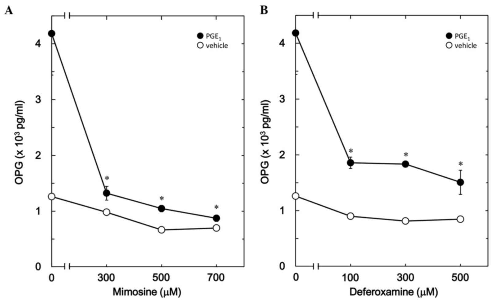
Effects of mimosine or deferoxamine on
PGE1-stimulated OPG release in MC3T3-E1 cells
It has previously been reported that PGE1
stimulates OPG synthesis in osteoblast-like MC3T3-E1 cells
(9). Therefore, the effects of
mimosine, an inducer of HIF (14),
on PGE1-stimulated OPG release in MC3T3-E1 cells were
investigated. Mimosine treatment significantly decreased
PGE1-stimulated OPG release for up to 48 h compared with
untreated cells (12 h, P=0.01; 24 h, P=0.001; 36 h, P=0.0001; 48 h,
P=0.000001; Fig. 1), and the
inhibitory effect was dose-dependent between 300 and 700 µM (300
µM, P=0.02; 500 µM, P=0.01; 700 µM, P=0.01 vs. untreated cells;
Fig. 2A). The maximum inhibitory
effect of mimosine was observed at 700 µM, which resulted in a ~90%
decrease in PGE1-stimulated OPG release compared with
the untreated group (Fig. 2A). In
addition, deferoxamine, another inducer of HIF-1α that exerts an
angiogenic action through stimulation of the HIF-1α pathway
(19), also significantly reduced
PGE1-stimulated OPG release compared with untreated
cells (12 h, P=0.02; 24 h, P=0.000003; 36 h, P=0.0001; 48 h,
P=0.000001; Fig. 1). The
suppressive effect of deferoxamine on OPG release was
dose-dependent between 100 and 500 µM (Fig. 2B). Deferoxamine at 500 mM induced a
~80% decrease in the PGE1-stimulated OPG release
(Fig. 2B).
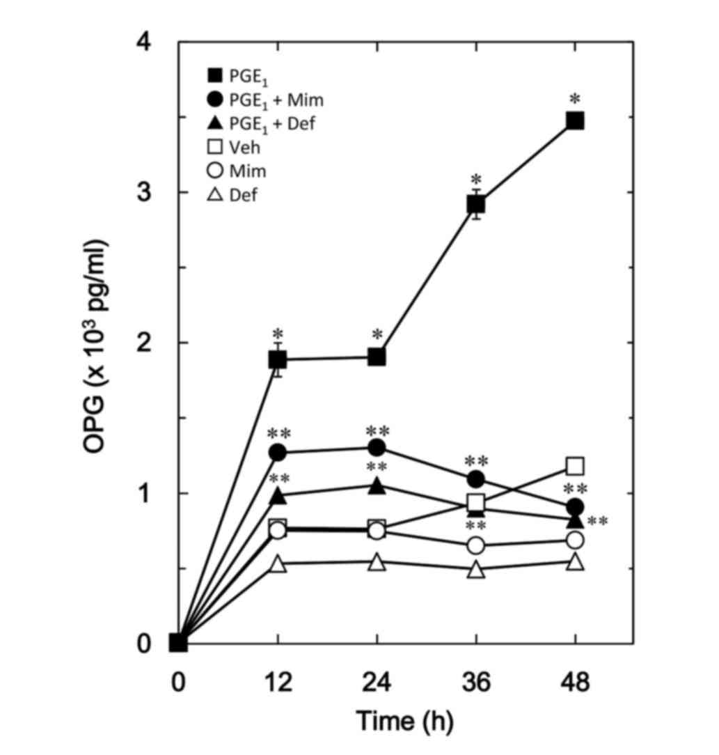 | Figure 1.Effects of mimosine or deferoxamine
on PGE1-stimulated OPG release in MC3T3-E1 cells.
Cultured cells were pretreated with 700 µM mimosine (●,○), 500 µM
deferoxamine (▲,Δ) or vehicle (■,□) for 60 min, and then stimulated
with 10 mM of PGE1 (●,▲,■) or vehicle (○,Δ,□) for the
indicated periods. Each value represents the mean ± standard error
of the mean, calculated from three independent cell preparations.
*P<0.05 vs. control. **P<0.05 vs. PGE1 alone.
PGE1, prostaglandin E1; OPG, osteoprotegerin;
Mim, mimosine: Def, deferoxamine; Veh, vehicle. |
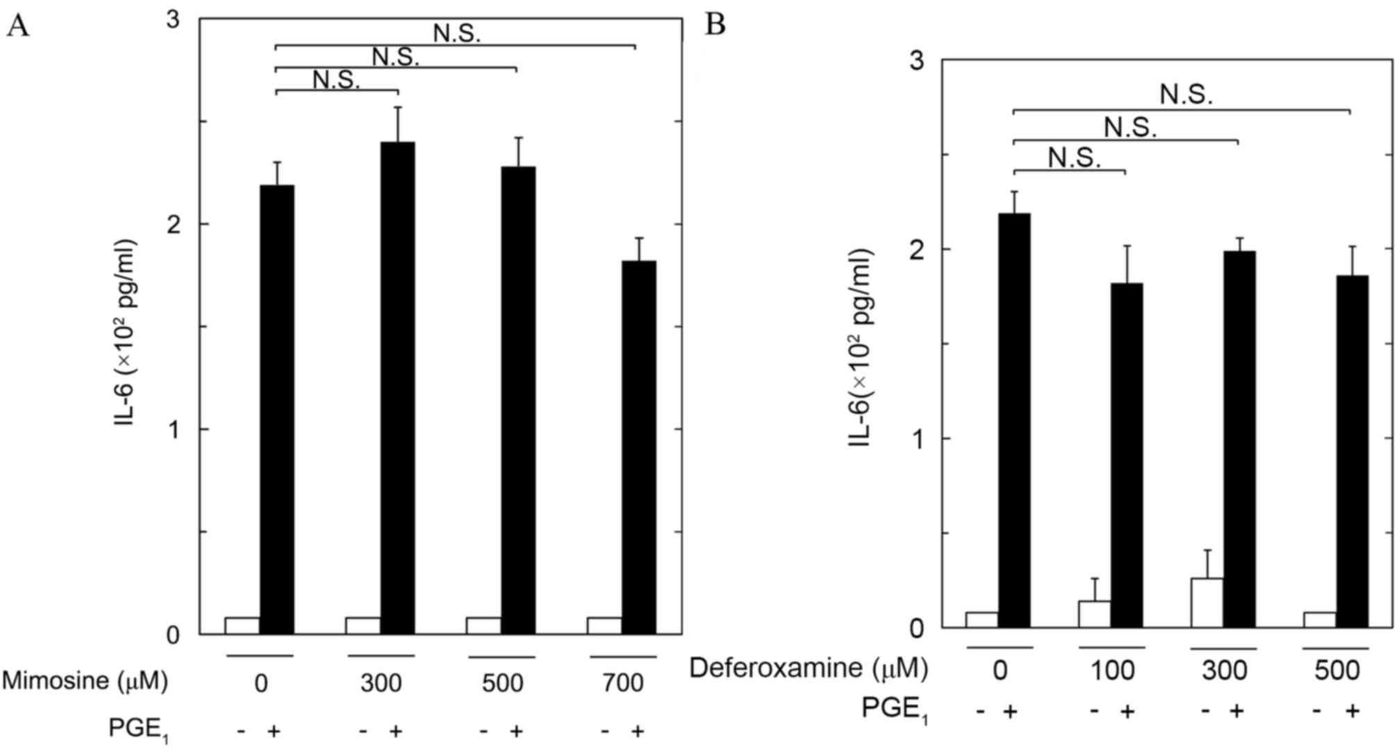
Effects of mimosine and deferoxamine
on PGE1-stimulated IL-6 release in MC3T3-E1 cells
It has previously been demonstrated that
PGE1 upregulates the synthesis of IL-6 in
osteoblast-like MC3T3-E1 cells (12). Therefore, the effects of mimosine
and deferoxamine on PGE1-stimulated IL-6 release were
investigated in these cells. Treatment with up to 700 µM mimosine
had little effect on PGE1-induced IL-6 release (Fig. 3A). In addition, treatment with up
to 500 µM deferoxamine failed to affect PGE1-induced
IL-6 release (Fig. 3B).
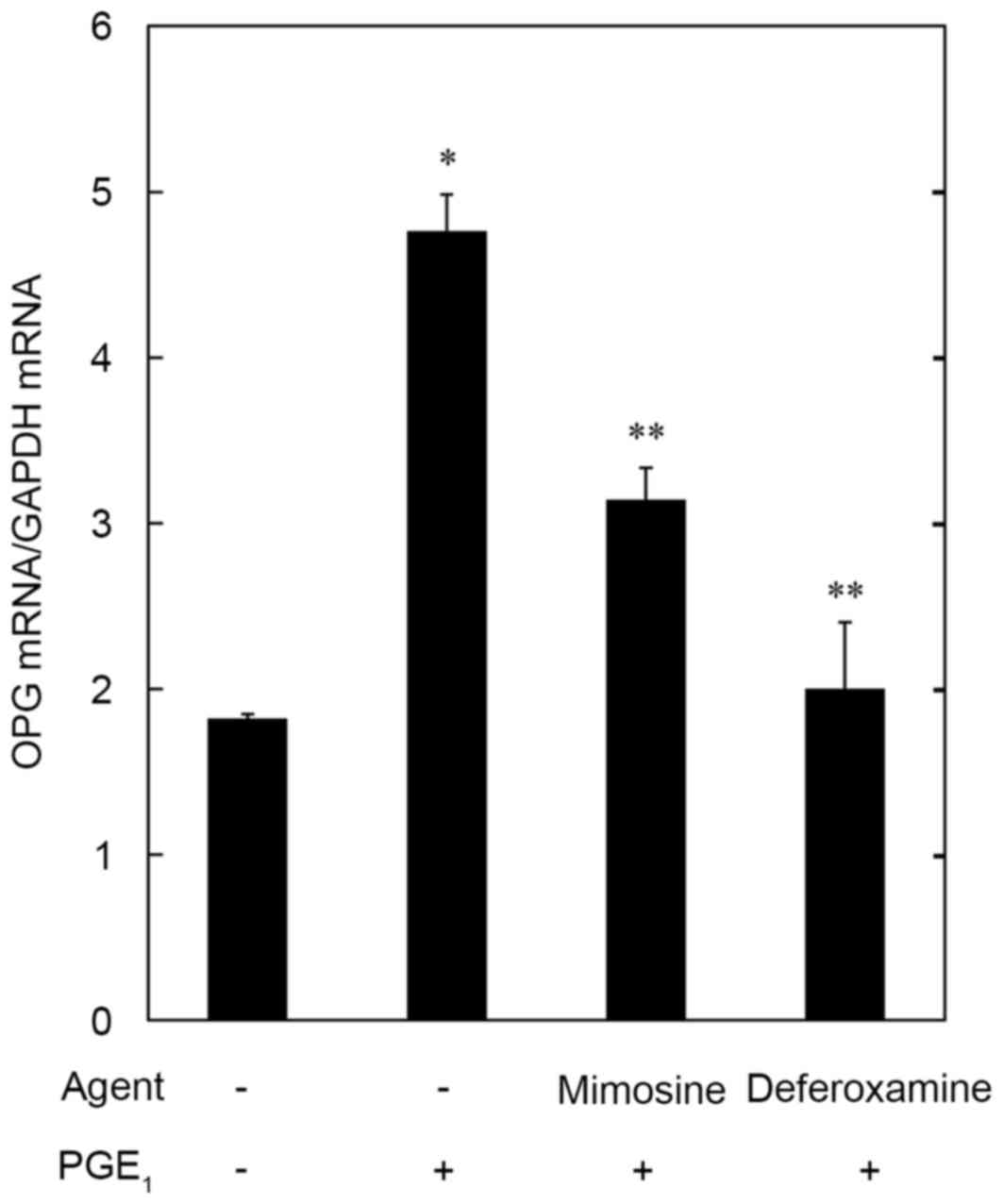
Effects of mimosine and deferoxamine
on PGE1-induced OPG mRNA expression levels in MC3T3-E1
cells
To clarify whether the suppressive effects of
mimosine and deferoxamine on PGE1-stimulated OPG release
were mediated via transcriptional events, the effects of mimosine
and deferoxamine on PGE1-induced mRNA expression levels
of OPG were examined using semi-quantitative RT-PCR. OPG mRNA
expression levels were downregulated by 700 µM mimosine and 500 µM
deferoxamine compared with in untreated cells (P=0.01 and P=0.004,
respectively; Fig. 4).
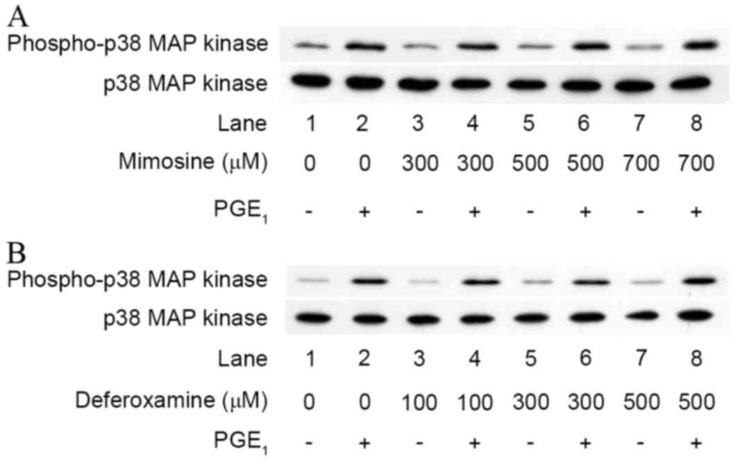
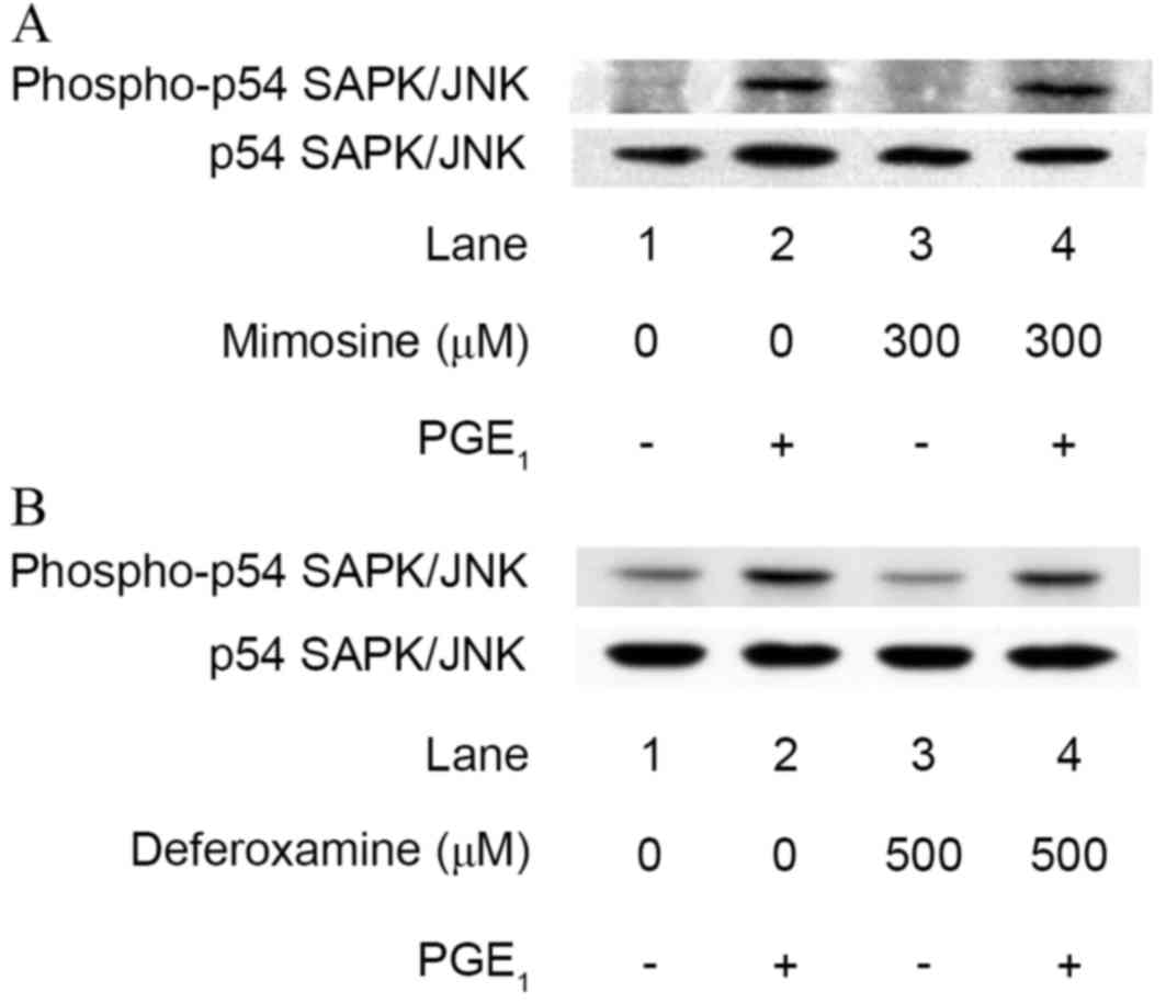
Effects of mimosine and deferoxamine
on PGE1-induced phosphorylation of p38 MAP kinase or
SAPK/JNK in MC3T3-E1 cells
A previous study demonstrated that PGE1
stimulates OPG synthesis via the activation of p38 MAP kinase and
SAPK/JNK in osteoblast-like MC3T3-E1 cells (9). In order to clarify whether the
inhibitory effects of mimosine and deferoxamine on
PGE1-stimulated OPG synthesis are exerted through the
modulation of p38 MAP kinase or SAPK/JNK, the effects of mimosine
and deferoxamine on PGE1-induced phosphorylation of p38
MAP kinase or SAPK/JNK were examined in MC3T3-E1 cells. Mimosine
failed to visibly affect PGE1-induced phosphorylation of
either p38 MAP kinase or SAPK/JNK (Figs. 5 and 6). Deferoxamine also demonstrated little
effect on the phosphorylation of p38 MAP kinase or SAPK/JNK
stimulated by PGE1 (Figs.
5 and 6).
Discussion
The present study demonstrated that
PGE1-induced OPG synthesis was suppressed by mimosine in
osteoblast-like MC3T3-E1 cells. Deferoxamine also reduced
PGE1-induced release of OPG in these cells. Mimosine is
recognized as an inhibitor of DNA replication, as well as prolyl
hydroxylase domain proteins, which are responsible for degrading
HIF-1α (14,25). It has been established that
deferoxamine, an iron chelator, exerts its angiogenic effects via
stimulation of the HIF-1α pathway (19), and it has previously been
demonstrated that mimosine and deferoxamine upregulate HIF-1α
protein levels in osteoblast-like MC3T3-E1 cells (18). Therefore, it is possible that the
inhibitory effects of mimosine and deferoxamine on
PGE1-induced OPG release are exerted via upregulation of
the HIF-1α-dependent pathway in MC3T3-E1 cells. In addition,
mimosine and deferoxamine significantly attenuated
PGE1-induced OPG mRNA expression levels. It seems
unlikely that the suppressive effects of mimosine or deferoxamine
on the PGE1-induced release of OPG are mediated through
a post-transcriptional regulatory event in these cells. Therefore,
these findings suggested that mimosine and deferoxamine suppress
the synthesis of OPG through upregulation of HIF-1α in response to
these agents in osteoblast-like MC3T3-E1 cells. HIF-1 consists of
HIF-1α and HIF-1β subunits (14,25),
and under normoxic conditions HIF-1α is immediately degraded by the
ubiquitin-proteasome system. Chemical hydroxylase inhibitors,
including mimosine and deferoxamine, attenuate the process of
HIF-1α degradation, resulting in stabilization (14,19,25).
Taking these findings into account, it is likely that
PGE1-induced OPG synthesis is downregulated by normoxic
HIF inducers in osteoblast-like MC3T3-E1 cells.
It has previously been demonstrated that
PGE1 stimulates the secretion of IL-6 in osteoblast-like
MC3T3-E1 cells (12). In the
present study, however, PGE1-induced release of IL-6 was
demonstrated to be unaffected by mimosine or deferoxamine in
MC3T3-E1 cells. This result indicated that the inhibitory effects
of mimosine and deferoxamine on PGE1-stimulation are OPG
synthesis-specific in osteoblast-like MC3T3-E1 cells.
The MAP kinase superfamily is involved in the
regulation of cell proliferation, differentiation and survival
(26). It is currently established
that three MAP kinases, including p38 MAP kinase, p44/p42 MAP
kinase and SAPK/JNK, are the most important elements of the
superfamily (27). With regards to
the effects of PGE1-intracellular signaling on OPG
synthesis, it has previously been reported that p38 MAP kinase and
SAPK/JNK act as positive regulators of PGE1-induced OPG
synthesis in osteoblast-like MC3T3-E1 cells, whereas p44/p42 MAP
kinase does not (9). The effects
of mimosine on PGE1-stimulated activation of p38 MAP
kinase and SAPK/JNK were investigated in these cells, and it was
revealed that PGE1-stimulated phosphorylation of p38 MAP
kinase or SAPK/JNK was unaffected by mimosine or deferoxamine.
Therefore, it seems unlikely that the suppressive effects of
mimosine and deferoxamine are mediated through modulation of MAP
kinase activity in MC3T3-E1 cells. In addition, it has previously
been reported that mimosine reduces PGF2α-stimulated
synthesis of OPG, but not IL-6, in MC3T3-E1 cells, and also fails
to affect PGF2α-stimulated activation of p38 MAP kinase,
p44/p42 MAP kinase and SAPK/JNK (18). It is generally recognized that the
intracellular signaling of PGE1 and PGF2α is
transduced through specific prostaglandin receptors: EP and FP,
respectively (28). Therefore, it
is likely that the suppressive effects of mimosine on
PGE1- or PGF2α-induced OPG synthesis are
exerted at the point between the action of MAP kinases and gene
transcription in osteoblast-like MC3T3-E1 cells. Further
investigations are required to elucidate the exact mechanism
underlying the inhibitory effects of normoxic HIF inducers on OPG
synthesis in osteoblasts.
It is well established that RANKL-mediated
osteoclastic bone resorption is the initial step of bone remodeling
(1). OPG, which is secreted by
osteoblasts, functions as a decoy receptor for RANKL and results in
the regulation of bone remodeling (3). Therefore, reduction of OPG secretion
may induce acceleration of bone resorption through upregulation of
osteoclastic bone resorption. Correct bone remodeling is essential
to ensure the removal of old, fragile bone and the renewal of the
skeleton, maintaining skeletal quality and quantity. It has
previously been demonstrated that mimosine induces the synthesis of
VEGF, a HIF-1 target gene considered to promote bone formation by
stimulating the generation of microvasculature (29) in osteoblast-like MC3T3-E1 cells
(18). Taking these findings into
account, the results of the present study, which demonstrated the
inhibitory effects of mimosine and deferoxamine on
PGE1-stimulated OPG synthesis in osteoblasts, may
provide novel insights into the hypoxic signaling pathway in bone
metabolism. However, further investigation is required to
understand the effects of hypoxic conditions on bone
metabolism.
In conclusion, the findings of the present study
strongly suggested that normoxic HIF inducers attenuate
PGE1-stimulated OPG synthesis without affecting IL-6
production in osteoblasts, providing novel insight into the
regulatory mechanisms underlying bone metabolism.
Acknowledgements
The authors are grateful to Mrs. Yumiko Kurokawa
(Gifu University Graduate School of Medicine, Gifu, Japan) for her
skillful technical assistance. This investigation was supported in
part by a Grant-in-Aid for Scientific Research (grant no. 19591042)
from the Ministry of Education, Culture, Sports, Science and
Technology of Japan, a Grant-in-Aid for Scientific Research
(H25-Aging-General-004) from the Ministry of Health, Labour and
Welfare of Japan, and the Research Funding for Longevity Sciences
(25–4,26–12)
from National Center for Geriatrics and Gerontology (NCGG),
Japan.
References
|
1
|
Karsenty G and Wagner EF: Reaching a
genetic and molecular understanding of skeletal development. Dev
Cell. 2:389–406. 2002. View Article : Google Scholar : PubMed/NCBI
|
|
2
|
Parfitt AM: Targeted and nontargeted bone
remodeling: Relationship to basic multicellular unit origination
and progression. Bone. 30:5–7. 2002. View Article : Google Scholar : PubMed/NCBI
|
|
3
|
Simonet WS, Lacey DL, Dunstan CR, Kelley
M, Chang MS, Lüthy R, Nguyen HQ, Wooden S, Bennett L, Boone T, et
al: Osteoprotegerin: A novel secreted protein involved in the
regulation of bone density. Cell. 89:309–319. 1997. View Article : Google Scholar : PubMed/NCBI
|
|
4
|
Mizuno A, Amizuka N, Irie K, Murakami A,
Fujise N, Kanno T, Sato Y, Nakagawa N, Yasuda H, Mochizuki S, et
al: Severe osteoporosis in mice lacking osteoclastogenesis
inhibitory factor/osteoprotegerin. Biochem Biophys Res Commun.
247:610–615. 1998. View Article : Google Scholar : PubMed/NCBI
|
|
5
|
Tat S Kwan, Padrines M, Théoleyre S,
Heymann D and Fortun Y: IL-6, RANKL, TNF-α/IL-1: Interrelations in
bone resorption pathophysiology. Cytokine Growth Factor Rev.
15:49–60. 2004. View Article : Google Scholar : PubMed/NCBI
|
|
6
|
Hikiji H, Takato T, Shimizu T and Ishii S:
The roles of prostanoids, leukotrienes, and platelet-activating
factor in bone metabolism and disease. Prog Lipid Res. 47:107–126.
2008. View Article : Google Scholar : PubMed/NCBI
|
|
7
|
Blackwell KA, Raisz LG and Pilbeam CC:
Prostaglandins in bone: Bad cop, good cop? Trends Endocrinol Metab.
21:294–301. 2010. View Article : Google Scholar : PubMed/NCBI
|
|
8
|
Agas D, Marchetti L, Hurley MM and
Sabbieti MG: Prostaglandin F2α: A bone remodeling mediator. J Cell
Physiol. 228:25–29. 2013. View Article : Google Scholar : PubMed/NCBI
|
|
9
|
Yamamoto N, Otsuka T, Kuroyanagi G, Kondo
A, Kainuma S, Nakakami A, Matsushima-Nishiwaki R, Kozawa O and
Tokuda H: Resveratrol reduces prostaglandin E1-stimulated
osteoprotegerin synthesis in osteoblasts: Suppression of
stress-activated protein kinase/c-Jun N-terminal kinase.
Prostaglandins Other Lipid Mediat. 116–117, 57–63. 2015.
|
|
10
|
Hirano T: Revisiting the 1986 molecular
cloning of interleukin 6. Front Immunol. 23:4562014.
|
|
11
|
Franchimont N, Wertz S and Malaise M:
Interleukin-6: An osteotropic factor influencing bone formation?
Bone. 37:601–606. 2005. View Article : Google Scholar : PubMed/NCBI
|
|
12
|
Watanabe-Tomita Y, Suzuki A, Oiso Y and
Kozawa O: Prostaglandin E1 stimulates interleukin-6 secretion via
protein kinase A in osteoblast-like cells. Cell Signal. 9:105–108.
1997. View Article : Google Scholar : PubMed/NCBI
|
|
13
|
Warnecke C, Griethe W, Weidemann A,
Jürgensen JS, Willam C, Bachmann S, Ivashchenko Y, Wagner I, Frei
U, Wiesener M and Eckardt KU: Activation of the hypoxia-inducible
factor-pathway and stimulation of angiogenesis by application of
prolyl hydroxylase inhibitors. FASEB J. 17:1186–1188.
2003.PubMed/NCBI
|
|
14
|
Schofield CJ and Ratcliffe PJ: Oxygen
sensing by HIF hydroxylases. Nat Rev Mol Cell Biol. 5:343–354.
2004. View
Article : Google Scholar : PubMed/NCBI
|
|
15
|
Knowles HJ, Cleton-Jansen AM, Korsching E
and Athanasou NA: Hypoxia-inducible factor regulates
osteoclast-mediated bone resorption: Role of angiopoietin-like 4.
FASEB J. 24:4648–4659. 2010. View Article : Google Scholar : PubMed/NCBI
|
|
16
|
Wang Y, Wan C, Deng L, Liu X, Cao X,
Gilbert SR, Bouxsein ML, Faugere MC, Guldberg RE, Gerstenfeld LC,
et al: The hypoxia-inducible factor alpha pathway couples
angiogenesis to osteogenesis during skeletal development. J Clin
Invest. 117:1616–1626. 2007. View
Article : Google Scholar : PubMed/NCBI
|
|
17
|
Wan C, Gilbert SR, Wang Y, Cao X, Shen X,
Ramaswamy G, Jacobsen KA, Alaql ZS, Eberhardt AW, Gerstenfeld LC,
et al: Activation of the hypoxia-inducible factor-1α pathway
accelerates bone regeneration. Proc Natl Acad Sci USA. 105:686–691.
2008. View Article : Google Scholar : PubMed/NCBI
|
|
18
|
Kuroyanagi G, Otsuka T, Yamamoto N,
Kainuma S, Ohguchi R, Fujita K, Matsushima-Nishiwaki R, Kozawa O
and Tokuda H: Mimosine suppresses the PGF2α-induced synthesis of
osteoprotegerin but not interleukin-6 in osteoblasts. Int J Mol
Med. 37:533–541. 2016.PubMed/NCBI
|
|
19
|
Donneys A, Deshpande SS, Tchanque-Fossuo
CN, Johnson KL, Blough JT, Perosky JE, Kozloff KM, Felice PA,
Nelson NS, Farberg AS, et al: Deferoxamine expedites consolidation
during mandibular distraction osteogenesis. Bone. 55:384–390. 2013.
View Article : Google Scholar : PubMed/NCBI
|
|
20
|
Sudo H, Kodama HA, Amagai Y, Yamamoto S
and Kasai S: In vitro differentiation and calcification in a new
clonal osteogenic cell line derived from newborn mouse calvaria. J
Cell Biol. 96:191–198. 1983. View Article : Google Scholar : PubMed/NCBI
|
|
21
|
Kozawa O, Tokuda H, Miwa M, Kotoyori J and
Oiso Y: Cross-talk regulation between cyclic AMP production and
phosphoinositide hydrolysis induced by prostaglandin E2 in
osteoblast-like cells. Exp Cell Res. 198:130–134. 1992. View Article : Google Scholar : PubMed/NCBI
|
|
22
|
Simpson DA, Feeney S, Boyle C and Stitt
AW: Retinal VEGF mRNA measured by SYBR green I fluorescence: A
versatile approach to quantitative PCR. Mol Vis. 6:178–183.
2000.PubMed/NCBI
|
|
23
|
Laemmli UK: Cleavage of structural
proteins during the assembly of the head of bacteriophage T4.
Nature. 227:680–685. 1970. View
Article : Google Scholar : PubMed/NCBI
|
|
24
|
Kato K, Ito H, Hasegawa K, Inaguma Y,
Kozawa O and Asano T: Modulation of the stress-induced synthesis of
hsp27 and alphaB-crystallin by cyclic AMP in C6 rat glioma cells. J
Neurochem. 66:946–950. 1996. View Article : Google Scholar : PubMed/NCBI
|
|
25
|
Shen X, Wan C, Ramaswamy G, Mavalli M,
Wang Y, Duvall CL, Deng LF, Guldberg RE, Eberhart A, Clemens TL and
Gilbert SR: Prolyl hydroxylase inhibitors increase neoangiogenesis
and callus formation following femur fracture in mice. J Orthop
Res. 27:1298–1305. 2009. View Article : Google Scholar : PubMed/NCBI
|
|
26
|
Kyriakis JM and Avruch J: Mammalian
mitogen-activated protein kinase signal transduction pathways
activated by stress and inflammation. Physiol Rev. 81:807–869.
2001.PubMed/NCBI
|
|
27
|
Widmann C, Gibson S, Jarpe MB and Johnson
GL: Mitogen-activated protein kinase: Conservation of a
three-kinase module from yeast to human. Physiol Rev. 79:143–180.
1999.PubMed/NCBI
|
|
28
|
Narumiya S, Sugimoto Y and Ushikubi F:
Prostanoid receptors: Structures, properties, and functions.
Physiol Rev. 79:1193–1226. 1999.PubMed/NCBI
|
|
29
|
Zelzer E and Olsen BR: Multiple roles of
vascular endothelial growth factor (VEGF) in skeletal development,
growth, and repair. Curr Top Dev Biol. 65:169–187. 2005. View Article : Google Scholar : PubMed/NCBI
|