Introduction
Intervertebral disc (ID) herniation is a common, yet
poorly understood disorder. Behavioral, hereditary and
environmental factors cumulatively affect the nutrition and
homeostasis of the disc, ultimately resulting in its structural
breakdown and in the occurrence of herniation. The incidence of
symptomatic lumbar ID herniation in the United States is estimated
at 1–2%, for which approximately 200,000 discectomies are performed
annually (1). The direct costs for
conservative or surgical treatment and he indirect costs deriving
from the loss of productivity are enormous (2).
However, ID herniation is considered to present a
favorable natural history. Sequential magnetic resonance imaging
(MRI) studies have documented that the majority of disc herniations
diminish in size (3). Saal et
al reported that almost 80% of disc herniations treated
non-operatively decreased by >50% in follow-up examinations
(4). In addition, a positive
correlation has been found between the regression of disc
herniation and the remission of symptoms (3,4).
Various molecules, such as pro-inflammatory cytokines, growth
factors (GFs) and matrix metalloproteinases (MMPs) have been
implicated in the mechanisms that regulate ID degradation and
herniation, as well as the spontaneous resorption of herniated disc
fragments (5).
GFs are polypeptides involved in the modulation of
cell growth and differentiation that exert their biologic effects
through endocrine, paracrine and autocrine pathways (6). Over 18 different GFs have been
identified, including vascular endothelial growth factor (VEGF),
transforming growth factor β1 (TGF-β1), basic fibroblast growth
factor (bFGF)-2, platelet derived growth factor (PDGF), epidermal
growth factor (EGF) and insulin growth factor-1 (IGF-1). The PDGF
family in particular is formed by the PDGF-A, -B, -C and -D
isoforms that are encoded by four genes located at different
chromosomes and may form homodimers, as well as heterodimers. These
molecules signal through two cell-surface receptors, PDGF-Rα and β
(7).
To date, the expression of various GFs in the
degenerate and herniated ID has been investigated in numerous
studies. The increased protein expression of IGF-1, FGF-2, VEGF,
PDGF and TGF-β has been found by means of immunohistochemistry
(8–11). The transcript levels of TGF-β and
VEGF have also been examined in human ID herniation specimens
(12). Consequently, preliminary
studies have examined the effect of administering different GFs on
ID physiology and degeneration in vitro and in vivo
(13,14). However, a simultaneous assessment
of the molecular profile of numerous GFs in human herniated IDs has
not been performed to date, at least to the best of our knowledge.
In addition, clinical and epidemiological determinants, such as the
extent of disc herniation, the duration and intensity of symptoms,
age and employment may considerably affect the pathophysiology of
ID degeneration and the potential for regression of herniated ID
fragments. The association between the clinical, epidemiological
and molecular profile of ID herniation would potentially provide
valuable insight into the biology and clinical course of the
disorder. Therefore, the purpose of the present study was to
simultaneously examine the molecular profiles of numerous GFs by
means of reverse transcriprtion-quantitative polymerase chain
reaction (RT-qPCR), and to investigate potential correlations with
the clinical and epidemiological characteristics of patients
suffering from lumbar ID herniation.
Materials and methods
Tissue samples
ID tissue samples were collected from a total of 63
patients who underwent posterior open discectomy for lumbar ID
herniation in our department during a recruitment period of one
year (2008). The patients reported symptoms of radiculopathy prior
to surgery. Patients suffering from spinal stenosis,
spondylolisthesis, scoliosis or systemic inflammatory disorders
were excluded from the study. After excision, an experienced
pathologist, in addition to the clinicians present, examined the
tissue samples and identified the pulposus sections in order to
limit the percentage of end-plates or annulus. Samples were then
immediately refrigerated at −80°C until RNA extraction. Ten
cadaveric ID tissue samples were also obtained within 12 h of the
patient succumbing to the disease, referring to various diseases
other than ID herniation. The samples were used as controls of
normal growth factor mRNA expression. Donors were aged between 20
and 46 years and were known to have a negative history of lower
back pain, spinal trauma or systemic inflammatory disease. All the
patients or relatives signed an informed consent form approved by
the University Ethics Committee to participate in the present
study. The study design was in accordance with the Declaration of
Helsinki Guidelines.
Prior to surgery, the patients routinely underwent
an MRI examination for the classification of ID herniation. The
recommendations of the combined task force from the North American
Spine Society, the American Society of Spine Radiology and the
American Society of Neuroradiology based on the imaging
characterization of Milette (15)
were used to determine the type of ID herniation. Pre-operative
pain intensity was measured subjectively using the visual analogue
scale score (VAS). Patient employment was defined as heavy if it
included manual labour with repetitive loading of the spine. A
summary of patient and control group baseline data is presented in
Table I. There were no significant
differences in terms of age, gender, body mass index (BMI),
employment and smoking habits between the control and ID herniation
groups (minimum P=0.24).
 | Table I.Patient clinical characteristics. |
Table I.
Patient clinical characteristics.
| Characteristic | No. of patients |
|---|
| ID herniation group
Age (years) |
| Mean
(range) | 47.3 (27–68) |
|
20–39 | 27 |
|
40–59 | 23 |
|
>60 | 12 |
| Gender |
|
|
Male/female | 33/30 |
| Body mass index
(BMI) |
|
| Mean
(range) | 27.2
(21.07–33.05) |
| Level of ID
herniation |
|
L3-L4 | 11 |
|
L4-L5 | 24 |
|
L5-S1 | 28 |
| Type of
herniation |
|
Protrusion | 23 |
|
Extrusion | 33 |
|
Sequestration | 7 |
| Duration of
symptoms |
| <3
months | 15 |
| 3–12
months | 20 |
| >12
months | 28 |
| Pain intensity
(VAS) |
|
0–5 | 18 |
|
5–7 | 14 |
|
8–10 | 31 |
| Employment |
|
Heavy/light | 36/27 |
| Smoking Habits |
|
Smokers/non-smokers | 26/37 |
| <10
cig per day | 6 |
| 10- 20
cig per day | 8 |
| >20
cig per day | 12 |
| Control group Age
(years) |
| Mean
(range) | 36.8 (20–46) |
| Gender |
|
Male/female | 6/4 |
| Body mass index
(BMI) |
| Mean
(range) | 27.0
(21.9–33.0) |
| Employment |
|
Heavy/light | 7/3 |
| Smoking Habits |
|
Smokers/non-smokers | 4/6 |
RNA extraction and RT-qPCR
Total RNA was isolated from fresh tissue and
homogenized with a power homogenizer using TRIzol reagent
(Invitrogen, Carlsbad, CA, USA). RNA concentration and purity were
determined on a UV spectrophotometer (Hitachi Instruments Inc., San
Jose, CA, USA) by absorbance measurements (260 nm absorbance and
260/280 nm absorbance ratio). RNA integrity was examined by 1%
agarose gel electrophoresis and ethidium bromide staining.
Reverse transcription reactions for the preparation
of first-strand cDNA from 2 µg of total RNA were performed using
the AffinityScript™ Multi Temperature cDNA synthesis kit
(Stratagene, La Jolla, CA, USA). Random hexamers were used as
amplification primers. Quantitative PCR (qPCR) reactions were
performed using the Mx3000P real-time PCR system with
SYBR®-Green I Master Mix (both from Stratagene). Data
were collected and analyzed using the Mx3000P real-time PCR
software version 2.00, Build 215 Schema 60 (Stratagene).
Glyceraldehyde-3-phosphate dehydrogenase (GAPDH) was used as an
internal control to normalize the mRNA expression levels of the GFs
examined. The primer pair sequences used are listed in Table II. PCR products were analyzed by
electrophoresis on 2% agarose gels, stained with ethidium bromide
and photographed on a UV light transilluminator. GF transcription
levels were calculated using the following formula: normalized
sample or control = (1 + EGF)−∆∆CqGF/(1 +
EGAPDH)−∆∆CqGAPDH. Procedures were repeated
with a cDNA template, synthesized three times from the same RNA.
The mRNA levels of each sample for each gene tested represent the
mean value of data acquired from three independent RT-qPCR
experiments. The reproducibility of the RT-qPCR results for the
same samples was 99%.
| Table II.Sequences of primers used for
RT-qPCR. |
Table II.
Sequences of primers used for
RT-qPCR.
| Gene | Primer pair
sequence (5′→3′) | Annealing
temperature (°C) | Product size
(bp) |
|---|
| IGF-1 |
CCTCCTCGCATCTCTTCTACCTGC | 60 | 166 |
|
|
TGCTGGAGCCATACCCTGTG |
|
|
| FGF-2 |
CTGGCTATGAAGGAAGATGGA | 55 | 149 |
|
|
TGCCCAGTTCGTTTCAGTG |
|
|
| TGF-Β |
AAGGACCTCGGCTGGAAGTGC | 62 | 137 |
|
|
CCGGGTTATGCTGGTTGTA |
|
|
| VEGF |
ATGACGAGGGCCTGGAGTGTG | 60 | 91 |
|
|
CCTATGTGCTGGCCTTGGTGAG |
|
|
| PDGF-A |
ACACGAGCAGTGTCAAGTGC | 55 | 76 |
|
|
CCTGACGTATTCCACCTTGG |
|
|
| PDGF-B |
AGATCGAGATTGTGCGGAAG | 52 | 94 |
|
|
CAGCTGCCACTGTCTCACAC |
|
|
| PDGF-C |
GCCAGGTTGTCTCCTGGTTA | 52 | 86 |
|
|
TGCTTGGGACACATTGACAT |
|
|
| PDGF-D |
CCCAGGAATTACTCGGTCAA | 52 | 105 |
|
|
ACAGCCACAATTTCCTCCAC |
|
|
| PDGF rec-A |
TGGGAGTTTCCAAGAGATGG | 52 | 78 |
|
|
TGTTCCTTCAACCACCTTCC |
|
|
| PDGF rec-B |
GTGCTCACCATCATCTCCCT | 52 | 85 |
|
|
ACTCAATCACCTTCCATCGG |
|
|
| GAPDH |
GGAAGGTGAAGGTCGGAGTCA | 60 | 101 |
|
|
GTCATTGATGGCAACAATATCCACT |
|
|
Statistical analysis
The one-sample Kolmogorov-Smirnov test was used to
assess the normality of the distribution of the mRNA expression
values for the genes studied. Accordingly, the mRNA expression
levels of the GFs examined in the control and herniated groups were
compared using parametric and non-parametric procedures,
respectively. Differences in GF expression between herniated and
control IDs and between subgroups of different clinical
characteristics were examined using parametric (one-way and
repeated measures ANOVA) and non-parametric (Kruskal-Wallis and
Mann-Whitney) tests accordingly. Pairwise comparisons were
performed to further explore the interaction. Categorical variables
were compared with the use of χ2 test. The Spearman rank
correlation test (non-parametric) was also employed to examine
pair-wise correlations in growth factor mRNA expression in control
and herniated discs. The significance level was set at P<0.05.
Statistical calculations were performed using SPSS software,
version 15 (SPSS, Inc., Chicago, IL, USA).
Results
GF mRNA levels in normal and herniated
discs
The transcript levels of the GFs examined were not
significant between the control and ID herniation groups (minimum
P=0.26, Mann-Whitney test, Table
III). With the exception of EGF that was not expressed in
neither of the 2 groups, all the GFs examined were expressed in the
two study groups. Specifically, the mRNA expression status
(expressed or not) of the genes examined ranged from 40 to 100% in
the controls and 35 to 100% in the ID herniation groups (Table III). PDGF-C and -D mRNA
expression was observed in all the herniated IDs, while IGF was the
least commonly expressed in the same group (34.9%).
| Table III.Growth factor mRNA expression. |
Table III.
Growth factor mRNA expression.
| Expressed Gene | Control discs
(n=10) | Herniated discs
(n=63) |
P-valuea | Control discs
(n=10) | Herniated discs (%)
(n=63) |
P-valueb |
|---|
| IGF | 1.2637±0.961 |
11256.0652±1.12 | NS | 5/10 | 22/63 (34.9) | NS |
| FGF | 0.0431±0.032 | 46.0385±38.749 | NS | 4/10 | 30/63 (47.6) | NS |
| TGF | 1.1329±0.616 | 36.8798±13.531 | NS | 9/10 | 56/63 (88.8) | NS |
| VEGF | 0.0003±0.001 | 9.9859±9.7546 | NS | 5/10 | 33/63 (52.3) | NS |
| PDGF-A | 0.6554±0.613 | 24.7222±10.603 | NS | 4/10 | 31/63 (49.2) | NS |
| PDGF-B | 4.5424±2.395 | 65.3348±52.903 | NS | 10/10 | 62/63 (98.4) | NS |
| PDGF-C | 1.7382±0.427 |
121.2165±68.593 | NS | 10/10 | 63/63 (100) | NS |
| PDGF-D | 3.2749±0.969 |
153.5597±97.695 | NS | 10/10 | 63/63 (100) | NS |
| PDGF rec-A | 0.1241±0.052 | 25.8371±13.227 | NS | 6/10 | 32/63 (50.7) | NS |
| PDGF rec-B | 0.0823±0.053 | 12.5163±5.361 | NS | 4/10 | 29/63 (46) | NS |
Pair-wise GF mRNA co-expression
analysis
Pair-wise correlations between the genes examined in
the ID herniation groups and the controls are presented in Tables IV and V, respectively (Spearman correlations).
Compared with the controls, an increased number of positive
correlations was observed in the ID herniation group between most
of the genes examined.
| Table IV.Spearman correlation tables
demonstrating the co-expression profile of GFs in the group of
herniated lumbar discs. |
Table IV.
Spearman correlation tables
demonstrating the co-expression profile of GFs in the group of
herniated lumbar discs.
| Factor | Method |
bFGF | IGF | PDGF-A | PDGF-B | PDGF-C | PDGF-D | PDGFRA | PDGFRB | TGF-b | VEGF |
|---|
| bFGF | Spearman's rho | 1.000 |
|
|
|
|
|
|
|
|
|
|
| Sig. 2-tailed | . |
|
|
|
|
|
|
|
|
|
| IGF | Spearman's rho | 0.644 | 1.000 |
|
|
|
|
|
|
|
|
|
| Sig. 2-tailed | 0.000 | . |
|
|
|
|
|
|
|
|
| PDGF-A | Spearman's rho | 0.136 | 0.181 | 1.000 |
|
|
|
|
|
|
|
|
| Sig. 2-tailed | 0.294 | 0.159 | . |
|
|
|
|
|
|
|
| PDGF-B | Spearman's rho | −0.175 | −0.132 | 0.038 | 1.000 |
|
|
|
|
|
|
|
| Sig. 2-tailed | 0.171 | 0.301 | 0.767 | . |
|
|
|
|
|
|
| PDGF-C | Spearman's rho | −0.119 | −0.050 | −0.101 | 0.546 | 1.000 |
|
|
|
|
|
|
| Sig. 2-tailed | 0.354 | 0.700 | 0.436 | 0.000 | . |
|
|
|
|
|
| PDGF-D | Spearman's rho | −0.151 | −0.069 | −0.091 | 0.536 | 0.728 | 1.000 |
|
|
|
|
|
| Sig. 2-tailed | 0.239 | 0.590 | 0.480 | 0.000 | 0.000 | . |
|
|
|
|
| PDGFRA | Spearman's rho | 0.744 | 0.572 | 0.155 | −0.031 | 0.002 | −0.082 | 1.000 |
|
|
|
|
| Sig. 2-tailed | 0.000 | 0.000 | 0.229 | 0.807 | 0.987 | 0.524 | . |
|
|
|
| PDFGRB | Spearman's rho | 0.845 | 0.654 | 0.212 | −0.095 | −0.057 | −0.199 | 0.822 | 1.000 |
|
|
|
| Sig. 2-tailed | 0.000 | 0.000 | 0.098 | 0.457 | 0.655 | 0.118 | 0.000 | . |
|
|
| TGF-b | Spearman's rho | 0.376 | 0.325 | 0.164 | 0.060 | 0.201 | 0.168 | 0.287 | 0.365 | 1.000 |
|
|
| Sig. 2-tailed | 0.002 | 0.009 | 0.202 | 0.638 | 0.113 | 0.188 | 0.022 | 0.003 | . |
|
| VEGF | Spearman's rho | 0.886 | 0.557 | 0.077 | −0.263 | 0.005 | −0.054 | 0.680 | 0.770 | 0.477 | 1.000 |
|
| Sig. 2-tailed | 0.000 | 0.000 | 0.552 | 0.039 | 0.967 | 0.676 | 0.000 | 0.000 | 0.000 | . |
| Table V.Spearman correlation tables
demonstrating the co- expression profile of GFs in the group of
control lumbar discs. |
Table V.
Spearman correlation tables
demonstrating the co- expression profile of GFs in the group of
control lumbar discs.
| Factor | Method | bFGF | IGF | PDGF-A | PDGF-B | PDGF-C | PDGF-D | PDGFRA | PDGFRB | VEGF | TGF-b |
|---|
| bFGF | Spearman's rho | 1.000 |
|
|
|
|
|
|
|
|
|
|
| Sig. 2-tailed | . |
|
|
|
|
|
|
|
|
|
| IGF | Spearman's rho | −0.076 | 1.000 |
|
|
|
|
|
|
|
|
|
| Sig. 2-tailed | 0.886 | . |
|
|
|
|
|
|
|
|
| PDGF-A | Spearman's rho | −0.271 | 0.981 | 1.000 |
|
|
|
|
|
|
|
|
| Sig. 2-tailed | 0.604 | 0.001 | . |
|
|
|
|
|
|
|
| PDGF-B | Spearman's rho | −0.475 | 0.388 | 0.469 | 1.000 |
|
|
|
|
|
|
|
| Sig. 2-tailed | 0.341 | 0.447 | 0.348 | . |
|
|
|
|
|
|
| PDGF-C | Spearman's rho | −0.636 | 0.644 | 0.749 | 0.632 | 1.000 |
|
|
|
|
|
|
| Sig. 2-tailed | 0.175 | 0.168 | 0.087 | 0.178 | . |
|
|
|
|
|
| PDGF-D | Spearman's rho | −0.86 | 0.007 | 0.178 | 0.631 | 0.745 | 1.000 |
|
|
|
|
|
| Sig. 2-tailed | 0.027 | 0.989 | 0.735 | 0.179 | 0.089 | . |
|
|
|
|
| PDGFRA | Spearman's rho | 0.378 | −0.422 | −0.482 | −0.677 | −0.394 | −0.310 | 1.000 |
|
|
|
|
| Sig. 2-tailed | 0.460 | 0.405 | 0.333 | 0.140 | 0.440 | 0.550 | . |
|
|
|
| PDFGRB | Spearman's rho | 0.338 | −0.252 | −0.308 | −0.422 | −0.057 | −0.065 | 0.884 | 1.000 |
|
|
|
| Sig. 2-tailed | 0.512 | 0.630 | 0.553 | 0.405 | 0.914 | 0.903 | 0.020 | . |
|
|
| VEGF | Spearman's rho | 0.096 | −0.360 | −0.367 | −0.462 | −0.214 | −0.075 | 0.935 | 0.809 | 1.000 |
|
|
| Sig. 2-tailed | 0.857 | 0.483 | 0.474 | 0.356 | 0.683 | 0.887 | 0.006 | 0.051 | . |
|
| TGF-b | Spearman's rho | −0.238 | 0.968 | 0.982 | 0.580 | 0.789 | 0.237 | −0.504 | −0.263 | −0.392 | 1.000 |
|
| Sig. 2-tailed | 0.650 | 0.002 | 0.000 | 0.227 | 0.062 | 0.652 | 0.307 | 0.614 | 0.442 | . |
Correlation of transcript levels with
clinicopathological characteristics
Age and gender
Age did not significantly affect the mRNA expression
levels of the genes examined (minimum P=0.07, Kruskal-Wallis test).
Male patients exhibited increased mRNA expression levels of PDGF-C
and -D compared with female patients (P=0.007 and 0.024,
respectively, Mann-Whitney test) (absolute values not shown).
BMI
Significant negative correlations were observed
between the patient BMI values and the transcript levels of VEGF,
PDGFRA and PDGFRB (P=0.036, 0.037 and 0.02, respectively, Spearman
correlation).
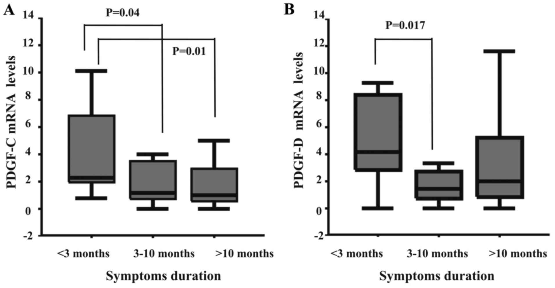
Duration of symptoms
The transcript levels of PDGF-C and -D were
significantly affected by the duration of patient symptoms (P=0.02
and 0.05, respectively, Kruskal-Wallis test). Specifically,
patients suffering from acute pain (<3 months) exhibited an
increased mRNA expression of PDGF-C compared with those suffering
for 3–10 months and for >10 months (P=0.04 and 0.01,
respectively, Mann-Whitney test). Similarly, the PDGF-D expression
levels were also significantly higher in patients suffering from
acute (<3 months) compared with chronic pain (P=0.017,
Mann-Whitney test, Fig. 1).
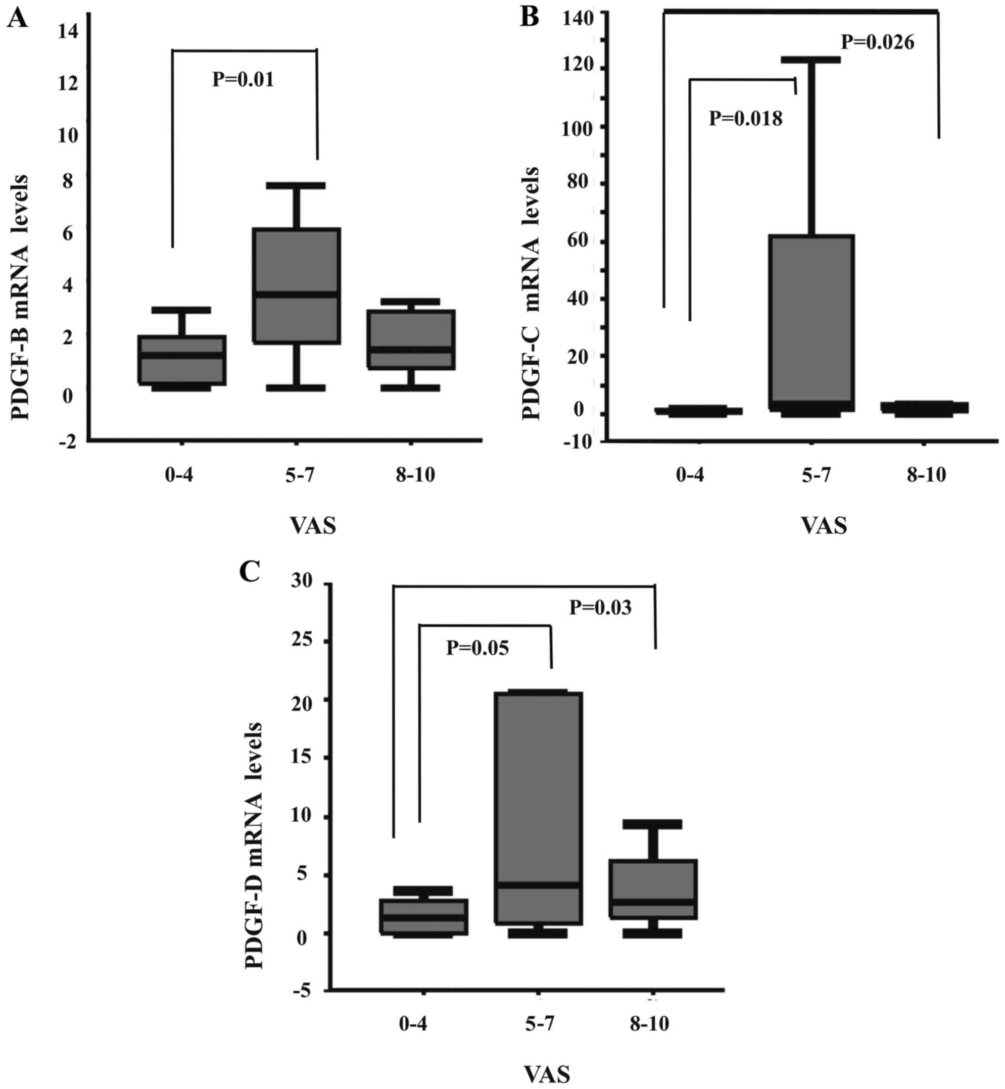
Pain intensity
The intensity of patient pain significantly affected
the expression levels of PDGF-B, -C and -D (P=0.044, 0.024 and
0.05, Kruskal-Wallis test). Specifically, patients experiencing
moderate pain (VAS 5–7) exhibited an increased mRNA expression of
PDGF-B, -C and -D compared with those with low intensity pain (VAS
0–4) (P=0.01, 0.018 and 0.05, respectively, Mann-Whitney test). In
addition, patients with intense pain (VAS 8–10) also exhibited a
significantly increased expression of PDGF-C and -D compared with
those with low intensity pain (P=0.026 and 0.03, respectively,
Mann-Whitney test, Fig. 2).
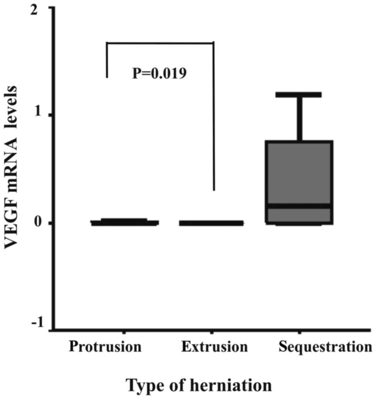
Type of ID herniation
The transcript levels of VEGF significantly
correlated with the type of ID herniation (P=0.023, Kruskal-Wallis
test). Specifically, ID herniation samples with protrusion
exhibited an increased mRNA expression of VEGF compared with the
extruded IDs (P=0.019, Mann-Whitney test, Fig. 3).
Employment
The transcript levels of the genes examined did not
differ significantly between patients with either light or heavy
employment (minimum P=0.15, Mann-Whitney test) (absolute values not
shown).
Smoking
The mRNA expression levels of the GFs examined did
not differ significantly between smoking and non-smoking patients
(minimum P=0.087, Mann-Whitney test). Accordingly, the amount of
cigarette smoking did not significantly affect the transcript
levels of any of the genes studied (minimum P=0.29, Kruskal-Wallis
test) (absolute values not shown).
Discussion
The current study examined the molecular profiles of
numerous GFs by means of RT-qPCR in surgically resected lumbar ID
herniation specimens and matched controls. Correlations of GF
transcript levels with the patient clinical and epidemiological
characteristics were also explored. Multiple positive correlations
between the GFs were identified in the group of herniated discs.
The VEGF transcript levels were significantly higher in protruding
compared with the extruded discs. The duration and intensity of
patient symptoms significantly affected the PDGF transcript levels.
BMI exhibited significant negative correlations with the transcript
levels of VEGF and both PDGF-receptor isotypes (data not
shown).
There have been limited reports in the literature
examining the molecular profiles of GFs in ID herniation. In lumbar
herniated IDs, Ahn et al detected TGF-β mRNA expression in 5
out of 10 specimens examined (16), whereas Kontinnen et al found
a decreased mRNA expression of the same factor (12). Control groups were not included in
these studies. Kontinnen et al also failed to identify the
mRNA or protein expression of EGF, which they attributed to the
reduced anabolic potential of the disc. The findings of the present
study did not identify any differences in the mRNA expression of
GFs in a substantial number of herniated and control ID specimens.
By contrast, immunohistochemistry, has produced more variable
results. Specchia et al reported stronger staining for IGF-1
in chondrocytes of herniated compared with control discs (10). Similarly, Tolonen et al
observed significantly stronger staining for PDGF and TGF-β in
human herniated ID tissue (8,9).
Given that no differences in GF expression are evident at the mRNA
level compared with the controls, these findings suggest that
post-transcriptional modifications may occur in the setting of ID
herniation.
The transcript levels of the GFs examined
significantly correlated with each other in herniated IDs. In
vitro experiments have demonstrated a synergistic effect of
VEGF and TGF-β1 in the induction of angiogenesis (17). In addition, PDGF has been found to
upregulate VEGF in mural cells, thereby further enhancing
angiogenesis (18). IGF-I has also
been shown to induce VEGF mRNA and protein expression in cultured
colorectal carcinoma cells (19).
Most of these GFs are also under the regulatory effect of
inflammatory cytokines, such as interleukin-1 (IL-1) and tumor
necrosis factor-α (TNF-α). Both TNF-α and IL-1 have been shown to
interact with numerous GFs in human degenerated discs and trigger a
sequence of macrophage and MMP activation that potentiates the
spontaneous resorption of herniated ID fragments (20–22).
Multiple positive correlations between numerous MMPs have also been
demonstrated in human ID herniation (5). Our findings, therefore, confirm a
synergistic activation of GFs in ID herniation as part of a
metabolic cascade that ultimately promotes the regression process
of herniated discs.
Our results demonstrated that the transcript levels
of VEGF were significantly higher in protruding compared with
extruded IDs. VEGF is a potent mitogen for vascular endothelial
cells. It induces new vessel formation through cell proliferation
and luminal formation and has also been found to increase blood
vessel permeability (23). Using
immunohistochemistry, Tolonen et al and Koike et al
confirmed the expression of VEGF in extruded and sequestrated human
IDs (9,24). A significant correlation was also
observed between the expression of VEGF and the presence of
vascular endothelial cells (24).
Protruding ID herniation samples were not included in these
studies. Ozaki et al reported the presence of small blood
vessels in 73.4% of herniated ID tissues examined
histopathologically, regardless of hernia type (25). They also suggested that
neovascularization was present in the outermost annulus layers,
even in protruding herniations. To the best of our knowledge, the
present study is the first to confirm the expression of VEGF in
protruding hernias at the mRNA level, thereby supporting the
hypothesis that VEGF upregulation plays a key role even in the
earliest stages of the neovascularization process that follows ID
herniation.
PDGF is considered a key mediator in the tissue
repair process. It stimulates mitogenicity and chemotaxis of
neutrophils and macrophages in a variety of mesenchymal-derived
cells, such as osteoblasts, chondrocytes and fibroblasts (7). It has been shown that PDGF exerts its
effect on the repair process, not by altering its normal sequence,
but by increasing its rate (26).
It has been hypothesized that PDGF induces the production of
extracellular matrix components and cellular proliferation in the
ID in a similar mechanism as in the articular cartilage (9). It has also been shown to participate
in the resorption process of herniated ID fragments by upregulating
MMP expression (22). The findings
of the present study indicated that the PDGF transcript levels were
significantly higher in patients suffering from intense and acute
pain. These findings support the role of PDGF as a key modulator of
the repair process of damaged connective tissue, and indicate that
it is primarily expressed in the acute phase of sciatica, during
which it participates in a metabolic attempt to repair the
herniated ID tissue.
Notably, PDGF isoforms -C and -D were the ones
principally upregulated in patients with intense and acute pain.
Among the PDGF isoforms, PDGF-BB is considered ‘universal’, mainly
due to its ability to bind to both known forms of PDGF-receptor
isotypes. In the musculoskeletal system, recombinant human PDGF-BB
injection therapy has already been employed in experimental and
clinical studies to promote fracture healing and foot and ankle
fusion rates (27,28). It is considered important in
upregulating cytokines that enhance osseous and soft-tissue healing
and regeneration. However, novel information on the biological
actions of PDGF-C and -D isoforms has recently been added. PDGF-D
was found to strongly attract macrophages to its expression sites
and to inhibit the vascular leakage that results from growth
factor-induced angiogenesis. When co-expressed with VEGF-E, it
promotes the in vitro stability of the newly generated
vessels induced by VEGF-E alone, leading to enlarged and less leaky
vessels (29). Both
neovascularization and subsequent macrophage infiltration are
critical stages of the acute repair response following ID
herniation. Our findings therefore suggest a more favorable effect
of PDGF-CC and -DD isoforms in the acute phase of disc herniation
and potentially provide an alternative target for growth factor
therapy research. PDGF-CC isoform is known to bind to PDGF-R A/A
and A/B dimers, whereas PDGF-DD binds to B/B dimers. B-receptor
expression was found to be low in connective tissue cells and to
increase during inflammation (30). In the present study, the expression
of each receptor isotype was detected in approximately 50% of the
herniated specimens. However, it was not significant compared with
the controls.
BMI exhibited significant negative correlations with
the transcript levels of VEGF and both PDGF-receptor isotypes. To
the best of our knowledge, there is a paucity of data available on
patient BMI in studies evaluating the molecular mechanisms
implicated in ID herniation. As a consequence, BMI has not been
previously associated with the expression of GFs in ID herniation.
In a recent histo-morphological study, Weiler et al
correlated histological disc alterations in surgical lumbar ID
specimens with patient BMI, suggesting an accelerated course of ID
degeneration in obese individuals (31). Occupational and lifestyle factors,
including obesity, have been implicated in the pathogenesis of ID
herniation, mainly on a biomechanical basis focusing on increased
loading and structural failure of the disc (32,33).
However, a metabolic pathway may also exist. The findings of this
study indicated that, in the obese population, a decrease in the
expression of GFs directly associated with the acute repair process
and regression of herniated IDs through peripheral
neovascularization and chemotaxis. A previous study demonstrated
the overexpression of TNF-α in the adipose tissue of obese
individuals, as well as a consequent decrease with weight loss
(34). In the ID, TNF-α has been
shown to play a central signaling role in the metabolic cascade
that regulates the natural history of degeneration and herniation
by promoting the expression of other cytokines, MMPs and various
GFs (16). Although a clear
association cannot be established solely on the available evidence,
these findings imply a molecular effet, in addition to the
biomechanical effect of obesity in the pathogenesis and natural
history of ID herniation.
In conclusion, the present study provides evidence
of the simultaneous molecular profile of numerous GFs in herniated
lumbar ID surgical specimens supporting the hypothesis of growth
factor involvement in the natural history of ID herniation. GFs
synergistically act in herniated IDs. Increased VEGF expression
possibly induces the neovascularization process in the earliest
stages of ID herniation. PDGF isoforms -C and -D potentially play a
role in the acute phase of radiculopathy in a metabolic response
for tissue healing. A molecular effect, in addition to the
biomechanical effect of obesity in the pathogenesis and natural
history of ID herniation is also suggested.
References
|
1
|
Taylor VM, Deyo RA, Cherkin DC and Kreuter
W: Low back pain hospitalization. Recent United States trends and
regional variations. Spine. 19:1207–1212; discussion 13. 1994.
View Article : Google Scholar : PubMed/NCBI
|
|
2
|
Katz JN: Lumbar disc disorders and
low-back pain: Socioeconomic factors and consequences. J Bone Joint
Surg Am. 88:21–24. 2006. View Article : Google Scholar : PubMed/NCBI
|
|
3
|
Komori H, Shinomiya K, Nakai O, Yamaura I,
Takeda S and Furuya K: The natural history of herniated nucleus
pulposus with radiculopathy. Spine. 21:225–229. 1996. View Article : Google Scholar : PubMed/NCBI
|
|
4
|
Saal JA, Saal JS and Herzog RJ: The
natural history of lumbar intervertebral disc extrusions treated
nonoperatively. Spine. 15:683–686. 1990. View Article : Google Scholar : PubMed/NCBI
|
|
5
|
Tsarouhas A, Soufla G, Katonis P, Pasku D,
Vakis A and Spandidos DA: Transcript levels of major MMPs and
ADAMTS-4 in relation to the clinicopathological profile of patients
with lumbar disc herniation. Eur Spine J. 20:781–790. 2010.
View Article : Google Scholar : PubMed/NCBI
|
|
6
|
Masuda K, Oegema TR Jr and An HS: Growth
factors and treatment of intervertebral disc degeneration. Spine.
29:2757–2769. 2004. View Article : Google Scholar : PubMed/NCBI
|
|
7
|
Alvarez RH, Kantarjian HM and Cortes JE:
Biology of platelet-derived growth factor and its involvement in
disease. Mayo Clin Proc. 81:1241–1257. 2006. View Article : Google Scholar : PubMed/NCBI
|
|
8
|
Tolonen J, Grönblad M, Virri J, Seitsalo
S, Rytömaa T and Karaharju E: Transforming growth factor beta
receptor induction in herniated intervertebral disc tissue: An
immunohistochemical study. Eur Spine J. 10:172–176. 2001.
View Article : Google Scholar : PubMed/NCBI
|
|
9
|
Tolonen J, Grönblad M, Virri J, Seitsalo
S, Rytömaa T and Karaharju EO: Platelet-derived growth factor and
vascular endothelial growth factor expression in disc herniation
tissue: And immunohistochemical study. Eur Spine J. 6:63–69. 1997.
View Article : Google Scholar : PubMed/NCBI
|
|
10
|
Specchia N, Pagnotta A, Toesca A and Greco
F: Cytokines and growth factors in the protruded intervertebral
disc of the lumbar spine. Eur Spine J. 11:145–151. 2002. View Article : Google Scholar : PubMed/NCBI
|
|
11
|
Tolonen J, Grönblad M, Virri J, Seitsalo
S, Rytömaa T and Karaharju E: Basic fibroblast growth factor
immunoreactivity in blood vessels and cells of disc herniations.
Spine. 20:271–276. 1995. View Article : Google Scholar : PubMed/NCBI
|
|
12
|
Konttinen YT, Kemppinen P, Li TF, Waris E,
Pihlajamäki H, Sorsa T, Takagi M, Santavirta S, Schultz GS and
Humphreys-Beher MG: Transforming and epidermal growth factors in
degenerated intervertebral discs. J Bone Joint Surg Br.
81:1058–1063. 1999. View Article : Google Scholar : PubMed/NCBI
|
|
13
|
Thompson JP, Oegema TR Jr and Bradford DS:
Stimulation of mature canine intervertebral disc by growth factors.
Spine. 16:253–260. 1991. View Article : Google Scholar : PubMed/NCBI
|
|
14
|
Masuda K: Biological repair of the
degenerated intervertebral disc by the injection of growth factors.
Eur Spine J (Suppl 17). 4:441–451. 2008. View Article : Google Scholar
|
|
15
|
Milette PC: Classification, diagnostic
imaging, and imaging characterization of a lumbar herniated disk.
Radiol Clin North Am. 38:1267–1292. 2000. View Article : Google Scholar : PubMed/NCBI
|
|
16
|
Ahn SH, Cho YW, Ahn MW, Jang SH, Sohn YK
and Kim HS: mRNA expression of cytokines and chemokines in
herniated lumbar intervertebral discs. Spine. 27:911–917. 2002.
View Article : Google Scholar : PubMed/NCBI
|
|
17
|
Pepper MS, Ferrara N, Orci L and Montesano
R: Potent synergism between vascular endothelial growth factor and
basic fibroblast growth factor in the induction of angiogenesis in
vitro. Biochem Biophys Res Commun. 189:824–831. 1992. View Article : Google Scholar : PubMed/NCBI
|
|
18
|
Guo P, Hu B, Gu W, Xu L, Wang D, Huang HJ,
Cavenee WK and Cheng SY: Platelet-derived growth factor-B enhances
glioma angiogenesis by stimulating vascular endothelial growth
factor expression in tumor endothelia and by promoting pericyte
recruitment. Am J Pathol. 162:1083–1093. 2003. View Article : Google Scholar : PubMed/NCBI
|
|
19
|
Warren RS, Yuan H, Matli MR, Ferrara N and
Donner DB: Induction of vascular endothelial growth factor by
insulin-like growth factor 1 in colorectal carcinoma. J Biol Chem.
271:29483–29488. 1996. View Article : Google Scholar : PubMed/NCBI
|
|
20
|
Kato T, Haro H, Komori H and Shinomiya K:
Sequential dynamics of inflammatory cytokine, angiogenesis inducing
factor and matrix degrading enzymes during spontaneous resorption
of the herniated disc. J Orthop Res. 22:895–900. 2004. View Article : Google Scholar : PubMed/NCBI
|
|
21
|
Pratsinis H, Constantinou V, Pavlakis K,
Sapkas G and Kletsas D: Exogenous and autocrine growth factors
stimulate human intervertebral disc cell proliferation via the ERK
and Akt pathways. J Orthop Res. 30:958–964. 2012. View Article : Google Scholar : PubMed/NCBI
|
|
22
|
Smith RJ, Justen JM, Sam LM, Rohloff NA,
Ruppel PL, Brunden MN and Chin JE: Platelet-derived growth factor
potentiates cellular responses of articular chondrocytes to
interleukin-1. Arthritis Rheum. 34:697–706. 1991. View Article : Google Scholar : PubMed/NCBI
|
|
23
|
Ferrara N and Davis-Smyth T: The biology
of vascular endothelial growth factor. Endocr Rev. 18:4–25. 1997.
View Article : Google Scholar : PubMed/NCBI
|
|
24
|
Koike Y, Uzuki M, Kokubun S and Sawai T:
Angiogenesis and inflammatory cell infiltration in lumbar disc
herniation. Spine. 28:1928–1933. 2003. View Article : Google Scholar : PubMed/NCBI
|
|
25
|
Ozaki S, Muro T, Ito S and Mizushima M:
Neovascularization of the outermost area of herniated lumbar
intervertebral discs. J Orthop Sci. 4:286–292. 1999. View Article : Google Scholar : PubMed/NCBI
|
|
26
|
Heldin CH and Westermark B: Mechanism of
action and in vivo role of platelet-derived growth factor. Physiol
Rev. 79:1283–1316. 1999.PubMed/NCBI
|
|
27
|
Digiovanni CW, Baumhauer J, Lin SS,
Berberian WS, Flemister AS, Enna MJ, Evangelista P and Newman J:
Prospective, randomized, multi-center feasibility trial of
rhPDGF-BB versus autologous bone graft in a foot and ankle fusion
model. Foot Ankle Int. 32:344–354. 2011. View Article : Google Scholar : PubMed/NCBI
|
|
28
|
Hee CK, Dines JS, Dines DM, Roden CM,
Wisner-Lynch LA, Turner AS, McGilvray KC, Lyons AS, Puttlitz CM and
Santoni BG: Augmentation of a rotator cuff suture repair using
rhPDGF-BB and a type I bovine collagen matrix in an ovine model. Am
J Sports Med. 39:1630–1639. 2011. View Article : Google Scholar : PubMed/NCBI
|
|
29
|
Uutela M, Wirzenius M, Paavonen K,
Rajantie I, He Y, Karpanen T, Lohela M, Wiig H, Salven P, Pajusola
K, et al: PDGF-D induces macrophage recruitment, increased
interstitial pressure, and blood vessel maturation during
angiogenesis. Blood. 104:3198–3204. 2004. View Article : Google Scholar : PubMed/NCBI
|
|
30
|
Rubin K, Tingström A, Hansson GK, Larsson
E, Rönnstrand L, Klareskog L, Claesson-Welsh L, Heldin CH,
Fellström B and Terracio L: Induction of B-type receptors for
platelet-derived growth factor in vascular inflammation: Possible
implications for development of vascular proliferative lesions.
Lancet. 1:1353–1356. 1988. View Article : Google Scholar : PubMed/NCBI
|
|
31
|
Weiler C, Lopez-Ramos M, Mayer HM, Korge
A, Siepe CJ, Wuertz K, Weiler V, Boos N and Nerlich AG:
Histological analysis of surgical lumbar intervertebral disc tissue
provides evidence for an association between disc degeneration and
increased body mass index. BMC Res Notes. 4:4972011. View Article : Google Scholar : PubMed/NCBI
|
|
32
|
Pye SR, Reid DM, Adams JE, Silman AJ and
O'Neill TW: Influence of weight, body mass index and lifestyle
factors on radiographic features of lumbar disc degeneration. Ann
Rheum Dis. 66:426–427. 2007. View Article : Google Scholar : PubMed/NCBI
|
|
33
|
Hangai M, Kaneoka K, Kuno S, Hinotsu S,
Sakane M, Mamizuka N, Sakai S and Ochiai N: Factors associated with
lumbar intervertebral disc degeneration in the elderly. Spine J.
8:732–740. 2008. View Article : Google Scholar : PubMed/NCBI
|
|
34
|
Kern PA, Saghizadeh M, Ong JM, Bosch RJ,
Deem R and Simsolo RB: The expression of tumor necrosis factor in
human adipose tissue. Regulation by obesity, weight loss, and
relationship to lipoprotein lipase. J Clin Invest. 95:2111–2119.
1995. View Article : Google Scholar : PubMed/NCBI
|