Introduction
Pathological scar is a hyperplastic disease of
connective tissue in the skin. At present, many studies in China
and other countries have focused on its pathogenesis, which has yet
to be completely elucidated, and no breakthrough has been obtained
for the prevention and treatment of pathological scar. The
excessive proliferation of fibroblasts plays a critical role in the
formation process of pathological scar. As a polyphenol compound,
resveratrol (Res) has an inhibitory effect on the proliferation of
various tumor cells (1). In a
previous study, we showed that Res inhibited the proliferation of
pathological scar fibroblasts (2),
although the specific mechanism has not been completely clarified.
However, pathological scar had tumor-like characteristics. Various
studies have shown that many tumor-related genes are involved in
the formation of pathological scar (3,4),
indicating the great significance of studying pathological scar.
Mammalian target of rapamycin (mTOR) gene was closely related to
the abnormal proliferation of cells and the occurrence of tumor and
markedly enhanced its expression in various tumors, including
glioma, esophageal and colorectal cancer (5). Therefore, the mTOR signaling pathway
may be important in the process of the proliferation of
pathological scar fibroblasts.
Therefore, the present study was undertaken to
clarify the important role that mTOR plays in the signaling pathway
of pathological scar fibroblasts and to determine the effects of
Res intervention. Immunofluorescence, RT-PCR, western blot analysis
and other technologies were respectively used to detect the
expression of mTOR and its downstream molecule 70S6K in
pathological scar fibroblasts and the effects of Res in order to
clarify and provide a theoretical basis for the application of Res
in the clinical treatment of pathological scar.
Materials and methods
Materials
Main reagents
The main reagents used in the study were: Fetal
bovine serum (FBS; Hangzhou Sijiqing Biological Engineering
Material Co., Ltd., Hangzhou, China), Dulbecco's modified Eagle's
medium (DMEM) and phosphate-buffered saline (PBS) solution (Tianjin
Hao Yang Biological Technology Co., Ltd., Tianjin, China), D-Hank's
liquid (Beijing Hua Maike Biotechnology Co., Ltd., Beijing, China),
0.25% trypsin-EDTA solution (Beijing Solarbio Technology Co., Ltd.,
Beijing, China), TRIzol reagent and reverse transcription kit
superscript III (both from Invitrogen Life Technologies, Carlsbad,
CA, USA), rabbit anti-human mTOR, 70S6K monoclonal antibody (Santa
Cruz Biotechnology, Inc., Santa Cruz, CA, USA), goat anti-rabbit
IgG second antibody (BD Pharmingen, San Diego, CA, USA) labeled by
fluorescein isothiocyanate (FITC), and Res (Xi'an Rui Ying
Biological Technology Co., Ltd., Xi'an, China).
Main instruments
High-speed centrifuge (Changsha Pingfan Instrument
Co., Ltd., Changsha, China), CO2 incubator (Sanyo,
Tokyo, Japan), electrophoresis apparatus (Beijing Liuyi Instrument
Factory, Beijing, China), PCR instrument (Shanghai Shan Fu Bio-Tech
Co., Ltd., Shanghai, China), Quantity One gel image-forming
analysis system (Bio-Rad, Berkeley, CA, USA), fluorescence
microscope and digital CCD imaging device (Olympus, Tokyo, Japan),
Image-Pro Plus imaging analysis system (Media Cybernetics, Inc.,
Rockville, MD, USA), and Odyssey infrared fluorescence imaging
instrument (LI-COR Biosciences, Lincoln, NE, USA) were used in the
present study.
Specimen source
Experimental specimens were derived from patients
with pathological scar surgery in the Departments of Burns and
Plastic Surgery, and Dermatology (Jiangsu, China). The specimens
were obtained from the patients with signed informed consent.
Methods
Separation and culture of fibroblast cells
Tissue specimens of pathological scar and normal
skin near the scar were cut respectively, under aseptic conditions,
and epithelial and subcutaneous tissues were removed carefully with
ophthalmic scissors. The specimens were washed three times with
D-Hank's liquid and sheared into tissue masses of 1 mm3.
The specimens were placed in a sterile bottle, filtered after being
digested into cotton-shape with 20% trypsin-EDTA solution and
centrifuged at 650 × g for 5 min, and 20% DMEM liquid of 5 ml
containing FBS mixture was added to deposit cells. The specimens
were placed in a culture bottle of 25 ml to cultivate and were
cultivated in a 5% CO2 incubator at 37°C. The liquid was
replaced by 20% DMEM liquid containing FBS, and growth was observed
under microscope (Olympus Corp.). When the cell growth was close to
completion, the passage was conducted with a proportion of 1:3.
Expression of mTOR and 70S6K in fibroblast cells
detected by immunofluorescence
Fifth generation-cultured fibroblast cells were
taken, 4% paraformaldehyde was added, and fibroblast cells were
fixed for 30 min at 25°C, and washed three times with PBS buffer
liquid. The cells were then incubated for 20 min with 0.1% Triton
X-100 at 37°C, washed with PBS liquid, and sealed for 1 h with
non-immune goat serum. When the serum was absorbed completely,
primary antibody was added. Rabbit anti-human mTOR monoclonal
antibody (dilution: 1:500; catalog no. 8665T) and rabbit anti-human
70S6K monoclonal antibody (dilution: 1:500; catalog no. 2903T) were
obtained from Cell Signaling Technology (Danvers, MA, USA). The
cells were incubated overnight at 4°C, washed with PBS liquid the
following day, and FITC-labeled goat anti-rabbit secondary antibody
(dilution: 1:200; catalog no. 4412S; Cell Signaling Technology) was
added. Then they were incubated for 60 min at 37°C, washed with PBS
liquid, redyed with DAPI in the dark, and observed under a
fluorescence microscope (Olympus Corp.) after 10 min.
Expression of mTOR, 70S6K in pathological scar
fibroblast cells detected by RT-PCR
Vaccination cells in 96-well culture plate were
divided into the control group (group A) and 3 drug concentration
groups: group B (at a concentration of 10 µmol/l), group C (of 50
µmol/l) and group D (of 100 µmol/l), (concentration screening was
obtained in accordance with Res concentration when the
IC50 of cells in the experiment ranging from 1/3 to 2/3)
with 24 culture well in each group. The 5th generation of
fibroblast cells was transformed into single cell suspension with
DMEM solution containing 10% FBS, vaccinated in 96-well culture
plate with a density of 1.0×10⁴/ml with 200 µl per well,
placed into CO2 incubator for routine culture. When
cells adhered, the old culture solution in the well of the culture
plate was absorbed, and DMEM solution of 180 µl containing no FBS
was added to each well, followed by adding corresponding
concentration of Res 20 µl into the 3 groups, and DMEM solution (20
µl) containing 10% FBS was added into the control group.
After incubating for 48 h under the standard
environment, a TRIzol reagent total RNA extraction kit was used to
extract RNA, and the procedures were in strict accordance with the
instructions. Electrophoresis was used to determine the integrity
of RNA extraction. Through measurement,
A260/A280 (the proportion of RNA light
absorption value at 260 and 280 nm) values of all the samples were
>1.8, which indicated that sample purification was high without
obvious protein contamination. Total RNA purity was regulated
uniformly into 0.5 g/l and preserved at −70°C.
RNA (6 µl) was taken as reverse transcription
template whose reaction system was 20 µl, and the reaction
condition was 37°C 1 h, 95°C 3 min. Standard product of cDNA
template was prepared by standard curve quantitative method and
diluted with 10-fold gradient in turn to prepare 5-fold gradients
cDNA template quantitative standard product. mRNA list of gene was
obtained from GenBank database, and Primer Express software
(Applied Biosystems, Foster City, CA, USA) was used, with specific
primers being designed in CDS area. Table I shows the primer sequences.
 | Table I.mTOR and 70S6K gene primer sequences
and amplification fragment length of the product. |
Table I.
mTOR and 70S6K gene primer sequences
and amplification fragment length of the product.
| Gene | Primer sequences
(5′-3′) | Product size
(bp) |
|---|
| mTOR | U:
ACTCGCTTCTATGACCAACTGA | 494 |
|
| D:
TTTCCATGACAACTGGGTCATTG |
|
| 70S6K | U:
TACTTCGGGTACTTGGTAA | 604 |
|
| D:
GATGAAGGGATGCTTTACT |
|
The reaction system of PCA was 50 µl, and reaction
conditions were: 3 min initial denaturation at 93°C, 30 sec
denaturation at 93°C, 30 sec annealing at 57°C, repeated for 30
cycles, with a 5 min extension at 72°C; 6 µl PCR product was taken
for electrophoresis in 1.5% agarose gel. Quantity One gel imaging
analysis system was used to analyze the grey value of each band,
and the relative expression level of mRNA was expressed by the
ratio grey value of target gene to that of reference gene β-actin
grey.
Expression of mTOR and 70S6K protein in
pathological scar fibroblast cells detected by western blot
analysis
Cell culture and intervention methods were the same
as the above. Cultured fibroblast cells were collected, washed
twice with pre-cooled PBS, cell lysates were added, and the cells
were placed on ice for 10–20 min, after being scraped with a cell
homogenate. The cells were then centrifuged at at 8,500 × g for 20
min under 4°C and the supernatant was removed. The Lowry method was
used for supernatant protein quantification, 50 µg protein was
taken to 15% SDS-polyacrylamide gel electrophoresis. When
bromophenol blue entered the bottom of the gel, protein was
re-imprinted on the nitrocellulose filter, and the primary antibody
was added for inclubation overnight at 4°C. Corresponding secondary
antibody labeled by horseradish peroxidase (HRP) was added, and the
cells were incubated for 1 h at room temperature. After the
membrane was washed, Odyssey infrared fluorescence imaging was used
to scan them and form imaging, which was analyzed by Image-Pro Plus
imaging analysis system.
Statistical analysis
SPSS 13.0 software (SPSS, Inc., Chicago, IL, USA)
was used for data analysis. Measurement data were presented as the
mean ± standard deviation (mean ± SD). PSS homogeneity of variance
was tested by t-test. When variance was not homogeneous, the rank
sum test was used. Pearson's method was used for correlation
analysis. P<0.05 was considered to indicate a statistically
significant difference.
Results
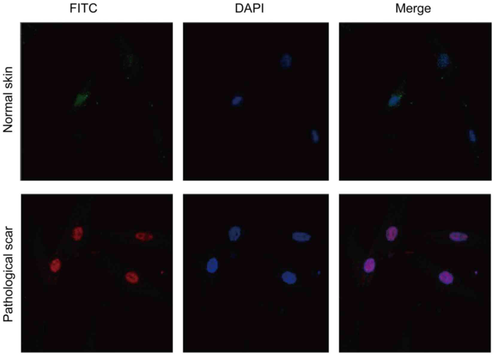
Immunofluorescence results
The immunofluorescence detection result of cells
showed that, mTOR was marked in red (Fig. 1). DAPI was marked in blue
fluorescence in the nucleus (Fig.
1). The merged image (Fig. 1)
indicated that mTOR expressed in the nucleus, while no obvious
expression was found in fibroblast cells of the normal skin tissue
(Fig. 1). Immunofluorescence
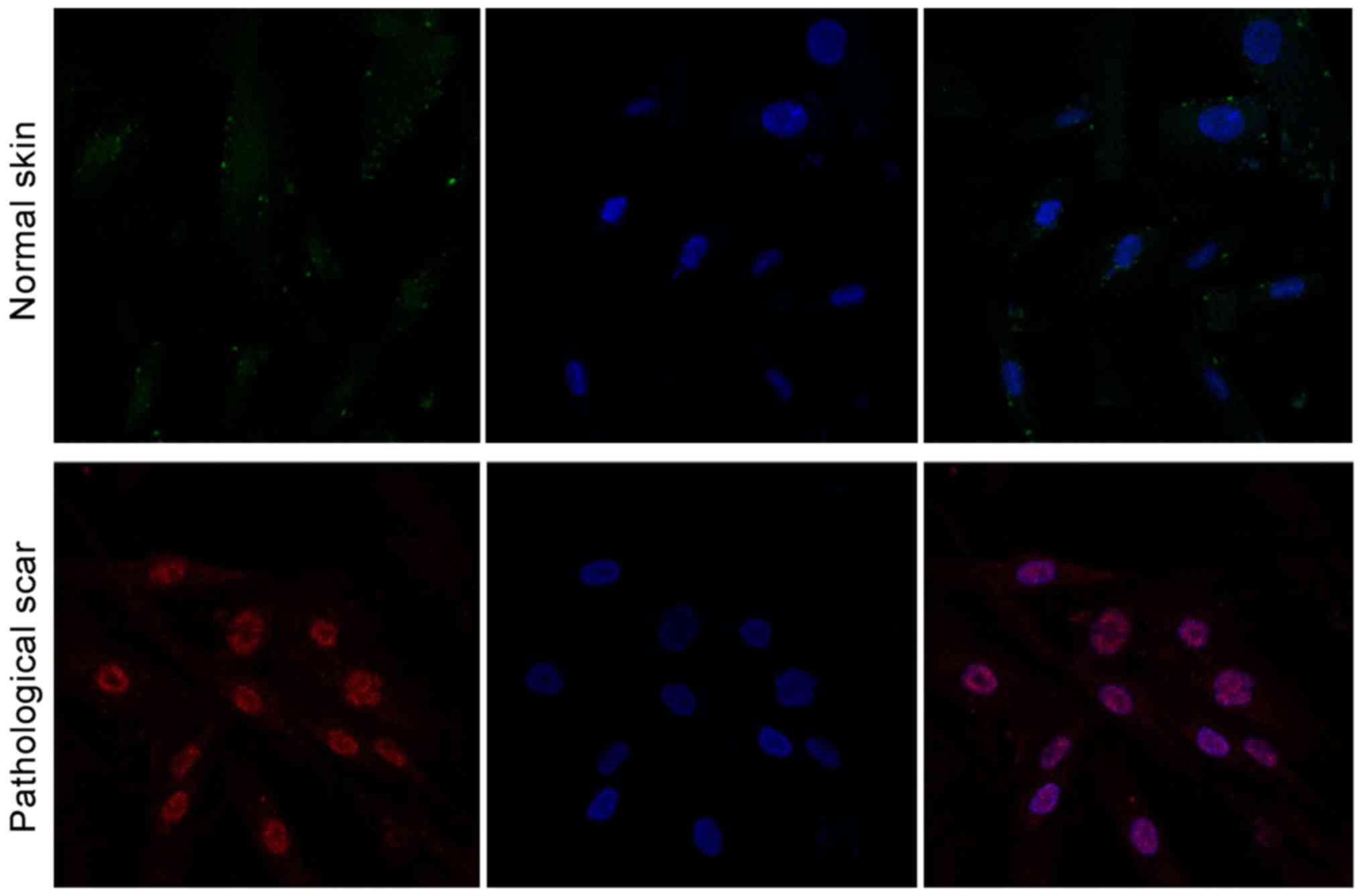
detection results showed that, 70S6K was expressed in the nucleus
in pathological scar fibroblast cells (Fig. 2), while no obvious expression was
found in normal skin tissue (Fig.
2).
RT-PCR results
After pathological scar fibroblast cells were
intervened by Res concentrations of 10, 50 and 100 µmol/l, the mTOR
and 70S6K mRNA relative expression levels decreased compared with
those of the control group. The differences were statistically
significant (P<0.05) (Table
II). Correlation analysis using Pearson's method indicated
that, relative expression of mTOR and 70S6K mRNA was negative with
Res intervention concentration (r=−0.872, P=0.02; r=−0.814,
P=0.03).
| Table II.Relative expression of mTOR and 70S6K
mRNA of various groups. |
Table II.
Relative expression of mTOR and 70S6K
mRNA of various groups.
| Groups | mTOR | 70S6K |
|---|
| A | 9.54±0.23 | 8.71±0.21 |
| B |
7.48±0.19a |
6.69±0.17a |
| C |
5.91±0.14b |
4.27±0.13b |
| D |
2.11±0.09c |
3.02±0.10c |
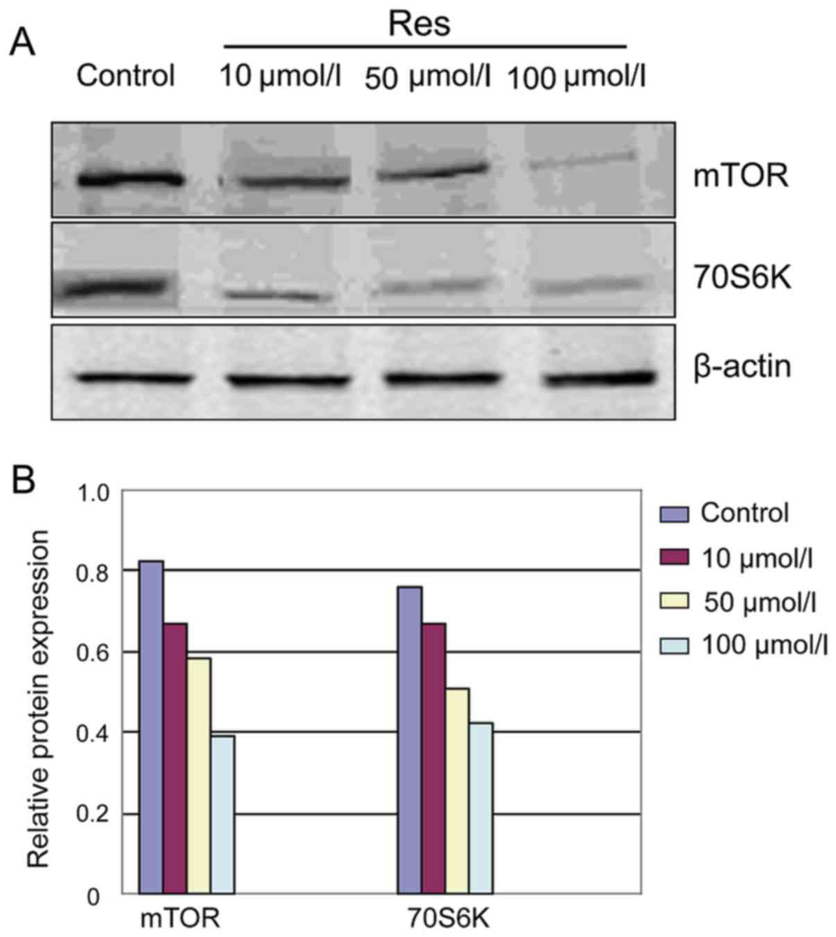
Western blot analysis results
After pathological scar fibroblast cells were
intervened by Res, the protein band ratio of mTOR and 70S6K in the
various concentration groups decreased significantly compared with
that of the control group (Fig.
3). The analysis of Image-Pro Plus imaging analysis system
showed that, relative expression of mTOR and 70S6K in the various
concentration groups was significantly lower than those of the
control group. The differences were statistically significant
(P<0.05) (Fig. 3 and Table III).
| Table III.Relative expression of mTOR, 70S6K
protein of various groups. |
Table III.
Relative expression of mTOR, 70S6K
protein of various groups.
| Groups | mTOR | 70S6K |
|---|
| A | 0.825±0.073 | 0.761±0.068 |
| B |
0.67±0.068a |
0.678±0.061a |
| C |
0.586±0.062b |
0.515±0.054b |
| D |
0.395±0.041c |
0.428±0.036c |
Discussion
Pathological scar includes hypertrophic scar and
keloid. As typical fiber hyperplastic disease, pathological scar is
the pathological product formed by the abnormal proliferation
during the process of wound repair and healing and its pathological
changes are mainly characterized by massive proliferation of
fibroblasts and excessive deposition of extracellular matrix
dominated by collagen (6). At
present, there are many studies on pathological scar in China and
other countries (7–9). However, its exact pathogenesis
remains to be elucidated and no breakthrough has been obtained for
its prevention and treatment. Despite the appearance of many
therapeutic methods, the curative effects are unsatisfactory.
Therefore, it is of vital practical significance to discuss the
pathogenesis of pathological scar from a new perspective and seek
safe and effective therapeutic methods and drugs
The pathogenesis of pathological scar remains
complex. The excessive proliferation of fibroblasts and its
increasing activity play a critical role in its formation. The
excessive proliferation of pathological scar fibroblasts is the
result of the integrated response of cells to the stimulation of
various extracellular signals (10,11).
Since this integrated response is complete in cells, it inevitably
involves the process of cell signal transduction. At present, more
studies have focused on typical TGF-β1/Smads signal pathway.
Nevertheless, the specific mechanism of the proliferation of
pathological scar fibroblasts was not revealed completely. Since
the proliferation of pathological scar fibroblasts involved many
multiple genes and multiple signal pathways, this process is
extremely complex. In recent years, an increasing number of studies
have shown that many types of independent Smad pathways, in
addition to the typical Smad pathway, were also involved in the
transduction process of TGF-β1 signals. Some of them directly or
indirectly were involved in tumor formation and were closely
related to proliferation, invasion and transfer of tumor (12–15).
As the main signal pathway to regulate and control
protein synthesis, mTOR signal pathway is involved in cell
proliferation, differentiation and other regulations, making it a
research hotspot, especially with regard to increasingly in-depth
studies on tumor (16–18). Activated mTOR can act on downstream
target protein, ribosomal protein S6 kinase (70S6K), while 70S6K is
the key regulating factor of protein translation (19–21).
Studies in recent years have demonstrated that mTOR gene was
closely related to abnormal proliferation of cells and the
occurrence of tumor and markedly enhanced its expression in various
tumors, including glioma, esophageal cancer and colorectal cancer
(16,18). Pathologically, pathological scar
belongs to benign solid tumor and can grow to surrounding normal
tissues invasively and its pathological characteristics are very
similar to those of tumor, with tumor-like characteristics. This is
an indication that mTOR signal pathway should also play an
important role in the process of the proliferation of pathological
scar fibroblasts.
Traditional Chinese medicine has a long history in
the prevention and treatment of scars. The present research group
has been engaged in the study of prevention and treatment of
Chinese medicine on pathological scar and the undertaken subjects
are all related to Res. As a non-flavonoid polyphenolic compound
existing in Polygonum Cuspidatum, Veratrum nigrum,
Cassia tora and other plants, Res belongs to a phytoalexin
generated by plants in resistance to external stimulations, such as
ultraviolet ray, fungus, virus infection and mechanical injury
(22). It has multiple biological
activities and pharmacological action, such as anti-inflammatory,
anti-shock, antioxidation, inhibition of platelet aggregation,
improving microcirculation, protection of mitochondrial functions
and estrogenic effects (23–25).
Our preliminary study also proved that Res inhibited the
proliferation of pathological scar fibroblasts, but the specific
mechanism of action has not been elucidated completely. It remains
to be determined whether Res exerts an effect on the proliferation
of pathological scar fibroblasts by regulating and controlling the
mTOR/70S6K signal pathway. At present, there is no domestic and
foreign literature to support this hypothesis. Therefore, our
research group implemented some explorations on it. The present
study applied cellular immunofluorescence to detect the expression
of mTOR and 70S6K and found a clearly strengthened expression of
mTOR and 70S6K in pathological scar fibroblasts, mainly in the cell
nucleus, without obvious expression in fibroblasts of normal skin
tissue. The results of RT-PCR and western blot analysis detection
revealed that after pathological scar fibroblasts being intervened
by Res with different concentrations, the expression of mTOR and
70S6K mRNA and protein decreased as compared with that of the
control group, with statistically significant differences
(P<0.05). Correlation analysis using the Pearson's method showed
that the expression of mTOR and 70S6K mRNA showed a negative
relationship with the intervention concentration of Res. This
showed that the mTOR/70S6K pathway was involved in the formation of
pathological scar, while Res inhibited the proliferation of
pathological scar fibroblasts, which may be associated with
reduction of the expression of mTOR and 70S6K.
References
|
1
|
Tinhofer I, Bernhard D, Senfter M, Anether
G, Loeffler M, Kroemer G, Kofler R, Csordas A and Greil R:
Resveratrol, a tumor-suppressive compound from grapes, induces
apoptosis via a novel mitochondrial pathway controlled by Bcl-2.
FASEB J. 15:1613–1615. 2001.PubMed/NCBI
|
|
2
|
Ding J, Zai X and Tang Z: Effects of
resveratrol on fibroblasts of pathological scar and its
Tgf-β1/smads signaling pathway. J Hebei Med Univ. 35:37–41.
2014.(In Chinese).
|
|
3
|
Huang LP, Mao Z, Zhang L, Liu XX, Huang C
and Jia ZS: Screening of differentially expressed genes in
pathological scar tissues using expression microarray. Genet Mol
Res. 14:10743–10751. 2015. View Article : Google Scholar : PubMed/NCBI
|
|
4
|
Teofoli P, Barduagni S, Ribuffo M,
Campanella A, De Pita' O and Puddu P: Expression of Bcl-2, p53,
c-jun and c-fos protooncogenes in keloids and hypertrophic scars. J
Dermatol Sci. 22:31–37. 1999. View Article : Google Scholar : PubMed/NCBI
|
|
5
|
Steelman LS, Chappell WH, Abrams SL, Kempf
RC, Long J, Laidler P, Mijatovic S, Maksimovic-Ivanic D, Stivala F,
Mazzarino MC, et al: Roles of the Raf/MEK/ERK and
PI3K/PTEN/Akt/mTOR pathways in controlling growth and sensitivity
to therapy-implications for cancer and aging. Aging (Albany NY).
3:192–222. 2011. View Article : Google Scholar : PubMed/NCBI
|
|
6
|
van der Veer WM, Bloemen MC, Ulrich MM,
Molema G, van Zuijlen PP, Middelkoop E and Niessen FB: Potential
cellular and molecular causes of hypertrophic scar formation.
Burns. 35:15–29. 2009. View Article : Google Scholar : PubMed/NCBI
|
|
7
|
Bollero D, Malvasio V, Catalano F and
Stella M: Negative pressure surgical management after pathological
scar surgical excision: a first report. Int Wound J. 12:17–21.
2015. View Article : Google Scholar : PubMed/NCBI
|
|
8
|
Huang C and Ogawa R: The link between
hypertension and pathological scarring: does hypertension cause or
promote keloid and hypertrophic scar pathogenesis? Wound Repair
Regen. 22:462–466. 2014. View Article : Google Scholar : PubMed/NCBI
|
|
9
|
Zhai XX, Ding JC and Tang ZM: Resveratrol
inhibits proliferation and induces apoptosis of pathological scar
fibroblasts through the mechanism involving TGF-β1/Smads signaling
pathway. Cell Biochem Biophys. 71:1267–1272. 2015. View Article : Google Scholar : PubMed/NCBI
|
|
10
|
Susami T: Wound healing in the palatal
mucosa and the mechanism of scar formation. Kokubyo Gakkai Zasshi.
52:5961985.(In Japanese). View Article : Google Scholar : PubMed/NCBI
|
|
11
|
Lu SL: Mechanism of scar formation and
strategy of treatment. Zhonghua Shao Shang Za Zhi. 29:130–133.
2013.(In Chinese). PubMed/NCBI
|
|
12
|
Sun Q, Guo S, Wang CC, Sun X, Wang D, Xu
N, Jin SF and Li KZ: Cross-talk between TGF-β/Smad pathway and
Wnt/β-catenin pathway in pathological scar formation. Int J Clin
Exp Pathol. 8:7631–7639. 2015.PubMed/NCBI
|
|
13
|
Chen W, Fu X, Sun T, Sun X, Zhao Z and
Sheng Z: Change of gene expression of transforming growth
factor-beta1, Smad 2 and Smad 3 in hypertrophic scars skins.
Zhonghua Wai Ke Za Zhi. 40:17–19. 2002.(In Chinese). PubMed/NCBI
|
|
14
|
Brown KA, Pietenpol JA and Moses HL: A
tale of two proteins: differential roles and regulation of Smad2
and Smad3 in TGF-beta signaling. J Cell Biochem. 101:9–33. 2007.
View Article : Google Scholar : PubMed/NCBI
|
|
15
|
Yu R and Cen Y: Transforming growth factor
beta1/Smad3 signal transduction pathway and post-traumatic scar
formation. Zhongguo Xiu Fu Chong Jian Wai Ke Za Zhi. 26:330–335.
2012.(In Chinese). PubMed/NCBI
|
|
16
|
Zhang YJ, Dai Q, Sun DF, Xiong H, Tian XQ,
Gao FH, Xu MH, Chen GQ, Han ZG and Fang JY: mTOR signaling pathway
is a target for the treatment of colorectal cancer. Ann Surg Oncol.
16:2617–2628. 2009. View Article : Google Scholar : PubMed/NCBI
|
|
17
|
Laplante M and Sabatini DM: mTOR signaling
in growth control and disease. Cell. 149:274–293. 2012. View Article : Google Scholar : PubMed/NCBI
|
|
18
|
Petroulakis E, Mamane Y, Le Bacquer O,
Shahbazian D and Sonenberg N: mTOR signaling: implications for
cancer and anticancer therapy. Br J Cancer. 96:(Suppl). R11–R15.
2007.PubMed/NCBI
|
|
19
|
Shinojima N, Yokoyama T, Kondo Y and Kondo
S: Roles of the Akt/mTOR/p70S6K and ERK1/2 signaling pathways in
curcumin-induced autophagy. Autophagy. 3:635–637. 2007. View Article : Google Scholar : PubMed/NCBI
|
|
20
|
Heinonen H, Nieminen A, Saarela M,
Kallioniemi A, Klefström J, Hautaniemi S and Monni O: Deciphering
downstream gene targets of PI3K/mTOR/p70S6K pathway in breast
cancer. BMC Genomics. 9:3482008. View Article : Google Scholar : PubMed/NCBI
|
|
21
|
Hou G, Xue L, Lu Z, Fan T, Tian F and Xue
Y: An activated mTOR/p70S6K signaling pathway in esophageal
squamous cell carcinoma cell lines and inhibition of the pathway by
rapamycin and siRNA against mTOR. Cancer Lett. 253:236–248. 2007.
View Article : Google Scholar : PubMed/NCBI
|
|
22
|
Zhao KS: Biological characteristics and
effects of resveratrol. Chinese Journal of Pathophysiology.
28:1709–1711. 2012.(In Chinese).
|
|
23
|
Pangeni R, Sahni JK, Ali J, Sharma S and
Baboota S: Resveratrol: review on therapeutic potential and recent
advances in drug delivery. Expert Opin Drug Deliv. 11:1285–1298.
2014. View Article : Google Scholar : PubMed/NCBI
|
|
24
|
Wu JM, Hsieh TC and Wang Z:
Cardioprotection by resveratrol: a review of effects/targets in
cultured cells and animal tissues. Am J Cardiovasc Dis. 1:38–47.
2011.PubMed/NCBI
|
|
25
|
Athar M, Back JH, Tang X, Kim KH,
Kopelovich L, Bickers DR and Kim AL: Resveratrol: a review of
preclinical studies for human cancer prevention. Toxicol Appl
Pharmacol. 224:274–283. 2007. View Article : Google Scholar : PubMed/NCBI
|