Introduction
Glioma accounts for 30 to 40% of all intracranial
tumors. Glioblastoma, which is the most lethal primary brain tumor,
is the most common malignant glioma (1,2).
Treatment strategies include combinations of aggressive resection,
radiotherapy and temozolomide (TMZ) treatment and various novel
chemotherapeutics. However, the median survival time of patients
remains poor (3). Chemoresistant
glioma cell subgroups are the most common cause of recurrence.
Our previous study determined that the expression of
Nf-E2 related factor 2 (Nrf2), a traditional cytoprotective
transcription factor, was elevated in glioma (4–8).
Increased expression and continuous activation of Nrf2 in glioma
may contribute to proliferation, invasion and chemoresistance of
cancer cells. A previous study identified increased expression
levels of Nrf2 and its target proteins in doxorubicin-resistant
BEL-7402 hepatocellular carcinoma cells compared with parent cells
(9). Chemoresistance was reduced
in doxorubicin-resistant BEL-7402 cells following suppression of
Nrf2 expression by chrysin, a potent Nrf2 inhibitor. The present
study investigated the Nrf2-antioxidant response element (ARE)
signaling pathway in TMZ-resistant U251. Additionally, the effects
of valproic acid (VPA) and melatonin (MEL) on Nrf2 expression
levels and chemoresistance in U251-TMZ cells were investigated. The
present study determined that increased expression of components of
the Nrf2-ARE signaling pathway in U251-TMZ cells was reversed by
VPA and MEL treatment, which act as Nrf2 inhibitors. Additionally,
U251-TMZ cells restored chemosensitivity to TMZ following
co-treatment with VPA or MEL and exhibited increased levels of
reactive oxygen species (ROS) and apoptosis.
Materials and methods
Cell culture and TMZ resistance
Human U251 glioblastoma cells were obtained from the
American Type Culture Collection (Manassas, VA, USA) and cultured
in Dulbecco's modified Eagle's medium (HyClone; GE Healthcare Life
Sciences, Logan, UT, USA) with 10% fetal bovine serum (Wisent
Biotechnology Co., Ltd., Nanjing, China). The TMZ-resistant U251
cell line (termed U251-TMZ) was established by sustained low
concentration TMZ (cat. no. T2577; Sigma; Merck KGaA, Darmstadt,
Germany) stimulation of U251 cells, at an initial dose of 0.25 µM
for 2 weeks. Then, the concentration of TMZ was doubled every 2–4
weeks. Following this stepwise increase in TMZ concentration for ~8
months, the final U251-TMZ cell line was able to survive in 100 µM
TMZ. Subsequently, Cell Counting kit-8 (CCK-8; Dojindo Molecular
Technologies, Inc., Kumamoto, Japan) was used to evaluate the
survival of U251-TMZ cells and the parent U251 cell line following
treatment with TMZ.
Transient transfection with small
interfering (si)RNA
Nrf2 siRNA (si-Nrf2) (target sequence, 5′GCA
GTTCAATGAAGCTCAACT3′) and scramble siRNA (si-con; sequence,
5′GAGUACGAUCCGAGUUGAG3′) were purchased from Guangzhou RiboBio Co.,
Ltd. (Guangzhou, China). U251-TMZ cells (1×10⁶/well)
were seeded in 6-well plates and transfected with 2 µl
Lipofectamine® 2000 (Invitrogen; Thermo Fisher
Scientific, Inc., Waltham, MA, USA), 100 pmol si-Nrf2 or si-con,
and 2 ml reduced serum Opti-Minimal Essential Medium (Invitrogen;
Thermo Fisher Scientific, Inc.) according to the manufacturer's
protocol. Cells treated with Lipofectamine 2000 only served as the
blank control group. The success of the transfection was confirmed
by western blot analysis 48 h after transfection. All further
experiments using U251-TMZ/si-Nrf2 cells were performed within 48 h
of the transfection.
CCK-8 assay
Cells (100 µl) were seeded in 96-well culture plates
at a density of 2×10³ cells/well and cultured for 24 h.
TMZ at 0, 12.5, 25, 50, 100, 200 and 400 µM was used to check the
chemoresistance of U251-TMZ. Treatment with 200 µM TMZ + VPA at
0.5, 1, 2 and 5 mM or MEL at 0.1, 0.5, 1 and 2 mM were used to
check the effect of VPA and MEL on U251-TMZ chemosensitivity.
Viable cells were quantified at 0, 12, 24 and 48 h following
treatment, using a CCK-8 according to the manufacturer's protocol.
Briefly, 10 µl CCK-8 solution was added into every well and
incubated at 37°C for 1 h. Subsequently the optical density of each
well was measured at a wavelength of 450 nm using a Bio-Rad ELISA
microplate reader (Bio-Rad Laboratories, Inc., Hercules, CA, USA).
All measurements were performed six times. Data are presented as
the mean ± standard deviation (SD).
Reverse transcription-quantitative
polymerase chain reaction (RT-qPCR)
Total RNA was isolated with TRIzol®
(Invitrogen; Thermo Fisher Scientific, Inc.) and single-stranded
cDNA was synthesized from 2 ug of total RNA with the Transcriptor
First Strand cDNA Synthesis kit (Roche Applied Science, Penzberg,
Germany) according to the manufacturer's protocol. The
thermocycling conditions were as follows: Denaturation for 10 min
at 65°C, cDNA synthesis for 1 h at 50°C and inactivation for 5 min
at 85°C. The cDNA was stored at −20°C. Quantitative analysis of
Nrf2 mRNA expression levels was performed using SYBR®
Green PCR Core reagents (Thermo Fisher Scientific, Inc.) and an ABI
7700 Real-time system (Applied Biosystems; Thermo Fisher
Scientific, Inc.), according to the manufacturer's protocol. The
primer sequences were as follows: Forward,
5′-TTCCCGGTCACATCGAGAG-3′ and reverse,
5′-TCCTGTTGCATACCGTCTAAATC-3′ for Nrf2; and forward,
5′-CATCTTCTTTTGCGTCGCCA-3′ and reverse, 5′-TTAAAAGCAGCCCTGGTGACC-3′
for GAPDH. Each amplification cycle consisted of denaturation for
15 sec at 95°C, annealing for 20 sec at 60°C and extension for 40
sec at 72°C; a total of 40 cycles were performed. A melting curve
was used to distinguish specific from non-specific products and
primer-dimers. Each sample was analyzed in triplicate, and
expression of Nrf2 was normalized to GAPDH using the
2−∆∆Cq method (10).
Alterations in Nrf2 expression were reported as fold increases
(2−∆∆Cq) relative to the control group.
Protein preparation
Cytoplasmic and nuclear proteins were obtained using
the Nuclear and Cytoplasmic Protein Extraction kit (Beyotime
Institute of Biotechnology, Shanghai, China). Briefly, cells were
treated with cytoplasmic protein extraction reagent. Lysates were
subsequently incubated for 10 min on ice and centrifuged at 12,000
× g for 5 min at 4°C. The supernatant was collected as the
cytoplasmic protein. Pellets containing crude nuclei were
resuspended in nuclear protein extraction regent and agitated for 2
h on ice, followed by centrifugation at 12,000 × g for 10 min at
4°C to obtain supernatants containing nuclear protein. To obtain
total protein lysate, cells were harvested and homogenized using
radioimmunoprecipitation assay buffer (Beyotime Institute of
Biotechnology) containing 1 mM phenylmethylsulfonyl fluoride, and
centrifuged at 12,000 × g for 15 min at 4°C. Protein concentrations
were estimated by Coomassie Plus Protein assay reagent (Pierce;
Thermo Fisher Scientific, Inc.).
Western blot analysis
Protein extracts (50 mg), including cytoplasmic and
nuclear protein for Nrf2 detection, nuclear protein for H3
detection and total protein for detecting all other proteins, were
heat denatured in loading buffer, separated by 10% sodium dodecyl
sulfate-polyacrylamide gel electrophoresis and electroblotted onto
a nitrocellulose membrane. Membranes were blocked with 5% bovine
serum albumin (BSA; cat. no. B2064; Sigma; Merck KGaA) for 2 h at
room temperature. The following primary antibodies were used:
Rabbit anti-Nrf2 (68 kDa; 1:500; cat. no. sc-722; Santa Cruz
Biotechnology, Inc., Dallas, TX, USA), rabbit anti-histone H3 (H3;
17 kDa; 1:2,000; cat. no. 9715; Cell Signaling Technology, Inc.,
Danvers, MA, USA), rabbit anti-β-actin (43 kDa; 1:10,000; cat. no.
AP0060; Bioworld Technology, Inc., St. Louis Park, MN, USA), rabbit
anti-HO-1 (32 kDa; 1:1,000; cat. no. ab13243; Abcam, Cambridge,
UK), rabbit anti-NQO1 (31 kDa; 1:1,000; cat. no. ab34173; Abcam),
rabbit anti-cleaved caspase-3 (17 kDa; 1:1,000; cat. no. 9661; Cell
Signaling Technology, Inc.), rabbit anti-B-cell lymphoma 2 (Bcl-2;
26 kDa; 1:1,000; cat. no. sc-492; Santa Cruz Biotechnology, Inc.),
mouse anti-Bcl-2-associated X protein (Bax; 1:500; 23 kDa; cat. no.
sc-20067; Santa Cruz Biotechnology, Inc.), mouse
anti-phosphorylated (p)-insulin like growth factor 1 receptor
(IGF-IR; 97 kDa; 1:500; cat. no. sc-81499; Santa Cruz
Biotechnology, Inc.), mouse anti-IGF-IR (97 kDa; 1:500; cat. no.
sc-81167; Santa Cruz Biotechnology, Inc.), rabbit anti-protein
kinase B (AKT; 60 kDa; 1:1,000; cat. no. 9272; Cell Signaling
Technology, Inc.), rabbit anti-p-AKT (60 kDa; 1:2,000; cat. no.
4060; Cell Signaling Technology, Inc.), rabbit anti-mammalian
target of rapamycin (mTOR; 289 kDa; 1:1,000; cat. no. 2983; Cell
Signaling Technology, Inc.), rabbit anti-p-mTOR (289 kDa; 1:500;
cat. no. 5536; Cell Signaling Technology, Inc.) and rabbit anti-P2X
purinoceptor 7 (P2RX7; 75 kDa; 1:1,000; cat. no. ab109246; Abcam).
Each primary antibody was diluted appropriately in 5% BSA and
incubated overnight at 4°C. The blots were washed three times in
washing buffer and incubated with goat anti-rabbit IgG-horseradish
peroxidase (HRP; cat. no. BS13278; 1:10,000; Bioworld Technology,
Inc.) or goat anti-mouse IgG-HRP (cat. no. BS12478; 1:10,000;
Bioworld Technology, Inc.) for 1 h at room temperature. Protein
bands were visualized using an Enhanced Chemiluminescence Detection
system (EMD Millipore, Billerica, MA, USA) and exposed by Tanon
5200 Chemiluminescence Image Analytical system (TanonScience and
Technology Co., Ltd., Shanghai, China). β-actin served as a loading
control for total or cytoplasmic protein; H3 served as a loading
control for nuclear protein. Quantification of band density was
performed using UN-SCAN-IT gel digitizing software version 6.1
(Silk Scientific, Inc., Orem, UT, USA) and data were normalized to
β-actin or H3.
Flow cytometric analysis of
apoptosis
Apoptosis was quantified using an Annexin
V-Fluorescein Isothiocyanate (FITC) Apoptosis Detection kit
(Beyotime Institute of Biotechnology), according to the
manufacturer's protocol. The cells were washed once with incubation
buffer following the aforementioned treatments, collected by
centrifugation at 1,000 × g for 5 min at room temperature, and
resuspended in an Annexin V-FITC/propidium iodide (PI) reactive
solution. Following a 15 min incubation at room temperature, the
percentage of apoptotic cells was quantified using a flow cytometer
(Cytomics FC500; Beckman Coulter, Inc., Brea, CA, USA) and analyzed
using CellQuest Pro software version 3.3 (Mac OS X.1; BD
Biosciences, San Jose, CA, USA).
ROS detection with
2′,7′-dichlorofluorescein diacetate (DCFH-DA)
Intracellular ROS levels were quantified using the
fluorescent probe DCFH-DA (cat. no. S0033; Beyotime Institute of
Biotechnology), according to the manufacturer's protocol. After 24
h treatment, cells were incubated with DCFH-DA in Opti-Minimal
Essential Medium (Invitrogen; Thermo Fisher Scientific, Inc.) for
10 min at 37°C. Subsequently, cells were washed with PBS to remove
excess dye prior to quantification using the microplate reader
Fluoroscan Ascent (Thermo Fisher Scientific, Inc.) with excitation
and emission wavelengths of 485and 538 nm, respectively.
Statistical analysis
Data are expressed as the mean ± SD and evaluated
using one-way analysis of variance followed by Tukey's post hoc
test. P<0.05 was considered to indicate a statistically
significant difference. All analyses were performed using SPSS
version 18.0 (SPSS, Inc., Chicago, IL, USA).
Results
Establishment of the TMZ-resistant
U251 cell line and protein expression levels of Nrf2-ARE signaling
pathway components
The U251-TMZ cell line was established following
exposure of U251 cells to continuously increasing concentrations of
TMZ. CCK-8 results indicated that the survival rate was greater in
in U251-TMZ cells treated with TMZ, compared with parent U251 cells
(Fig. 1A).
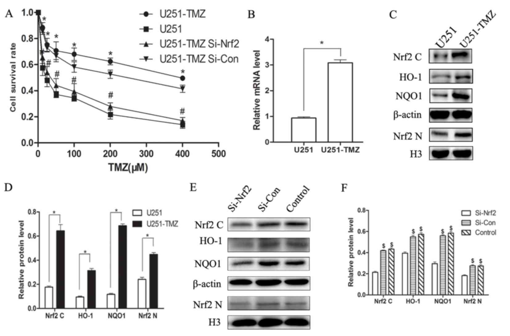 | Figure 1.Nrf2-ARE signaling pathway in U251-TMZ
and its association with chemoresistance. (A) Cell survival rates
following treatment with different concentrations of TMZ for 24 h.
(B) Reverse transcription-quantitative polymerase chain reaction
identified enhanced Nrf2 mRNA expression levels in U251-TMZ when
compared with U251 cells. (C) Western blotting demonstrated
elevated expression levels of Nrf2-ARE signaling pathway components
in U251-TMZ compared with parent U251 cells. (D) siRNA transfection
efficiency was determined by western blotting. (E) Grey scale
analysis results of target proteins in U251 or U251-TMZ when
compared with β-actin or H3. (F) Grey scale analysis results of
target proteins in U251-TMZ following siRNA transfection when
compared with β-actin or H3. *P<0.05 vs. U251;
#P<0.05 vs. U251-TMZ; $P<0.05 vs.
Si-Nrf2. Nrf2, Nf-E2 related factor 2; ARE, antioxidant response
element; U251-TMZ, temozolomide-resistant U251; TMZ, temozolomide;
siRNA, small interfering RNA; si-con, scramble control small
interfering RNA; Nrf2 C, cytoplasmic Nf-E2 related factor 2; Nrf2
N, nuclear Nf-E2 related factor 2; HO-1, heme oxygenease-1; NQO1,
NAD(P)H quinone dehydrogenase 1; H3, histone 3. |
Results from RT-qPCR (Fig. 1B) and western blotting (Fig. 1C and D) indicated that the
expression levels of Nrf2 and its downstream effectors, including
HO-1 and NQO1, were significantly increased in U251-TMZ cells when
compared with parent U251 cells.
Silencing Nrf2 in U251-TMZ cells
restores chemosensitivity to TMZ
The effect of Nrf2 silencing was determined 48 h
after siRNA transfection using western blotting. si-Nrf2
successfully reduced Nrf2 protein expression levels in U251-TMZ
cells (Fig. 1E and F). Protein
expression levels of HO-1 and NQO1, downstream effectors of the
Nrf2-ARE signaling pathway, were additionally decreased (Fig. 1E and F).
CCK-8 results indicated that the cell survival rate
in the U251-TMZ si-Nrf2 group was significantly reduced when
compared with the U251-TMZ si-con and blank control groups, almost
to the level of parent U251 cells (Fig. 1A).
VPA or MEL treatment restores
chemosensitivity of U251-TMZ cells
VPA and MEL have been identified as potent
chemotherapeutic sensitizers in various tumor cell lines (11–14).
The present study investigated whether these drugs may restore the
chemosensitivity of U251-TMZ cells to TMZ. The results of the CCK-8
assay demonstrated that when used in combination with 200 µM TMZ,
treatment with 2 or 5 mM VPA for 24 or 48 h significantly reduced
the survival rate of U251-TMZ cells, when compared with TMZ
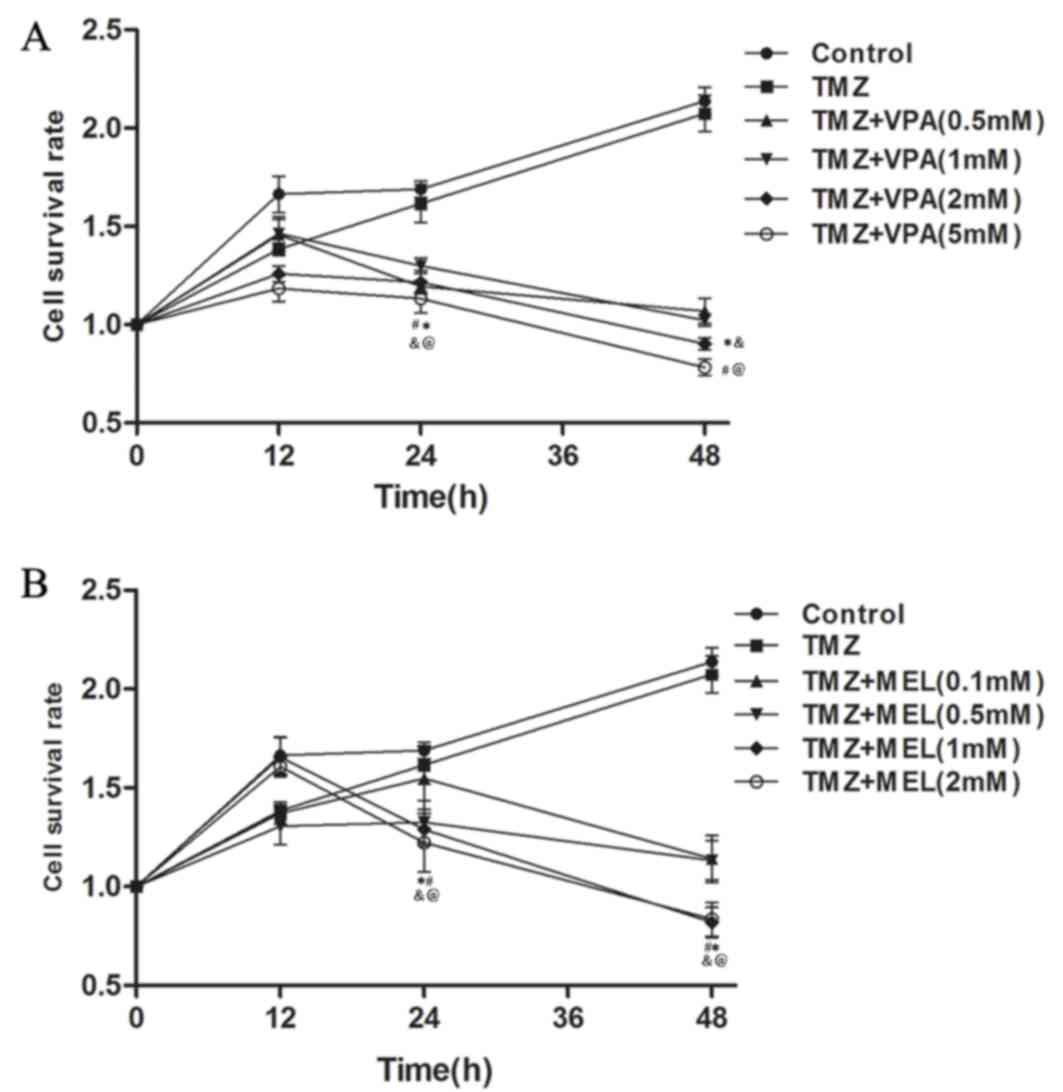
treatment alone (Fig. 2A).
Additionally, when U251-TMZ cells were treated with TMZ plus 1 or 2
mM MEL for 24 or 48 h, the survival rate was significantly reduced
when compared with TMZ treatment alone (Fig. 2B).
VPA and MEL treatment increases ROS
levels and apoptosis induced by TMZ in U251-TMZ cells
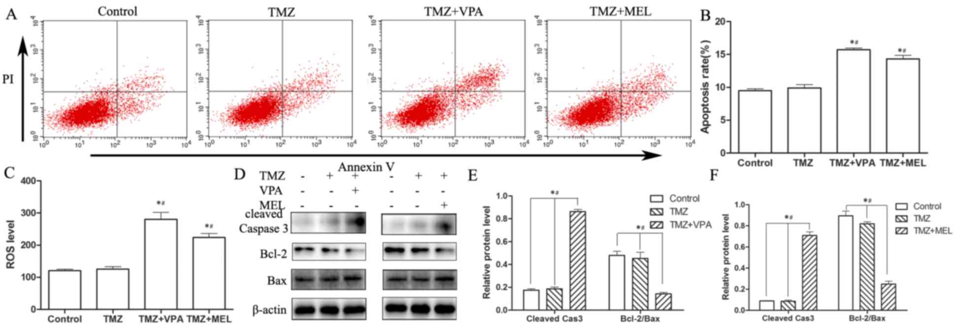
Apoptosis was investigated to determine the
molecular mechanism underlying the inhibitory effect of 2 mM VPA or
1 mM MEL on 200 µM TMZ-treated U251-TMZ cells for 24 h (Fig. 3A). The rate of apoptosis in Annexin
V + PI positive cells was increased in TMZ-treated U251-TMZ cells
co-treated with VPA or MEL, when compared with blank control
(untreated U251-TMZ cells) or TMZ only-treated cells (Fig. 3B). Following co-treatment with TMZ
and VPA/MEL, the protein expression levels of cleaved caspase-3 and
Bax were increased and those of Bcl-2 were reduced, as demonstrated
by western blotting (Fig. 3D-F).
Additionally, ROS levels were increased in U251-TMZ cells following
co-treatment with TMZ and VPA or MEL, compared with the TMZ
treatment alone or blank control groups (Fig. 3C).
VPA and MEL treatment may reduce Nrf2
expression via inhibition of the IGF-IR/AKT/mTOR signaling
pathway
VPA and MEL are effective chemotherapeutic
sensitizers in glioma (15–17).
However, this effect and its association with Nrf2 have not been
previously investigated in TMZ-resistant glioma cells. The present
study determined that co-treatment of 2 mM VPA or 1 mM MEL with 200
µM TMZ for 24 h significantly reduced Nrf2 protein expression
levels in U251-TMZ cell lines, when compared with cells treated
with TMZ only (Fig. 4A, C and D).
In addition, the protein expression levels of HO-1 and NQO1,
downstream effectors of the Nrf2-ARE signaling pathway, which
contribute to ROS clearance, were reduced following TMZ treatment
in combination with VPA or MEL, when compared with cells treated
with TMZ only (Fig. 4A, C and D).
Inhibition of the IGF-IR/AKT/mTOR signaling pathway by VPA or MEL
treatment was determined using western blotting (Fig. 4B, E and F). Furthermore, protein
expression levels of P2RX7, the receptor associated with ROS
generation, were increased following TMZ treatment with VPA or MEL,
compared with TMZ treatment only (Fig.
4B, E and F).
 | Figure 4.VPA or MEL and TMZ treatment may
reduceNrf2-ARE expression via inhibition of the IGF-IR/AKT/mTOR
signaling pathway and increase ROS levels via activation of P2RX7.
(A) VPA or MEL co-treatment with TMZ reduced the expression levels
of proteins involved in the Nrf2-ARE signaling pathway in U251-TMZ
cells compared with cells treated with TMZ only. (B) VPA or MEL
co-treatment with TMZ reduced phosphorylation of components of the
IGF-IR/AKT/mTOR signaling pathway and increased the protein
expression levels of P2RX7 in U251-TMZ cells compared with cells
treated with TMZ only. Grey scale analysis results of target
protein with (C) VPA and (D) MEL compared with β-actin or H3, and
grey scale analysis results of phosphorylation with (E) VPA and (F)
MEL compared with total protein in the IGF-IR/mTOR/AKT signaling
pathway or total P2RX7 protein compared with β-actin. *P<0.05
vs. TMZ. Control group, blank control with untreated U251-TMZ; TMZ,
temozolomide; VPA, valproic acid; MEL, melatonin; Nrf2, Nf-E2
related factor 2; ARE, antioxidant response element; U251-TMZ,
temozolomide-resistant U251; Nrf2 C, cytoplasmic Nf-E2 related
factor 2; Nrf2 N, nuclear Nf-E2 related factor 2; HO-1, heme
oxygenease-1; NQO1, NAD(P)H quinone dehydrogenase 1; H3, histone
H3; p, phosphorylated; IGF-IR, insulin like growth factor 1
receptor; AKT, protein kinase B; mTOR, mammalian target of
rapamycin; P2RX7, P2X purinoceptor 7. |
Discussion
Chemoresistance reduces the chances of survival in
patients with tumors, particularly in recurrent tumors. The present
study investigated the possible mechanism underlying glioma
chemoresistance. The findings of the current study indicated that
expression levels of components of the Nrf2-ARE signaling pathway
were increased in TMZ-resistant U251 cells compared with parent
U251 cells. Suppressing Nrf2 expression using siRNA or the Nrf2
inhibitors VPA or MEL, restored the chemosensitivity of U251-TMZ
cells to TMZ and increased the rate of apoptosis and ROS levels.
Additionally, the results of the present study suggested that VPA
and MEL treatment may reduce the expression levels of Nrf2-ARE
signaling pathway proteins via inhibition of the IGF-IR/AKT/mTOR
signaling pathway, and increase ROS levels via activation of
P2RX7.
Nrf2 was initially identified as a critical
transcription factor in the Kelch-like ECH-associated protein1
(Keap1)-Nrf2-ARE signaling pathway, which functions to resist
oxidative stress. Under normal conditions, low constitutive
quantities of Nrf2 protein are maintained by the Keap1-mediated
ubiquitination and proteasomal degradation system (18). Nrf2 is activated and dissociates
from Keap1 in response to oxidative stress. Following translocation
from the cytoplasm to the nucleus, Nrf2 forms a heterodimer with
MAF bZIP transcription factor, which binds to the ARE sequence to
increase the expression of various cytoprotective genes, such as
HO-1 and NQO1 (19). However, the
cell protective effect of Nrf2 may additionally be present in tumor
cells. Overexpression of Nrf2 accompanied by chemoresistance has
been identified in non-small-cell lung cancer, breast
adenocarcinoma, endometrial serous carcinoma and neuroblastoma
cells (20–22). Additionally, RNA
interference-mediated downregulation of Nrf2 expression in lung
cancer cells induced generation of ROS and resulted in increased
sensitivity to chemotherapeutic drugs in vitro and in
vivo (23). The findings of
the present study indicated increased Nrf2 expression in
TMZ-resistant U251 cells compared with parent U251 cells. The
protein expression levels of HO-1 and NQO1, Nrf2 targeted
cytoprotective and antioxidant genes, were additionally
significantly increased in U251-TMZ cells. HO-1 and NQO1 contribute
to ROS clearance (24–26), which may be an important mechanism
underlying TMZ-resistance. Silencing Nrf2 expression in U251-TMZ
cells through siRNA transfection increased chemosensitivity to TMZ.
The protein expression levels of HO-1 and NQO1 in U251-TMZ cells
were reduced following si-Nrf2 transfection.
A recent study identified that VPA, a traditional
antiepileptic drug, has chemotherapeutic effects (16). A meta-analysis revealed that
patients with glioblastoma may experience prolonged survival
following VPA administration. In addition, the benefit of VPA
treatment was confirmed in sub-group analysis compared with
non-anti-epileptic drug (AED) and other AED groups. These findings
indicated the benefits of TMZ and VPA combined treatment in
patients with glioblastoma. Previous studies have determined that
VPA may inhibit glioma cell proliferation in vitro and in
vivo by increasing apoptosis and inducing cell cycle arrest
(27–29). The molecular mechanism underlying
the effects of VPA in glioma treatment may in part rely on
inhibition of histone deacetylase, downregulation of
O-6-methylguanine-DNA methyltransferase, or activation of
mitogen-activated protein kinases or reversion inducing cysteine
rich protein with kazal motifs-matrix metalloproteinase pathways
and redox regulation. However, the association between VPA and Nrf2
expression in glioma remains to be fully elucidated. The present
study investigated the effect of VPA on Nrf2 expression in U251-TMZ
cells. The findings of the current study demonstrated that VPA
successfully downregulated the protein expression levels of
Nrf2-ARE signaling pathway effectors in U251-TMZ cells. The
suppression of the Nrf2-ARE signaling pathway may lead to an
imbalanced redox equilibrium in the cell, which may result in
elevation of ROS levels and induction of apoptosis. Therefore,
inhibition of the Nrf2-ARE signaling pathway may contribute to the
molecular mechanism underlying the effect of VPA treatment on
glioma cells.
MEL, an indoleamine hormone produced by the pineal
gland and a typical antioxidant, exerts antitumor activity in a
wide range of neoplasms in vitro and in vivo
(30). Martin et al
(31) initially determined that
MEL inhibited C6 glioma cell proliferation and induced cell cycle
arrest in vitro and in vivo. Similar findings were
published subsequently. Pharmacological concentrations of MEL (1
mM) may inhibit the ras oncogene at 85 D/protein kinase
C/AKT/nuclear factor-κB signaling pathway, which may reduce local
biosynthesis of estrogen and expression of the ABC transporter ATP
binding cassette subfamily G member 2 (32–35).
These mechanisms may participate in MEL inhibition of gliomas.
However, the association between the pharmacological concentration
of MEL and the Nrf2-ARE signaling pathway in glioma cells remains
to be fully elucidated. A previous study suggested that MEL at low
concentrations (up to 100 nM) may activate the Nrf2-ARE signaling
pathway in C6 glioma cells (36).
Additionally, MEL has been revealed to activate the Nrf2-ARE
signaling pathway in various neurological degeneration diseases and
traumatic brain injuries (37,38).
However, the findings of the present study indicated that 1 mM MEL
significantly reduced Nrf2 expression and that of downstream
effectors, including NQO1 and HO-1, when combined with TMZ
treatment in U251-TMZ cells. The differences in these findings may
be due to the different concentrations and cell types used.
Expression of Nrf2 is typically regulated by the
Keap1-Nrf2-ARE signaling pathway. However, previous studies
identified a role for the AKT/mTOR signaling pathway in the
regulation of Nrf2 expression. Phosphorylation of AKT has been
demonstrated to induce activation of Nrf2 and upregulate the
expression of HO-1 and NQO1. Nrf2 activators, including S-allyl
cysteine, berberine, oltipraz and sulforaphane, may induce the
activation of the AKT signaling pathway, thus influencing the
Nrf2-ARE signaling pathway (39–42).
The present study demonstrated that VPA and MEL inhibited the
phosphorylation of the IGF-IR/AKT/mTOR signaling pathway and
inhibited the Nrf2-ARE signaling pathway. Suppression of Nrf2-ARE
may cause an imbalance of the redox equilibrium, leading to
increased ROS levels and apoptosis following TMZ treatment. The
inhibitory effect of VPA and MEL on Nrf2 expression partly restored
chemosensitivity of U251-TMZ cells. To the best of our knowledge,
the present study is the first to identify that VPA and MEL maybe
potential therapeutic agents for the treatment of chemoresistant
glioblastoma. Further investigations are required to confirm this
effect of VPA and MEL, including in vivo experiments and
preclinical studies. Additionally, potential side effects due to
high doses of VPA or MEL should be considered.
Acknowledgements
The present study was supported by the National
Natural Science Foundation of China (grant no. 81402072), the
Natural Science Foundation of Jiangsu Province (grant no.
BK20140732) and the China Postdoctoral Science Foundation (grant
no. 2015M572716).
References
|
1
|
Schneider T, Mawrin C, Scherlach C, Skalej
M and Firsching R: Gliomas in adults. Dtsch Arztebl Int.
107:799–808. 2010.PubMed/NCBI
|
|
2
|
Ohgaki H and Kleihues P: Population-based
studies on incidence, survival rates, and genetic alterations in
astrocytic and oligodendroglial gliomas. J Neuropathol Exp. Neurol.
64:479–489. 2005.
|
|
3
|
DeAngelis LM: Brain tumors. N Engl J Med.
344:114–123. 2001. View Article : Google Scholar : PubMed/NCBI
|
|
4
|
Pan H, Wang H, Zhu L, Mao L, Qiao L and Su
X: The role of Nrf2 in migration and invasion of human glioma cell
U251. World Neurosurg. 80:363–370. 2013. View Article : Google Scholar : PubMed/NCBI
|
|
5
|
Ji XJ, Chen SH, Zhu L, Pan H, Zhou Y, Li
W, You WC, Gao CC, Zhu JH, Jiang K and Wang HD: Knockdown of
NF-E2-related factor 2 inhibits the proliferation and growth of
U251MG human glioma cells in a mouse xenograft model. Oncol Rep.
30:157–164. 2013.PubMed/NCBI
|
|
6
|
Pan H, Wang H, Zhu L, Wang X, Cong Z, Sun
K and Fan Y: The involvement of Nrf2-ARE pathway in regulation of
apoptosis in human glioblastoma cell U251. Neurol Res. 35:71–78.
2013. View Article : Google Scholar : PubMed/NCBI
|
|
7
|
Zhou Y, Wang HD, Zhu L, Cong ZX, Li N, Ji
XJ, Pan H, Wang JW and Li WC: Knockdown of Nrf2 enhances autophagy
induced by temozolomide in U251 human glioma cell line. Oncol Rep.
29:394–400. 2013.PubMed/NCBI
|
|
8
|
Ji X, Wang H, Zhu J, Zhu L, Pan H, Li W,
Zhou Y, Cong Z, Yan F and Chen S: Knockdown of Nrf2 suppresses
glioblastoma angiogenesis by inhibiting hypoxia-induced activation
of HIF-1α. Int J Cancer. 135:574–584. 2014. View Article : Google Scholar : PubMed/NCBI
|
|
9
|
Gao AM, Ke ZP, Shi F, Sun GC and Chen H:
Chrysin enhances sensitivity of BEL-7402/ADM cells to doxorubicin
by suppressing PI3K/Akt/Nrf2 and ERK/Nrf2 pathway. Chem Biol
Interact. 206:100–108. 2013. View Article : Google Scholar : PubMed/NCBI
|
|
10
|
Livak KJ and Schmittgen TD: Analysis of
relative gene expression data using real-time quantitative PCR and
the 2(−Delta Delta C(T)) Method. Methods. 25:402–408. 2001.
View Article : Google Scholar : PubMed/NCBI
|
|
11
|
Gong Y, Ni ZH, Zhang X, Chen XH and Zou
ZM: Valproic Acid Enhances the Anti-Tumor Effect of (−)-gossypol to
Burkitt Lymphoma Namalwa Cells. Biomed Environ Sci. 28:773–777.
2015.PubMed/NCBI
|
|
12
|
Fushida S, Kaji M, Oyama K, Hirono Y,
Nezuka H, Takeda T, Tsukada T, Fujimoto D, Ohyama S, Fujimura T and
Ohta T: Randomized Phase II trial of paclitaxel plus valproic acid
vs paclitaxel alone as second-line therapy for patients with
advanced gastric cancer. Onco Targets Ther. 8:939–941. 2015.
View Article : Google Scholar : PubMed/NCBI
|
|
13
|
Sookprasert A, Johns NP, Phunmanee A,
Pongthai P, Cheawchanwattana A, Johns J, Konsil J, Plaimee P,
Porasuphatana S and Jitpimolmard S: Melatonin in patients with
cancer receiving chemotherapy: A randomized, double-blind,
placebo-controlled trial. Anticancer Res. 34:7327–7337.
2014.PubMed/NCBI
|
|
14
|
Fan L, Sun G, Ma T, Zhong F and Wei W:
Melatonin overcomes apoptosis resistance in human hepatocellular
carcinoma by targeting survivin and XIAP. J Pineal Res. 55:174–183.
2013. View Article : Google Scholar : PubMed/NCBI
|
|
15
|
Thotala D, Karvas RM, Engelbach JA, Garbow
JR, Hallahan AN, DeWees TA, Laszlo A and Hallahan DE: Valproic acid
enhances the efficacy of radiation therapy by protecting normal
hippocampal neurons and sensitizing malignant glioblastoma cells.
Oncotarget. 6:35004–35022. 2015.PubMed/NCBI
|
|
16
|
Yuan Y, Xiang W, Qing M, Yanhui L, Jiewen
L and Yunhe M: Survival analysis for valproic acid use in adult
glioblastoma multiforme: A meta-analysis of individual patient data
and a systematic review. Seizure. 23:830–835. 2014. View Article : Google Scholar : PubMed/NCBI
|
|
17
|
Martín V, Sanchez-Sanchez AM, Herrera F,
Gomez-Manzano C, Fueyo J, Alvarez-Vega MA, Antolín I and Rodriguez
C: Melatonin-induced methylation of the ABCG2/BCRP promoter as a
novel mechanism to overcome multidrug resistance in brain tumour
stem cells. Br J Cancer. 108:2005–2012. 2013. View Article : Google Scholar : PubMed/NCBI
|
|
18
|
Cullinan SB, Gordan JD, Jin J, Harper JW
and Diehl JA: The Keap1-BTB protein is an adaptor that bridges Nrf2
to a Cul3-based E3 ligase: Oxidative stress sensing by a Cul3-Keap1
ligase. Mol Cell Biol. 24:8477–8486. 2004. View Article : Google Scholar : PubMed/NCBI
|
|
19
|
Lau A, Villeneuve NF, Sun Z, Wong PK and
Zhang DD: Dual roles of Nrf2 in cancer. Pharmacol Res. 58:262–270.
2008. View Article : Google Scholar : PubMed/NCBI
|
|
20
|
Jiang T, Chen N, Zhao F, Wang XJ, Kong B,
Zheng W and Zhang DD: High levels of Nrf2 determine chemoresistance
in type II endometrial cancer. Cancer Res. 70:5486–5496. 2010.
View Article : Google Scholar : PubMed/NCBI
|
|
21
|
Hu L, Miao W, Loignon M, Kandouz M and
Batist G: Putative chemopreventive molecules can increase
Nrf2-regulated cell defense in some human cancer cell lines,
resulting in resistance to common cytotoxic therapies. Cancer
Chemother Pharmacol. 66:467–474. 2010. View Article : Google Scholar : PubMed/NCBI
|
|
22
|
Wang XJ, Sun Z, Villeneuve NF, Zhang S,
Zhao F, Li Y, Chen W, Yi X, Zheng W, Wondrak GT, et al: Nrf2
enhances resistance of cancer cells to chemotherapeutic drugs, the
dark side of Nrf2. Carcinogenesis. 29:1235–1243. 2008. View Article : Google Scholar : PubMed/NCBI
|
|
23
|
Singh A, Boldin-Adamsky S, Thimmulappa RK,
Rath SK, Ashush H, Coulter J, Blackford A, Goodman SN, Bunz F,
Watson WH, et al: RNAi-mediated silencing of nuclear factor
erythroid-2-related factor 2 gene expression in non-small cell lung
cancer inhibits tumor growth and increases efficacy of
chemotherapy. Cancer Res. 68:7975–7984. 2008. View Article : Google Scholar : PubMed/NCBI
|
|
24
|
Kuroda H, Takeno M, Murakami S, Miyazawa
N, Kaneko T and Ishigatsubo Y: Inhibition of heme oxygenase-1 with
an epidermal growth factor receptor inhibitor and cisplatin
decreases proliferation of lung cancer A549 cells. Lung Cancer.
67:31–36. 2010. View Article : Google Scholar : PubMed/NCBI
|
|
25
|
Rushworth SA, Bowles KM, Raninga P and
MacEwan DJ: NF-kappaB-inhibited acute myeloid leukemia cells are
rescued from apoptosis by heme oxygenase-1 induction. Cancer Res.
70:2973–2983. 2010. View Article : Google Scholar : PubMed/NCBI
|
|
26
|
Kim YS, Zerin T and Song HY: Antioxidant
action of ellagic acid ameliorates paraquat-induced A549
cytotoxicity. Biol Pharm Bull. 36:609–615. 2013. View Article : Google Scholar : PubMed/NCBI
|
|
27
|
Ryu CH, Yoon WS, Park KY, Kim SM, Lim JY,
Woo JS, Jeong CH, Hou Y and Jeun SS: Valproic acid downregulates
the expression of MGMT and sensitizes temozolomide-resistant glioma
cells. J Biomed Biotechnol. 2012:9874952012. View Article : Google Scholar : PubMed/NCBI
|
|
28
|
Chen Y, Tsai YH and Tseng SH: Valproic
acid affected the survival and invasiveness of human glioma cells
through diverse mechanisms. J Neurooncol. 109:23–33. 2012.
View Article : Google Scholar : PubMed/NCBI
|
|
29
|
Chen CH, Chang YJ, Ku MS, Chung KT and
Yang JT: Enhancement of temozolomide-induced apoptosis by valproic
acid in human glioma cell lines through redox regulation. J Mol Med
(Berl). 89:303–315. 2011. View Article : Google Scholar : PubMed/NCBI
|
|
30
|
Srinivasan V, Spence DW, Pandi-Perumal SR,
Trakht I and Cardinali DP: Therapeutic actions of melatonin in
cancer: Possible mechanisms. Integr Cancer Ther. 7:189–203. 2008.
View Article : Google Scholar : PubMed/NCBI
|
|
31
|
Martín V, Herrera F, Carrera-Gonzalez P,
García-Santos G, Antolín I, Rodriguez-Blanco J and Rodriguez C:
Intracellular signaling pathways involved in the cell growth
inhibition of glioma cells by melatonin. Cancer Res. 66:1081–1088.
2006. View Article : Google Scholar : PubMed/NCBI
|
|
32
|
González A, Martinez-Campa C, Mediavilla
MD, Alonso-González C, Sánchez-Barceló EJ and Cos S: Inhibitory
effects of pharmacological doses of melatonin on aromatase activity
and expression in rat glioma cells. Br J Cancer. 97:755–760. 2007.
View Article : Google Scholar : PubMed/NCBI
|
|
33
|
Martín V, Herrera F, García-Santos G,
Antolín I, Rodriguez-Blanco J, Medina M and Rodriguez C:
Involvement of protein kinase C in melatonin's oncostatic effect in
C6 glioma cells. J Pineal Res. 43:239–244. 2007. View Article : Google Scholar : PubMed/NCBI
|
|
34
|
Martín V, García-Santos G,
Rodriguez-Blanco J, Casado-Zapico S, Sanchez-Sanchez A, Antolín I,
Medina M and Rodriguez C: Melatonin sensitizes human malignant
glioma cells against TRAIL-induced cell death. Cancer Lett.
287:216–223. 2010. View Article : Google Scholar : PubMed/NCBI
|
|
35
|
Martín V, Sanchez-Sanchez AM, Herrera F,
Gomez-Manzano C, Fueyo J, Alvarez-Vega MA, Antolín I and Rodriguez
C: Melatonin-induced methylation of the ABCG2/BCRP promoter as a
novel mechanism to overcome multidrug resistance in brain tumour
stem cells. Br J Cancer. 108:2005–2012. 2013. View Article : Google Scholar : PubMed/NCBI
|
|
36
|
Jumnongprakhon P, Govitrapong P, Tocharus
C, Pinkaew D and Tocharus J: Melatonin protects
methamphetamine-induced neuroinflammation through NF-κB and Nrf2
pathways in glioma cell line. Neurochem Res. 40:1448–1456. 2015.
View Article : Google Scholar : PubMed/NCBI
|
|
37
|
Ding K, Wang H, Xu J, Li T, Zhang L, Ding
Y, Zhu L, He J and Zhou M: Melatonin stimulates antioxidant enzymes
and reduces oxidative stress in experimental traumatic brain
injury: The Nrf2-ARE signaling pathway as a potential mechanism.
Free Radic Biol Med. 73:1–11. 2014. View Article : Google Scholar : PubMed/NCBI
|
|
38
|
Negi G, Kumar A and Sharma SS: Melatonin
modulates neuroinflammation and oxidative stress in experimental
diabetic neuropathy: Effects on NF-κB and Nrf2 cascades. J Pineal
Res. 50:124–131. 2011.PubMed/NCBI
|
|
39
|
Lee YJ, Jeong HY, Kim YB, Lee YJ, Won SY,
Shim JH, Cho MK, Nam HS and Lee SH: Reactive oxygen species and
PI3K/Akt signaling play key roles in the induction of Nrf2-driven
heme oxygenase-1 expression in sulforaphane-treated human
mesothelioma MSTO-211H cells. Food Chem Toxicol. 50:116–123. 2012.
View Article : Google Scholar : PubMed/NCBI
|
|
40
|
Tobón-Velasco JC, Vázquez-Victorio G,
Macías-Silva M, Cuevas E, Ali SF, Maldonado PD, González-Trujano
ME, Cuadrado A, Pedraza-Chaverrí J and Santamaría A: RETRACTED:
S-allyl cysteine protects against 6-hydroxydopamine-induced
neurotoxicity in the rat striatum: Involvement of Nrf2
transcription factor activation and modulation of signaling kinase
cascades. Free Radic Biol Med. 53:1024–1040. 2012. View Article : Google Scholar : PubMed/NCBI
|
|
41
|
Hsu YY, Tseng YT and Lo YC: Berberine, a
natural antidiabetes drug, attenuates glucose neurotoxicity and
promotes Nrf2-related neurite outgrowth. Toxicol Appl Pharmacol.
272:787–796. 2013. View Article : Google Scholar : PubMed/NCBI
|
|
42
|
Rao J, Qian X, Li G, Pan X, Zhang C, Zhang
F, Zhai Y, Wang X and Lu L: ATF3-mediated NRF2/HO-1 signaling
regulates TLR4 innate immune responses in mouse liver
ischemia/reperfusion injury. Am J Transplant. 15:76–87. 2015.
View Article : Google Scholar : PubMed/NCBI
|