Introduction
Cancer is one of the most serious diseases in the
worldwide. Based on the latest data from the International Agency
for Research on Cancer-World Health Organization GLOBOCAN Project
2012, there were ~14.1 million cancer cases and 8.2 million
cancer-related mortalities in 2012 (1). Data from the National Cancer Center
Registry of China indicated that there were ~4,292,000 new cancer
cases and ~2,814,000 cancer-related deaths in China in 2015
(2). A number of small chemical
molecules have been reported to target cancer cell growth and cell
signaling and exhibit good inhibition of tumor activity, which may
potentially be further developed to antitumor drugs (3).
Alkaloids are a type of alkaline organic compounds
that contain nitrogen and mostly occur in plants. Most alkaloids
have complex circular structures with heterocyclic nitrogen,
including indole, pyridine, quinoline and purine (4). Phenanthroindolizidine alkaloids (PAs)
have been isolated from various natural plants, mainly from genera
in the Asclepiadoideae family, such as Tylophora,
Vincetoxicum, Pergularia and Cynanchum
(5). Alkaloids have certain
biological functions, including cytotoxicity, antibacterial and
antiviral, and effect biochemical processes in plant and animal
cells (6,7). The biological function of PAs
primarily includes the mutagenicity, cytotoxicity and cell
biochemical processes. A previous report indicated that the
drawbacks of PAs include a low water solubility and central nervous
toxicity. Antofine, a representative PA, has been used as a
cytotoxicity agent that has low IC50 values in the
nanomolar range in multidrug-resistant and drug-sensitive cancer
cells (8).
Alkaloids extracted from plants exhibit potential
antitumor activity. However, most natural alkaloids are not useful
for humans owing to their poor stability and dissolubility, and the
potential adverse side effects. The present study aimed to develop
new derivatives of PAs to improve their specific anticancer
activities and cellular pharmaceutical effects on human cancer
cells.
Materials and methods
Chemical synthesis
The 12 different PA analogues were synthesized
primarily based on previous reports (9). The 12 PA analogues contain the same
phenanthrene ring with different functional groups at different
positions. Benzoic acid with different substituents were added in a
certain proportion for reaction with benzaldehyde derivatives with
different substituents, and finally 12 compounds were synthesized
through a series of organic chemistry experiments, including aldol
condensation, esterification, n-cyclohexylmaleimide of free
radicals, reduction reaction and amination reaction. The chemical
compounds were named S306, S307, S308, S206, S207, S208, S106b,
XS1, XS2, XS4, XS5 and S108, and their respective hydrochloride
forms were correspondingly named as YS306, YS307, YS308, YS206,
YS207, YS208, YS106b, YXS1, YXS2, YXS4, YXS5 and YS108.
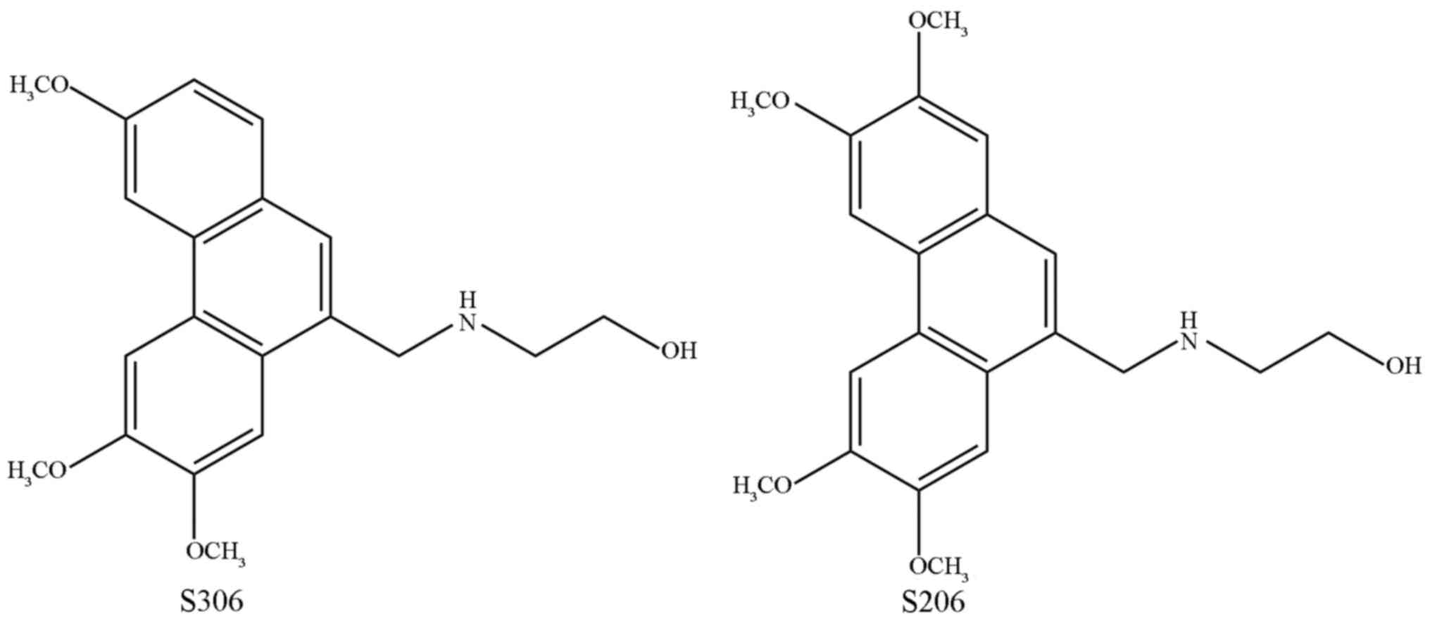
Representative structures of two compounds, S206 and S306, are
shown in Fig. 1.
The purity of all PAs used in cell experiments was
up to 99%, as measured by high performance liquid chromatography.
The anticancer drug paclitaxel (Nanjing Kangmanlin Chemical Co.,
Ltd., Nanjing, China) was used as a positive control when detecting
the anticancer activities of PAs. All PA compounds and
paclitaxelwere dissolved in 100% DMSO to make a stock solution, and
the final concentration of DMSO was adjusted to <0.1% with
Dulbecco's Modified Eagle's Medium (DMEM). All chemical compounds
were firstly dissolved in 100% DMSO, and then were diluted to 5
mg/ml stock liquor with DMEM media. Finally, the stock liquor was
further diluted to 0.5, 5 and 50 µg/ml with DMEM for subsequent
in vitro tests. All the chemical solutions were stored at
4°C, and operations were completed in a Class II biological safety
cabinet (NuAire, Inc., Plymouth, MN, USA). The hydrochloride
compounds had a higher solubility than their respective free auxin.
Therefore, the following cellular experiments were performed using
the hydrochloride compounds.
Cell culture
Human lung cancer A549 cells, liver cancer HepG2
cells and human colon cancer HT29 and HCT116 cells were purchased
from American Type Culture Collection (Manassas, VA, USA), and
normal human liver cell line LO2 was purchased from Cell Bank of
Shanghai Institute of Cell Biology, Chinese Academy of Sciences
(Shanghai, China) (10). Cells
were maintained in DMEM (Gibco; Thermo Fisher Scientific, Inc.,
Waltham, MA, USA) supplemented with 10% fetal bovine serum (FBS;
Gibco; Thermo Fisher Scientific, Inc.) at 37°C in humidified
atmosphere with 5% (v/v) CO2 and 95% (v/v) air (10).
MTT assay
Cell proliferation was measured by the MTT assay,
which was performed to rapidly detect the growth-inhibitory effects
of the chemical compounds on various human cancer cells in
vitro. Exponentially growing cells along with 200 µl culture
medium were seeded (8,000 cells/well) into a 96-well plate.
Different concentrations of the PAs (1, 2, 5,10, 20, 30, 40 and 50
µg/ml) dissolved in 100 µl medium was added to each well and the
plates were incubated for 48 h; 100 µl culture medium with 0.01%
DMSO was used as a blank control group. Following incubation, 10 µl
MTT solution was added to each well and incubated for 4 h, and the
absorbance at 570 nm was measured to calculate the average
inhibition rate. Each concentration gradient of PAs was detected at
least 3 times for each cell line. The average inhibition rate was
calculated according to the ratio of the blank group vs. the test
group.
Double staining for apoptosis
A double staining assay (Apoptosis Detection kit;
Nanjing KeyGen Biotech Co., Ltd., Nanjing, China) was used,
according to the manufacturer's protocol, to assess whether the
detected compounds were able to induce cell apoptosis.
Exponentially growing cells were seeded (3×10⁵
cells/well) in a 6-well plate, which was incubated with different
compounds for 24 h at 37°C. Cells were collected, washed twice with
phosphate buffered saline (PBS), and suspended in 500 µl binding
buffer. Cells were stained and incubated with 5µl Annexin
V-fluorescein isothiocyanate and 2 µl propidium iodide (PI)
solution homogeneously for 5–15 min in the dark at room
temperature. Cell suspensions were immediately measured by flow
cytometry and Novoexpress version 1.0.2 software (ACEA Biosciences
Inc., San Diego, CA, USA). All experiments for apoptosis were
repeated 3 times.
Cell cycle analysis
Cell cycle was analyzed with the KeyGenDNA Content
Quantitation Assay (Nanjing KeyGen Biotech Co., Ltd.), following
the manufacturer's protocol. A total of 3×10⁵ cells were
seeded into a well of a 6-well plate and incubated with test
chemicals or 0.01% DMSO as a negative control for 24 h. Following
incubation, cells were collected, washed twice with PBS and
1×10⁶ cells/ml were fixed with 500 µl 70% cold methanol
at 4°C overnight. Cells were then washed with PBS, 100 µl RNase
solution was added and the plate was incubated at 37°C for 30 min.
Cells were stained with 400 µl PI at 4°C in the dark for 30 min,
and fluorescence intensity was analyzed by flow cytometry at 488 nm
and using Novoexpress version 1.0.2 software (ACEA Biosciences
Inc.). All experiments for cell cycle detection were repeated 3
times.
Wound-healing assay
To further assess the effects of chemical compounds
on directional cell migration in vitro, a wound-healing
assay was used as previously described (11). Briefly, 3×10⁵ cells were
cultured in each well for a 6-well plate for 24 h at 37°C and then
a straight scratch was made using a pipette tip on the confluent
cell monolayer. Fresh media was added to remove the floating cells,
and the remaining cells were imaged immediately (at 0 h) using an
inverted microscope and cellSens version 1.12 (model CKX31; Olympus
Corporation, Tokyo, Japan). The media was removed and replaced with
fresh media containing different test compounds, except for the
control. Following 24 h incubation at 37°C, images were captured
and cell migration ability was calculated as a percentage of the
area covered by cells at 24 h compared to the same wound area at 0
h. Experiments were repeated 3 times.
Transwell migration assay
Cell migration was examined by a Transwell chamber
apparatus (Millipore; Merck KGaA, Darmstadt, Germany), as
previously described (10).
Briefly, the lower chamber was filled with 800 µl DMEM containing
10% FBS. A total of 1×10⁴ cells in 200 µl serum-free
DMEM were seeded in the upper well and were respectively incubated
with 1 µg/ml YS206 and YS306 for 24 h at 37°C. As a negative
control, 0.01% DMSO was used. Migrated cells were fixed with
methanol and stained with 1% crystal violet. Images were captured
using an inverted microscope and cellSens version 1.12 (model,
CKX31; Olympus Corporation) and the migrated cells were counted
manually. The number of migratory cells in the compound-treated
groups was calculated as a percentage of the control, and
experiments were repeated 3 times.
Results
PAs inhibit cancer cell
proliferation
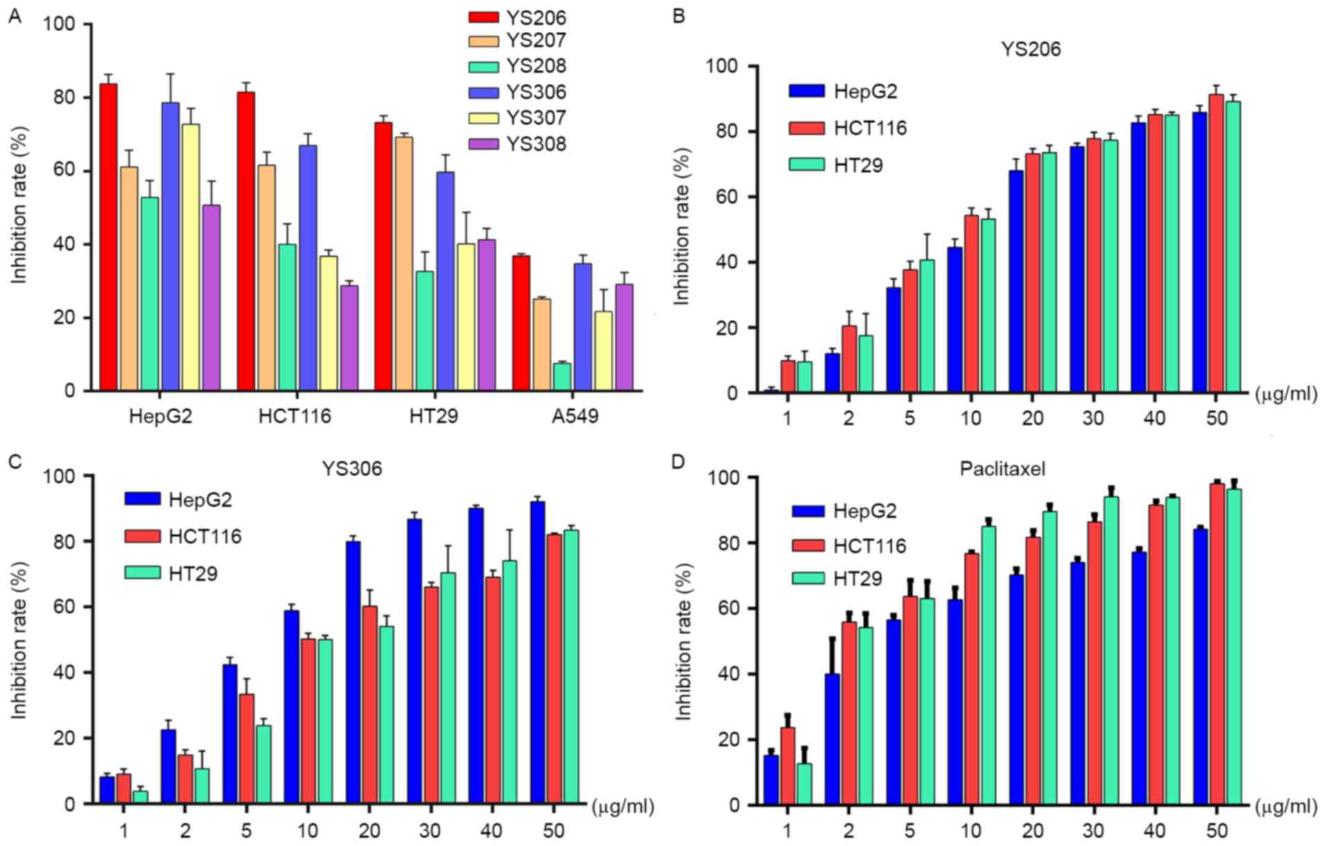
To quickly screen for inhibitory effects of PAs,
cancer cells were incubated with the compound for 24 h the status
of cell growth was observed. No inhibitory effect on cell growth
was observed using 0.5 or 5 µg/ml PA for HepG2, HCT116 or HT29
cells (data not shown). When incubated with 50 µg/ml PAs, increased
cell growth inhibition was noted for each of these three cancer
cell lines. A total of 6 out of the 12 PA analogues analyzed,
including YS206, YS207, YS208, YS306, YS307 and YS308, exhibited
in vitro anticancer activity (Fig. 2A). From the primary experimental
results, it was clear that 50 µg/ml PA compounds exhibited the most
effective anticancer activity on HepG2, HCT116 and HT29 cells
(Fig. 2A), whereas none of the
tested chemicals exhibited anticancer effects on A549 cells.
Among the six PA compounds, YS206 and YS306
exhibited the most efficient growth inhibition effects on colon and
liver cancer cells. Dose-response histograms for YS206 and YS306,
at concentrations of between 1 and 50 µg/ml, on HepG2, HCT116 and
HT29 cells suggested that their biological effects may be
concentration dependent (Fig. 2B and
C). The average half-maximal inhibitory concentration
(IC50) value of the YS206 against HepG2, HCT116 and HT29
cells were 10.26, 9.528 and 8.15 µg/ml, respectively. The
IC50 of YS306 on these cells was 6.826, 8.483 and 12.35
µg/ml, respectively. Compared with the IC50 of
paclitaxel for HepG2, HCT116 and HT29 cells (3.26, 1.89 and 1.91
µg/ml, respectively; Fig. 2D), the
in vitro antitumor activity of YS206 and YS306 were slightly
lower than paclitaxel. The compound YS206 appeared to exhibit a
stronger growth inhibition against the colon cancer HCT116 and HT29
cells compared with YS306 on the same cells, whereas YS306 appeared
to have a more effective inhibitory effect on HepG2 liver cancer
cells.
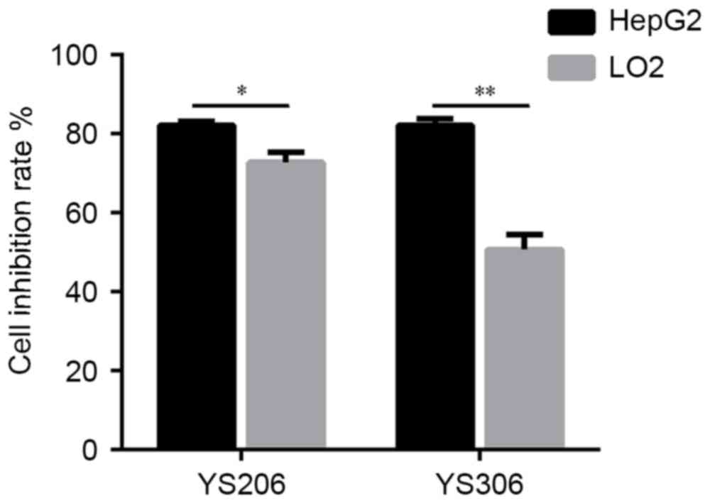
The cytotoxicity of YS206 and YS306 was examined on
LO2 normal liver cells and HepG2 cells. HepG2 cells appeared to be
more sensitive to the treatment with 50 µg/ml YS206 and YS306
compared with LO2 cells (Fig. 3).
The cell proliferation rate was 72.76 and 50.68% for HepG2 cells
incubated with YS206 and YS306, respectively, whereas LO2 cells
exhibited ~80% proliferation rate when incubated with either
compound (Fig. 3). These results
indicated that compounds YS206 and YS306, particularly YS306, may
target liver cancer cells with lower cytotoxicity for normal liver
cells.
Based on these primarily experiments with several
cancer cell lines, the potential biological effects of YS206 were
further examined on the colon cancer HCT116 and HT29 cells, and the
effects of YS306 were further investigated on HepG2 liver cancer
cells.
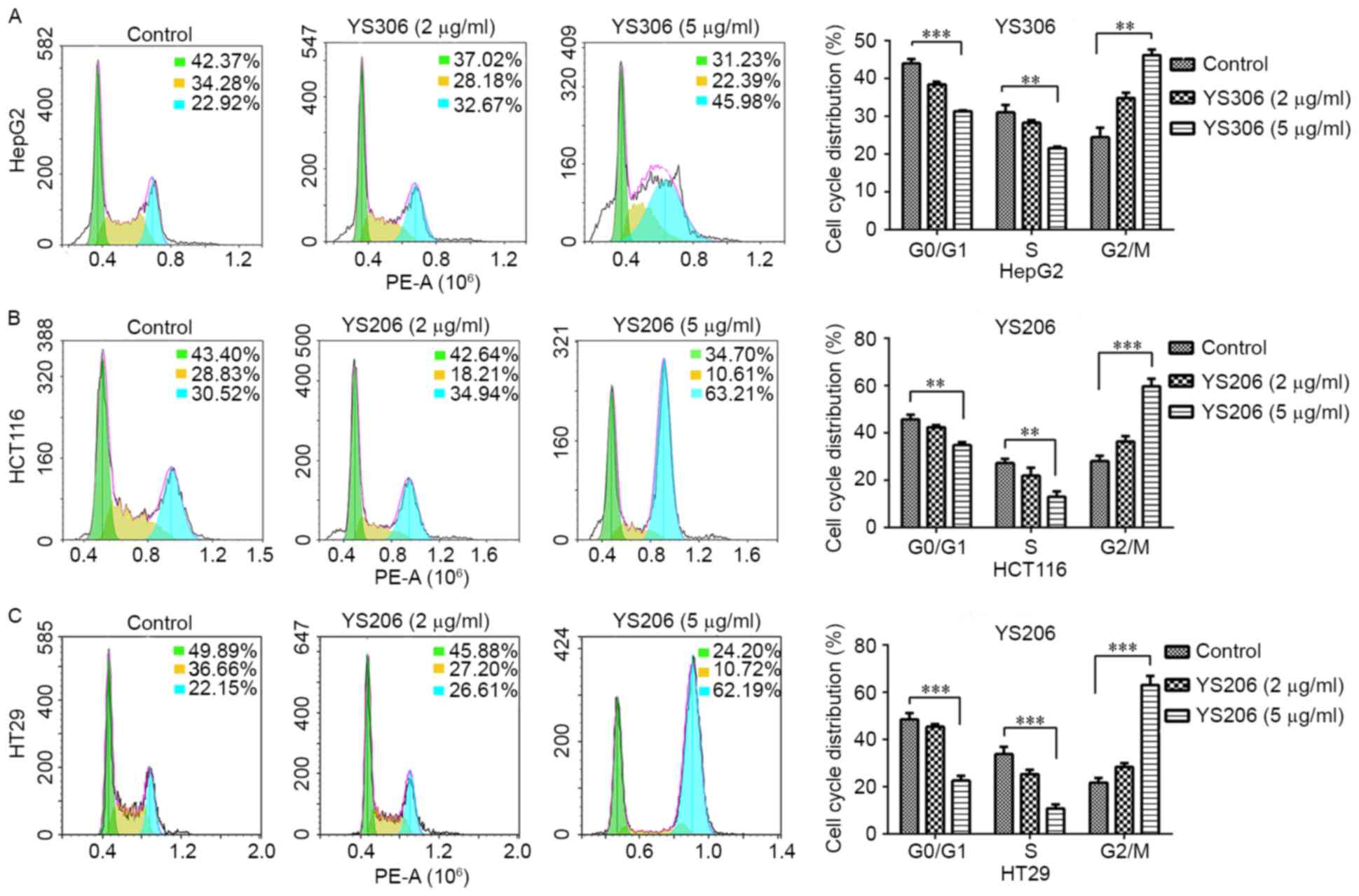
YS206 and YS306 induce cell cycle
arrest at G2/M phase
To investigate whether compounds YS206 and YS306
inhibit cell proliferation by regulating cell cycle progression,
changes to the cell cycle were detected following chemical
treatment for 24 h by PI staining and flow cytometry analysis.
YS206 and YS306 (5 µg/ml) both significantly induced cell cycle
arrest at G2/M phase (P<0.01; Fig.
4), and the distribution of cells in G0/G1 and S phases were
notably decreased compared with the control cells. In the HepG2
cells treated with YS306 (5 µg/ml) the percentage of cells at G2-M
increased to 45.98%, which was twice that of the control group
(22.92%; Fig. 4A). Compared with
the untreated control, in which 42.37 and 34.28% of cells were at
G0-G1 and S phase, respectively, HepG2 treated with YS306 had 31.23
and 22.39% of cells at the respective phases. However, no
significant differences were identified when these cells were
treated with 2 µg/ml YS306.
Both of the colon cancer cell lines treated with
YS206 (5 µg/ml) exhibited a significantly increased number of cells
arrested at the G2-M phase (Fig. 4B
and C). The percentage of HCT116 cells treated with YS206
arrested at G2-M increased ~2.07-fold, from 30.52% in the untreated
cells to 63.21% (Fig. 4B).
Additionally, the number of HCT116 cells at G0-G1 and S were
respectively decreased from 43.40 and 28.83% in the control to
34.70 and 10.61% with 5 µg/ml YS206. Similar results were observed
in HT29 cells incubated with 5 µg/ml YS206 (Fig. 4C). However, no significant
differences were identified in either HCT116 or HT29 cells treated
with 2 µg/ml of YS306.
YS306 and YS206 inhibit cell
migration
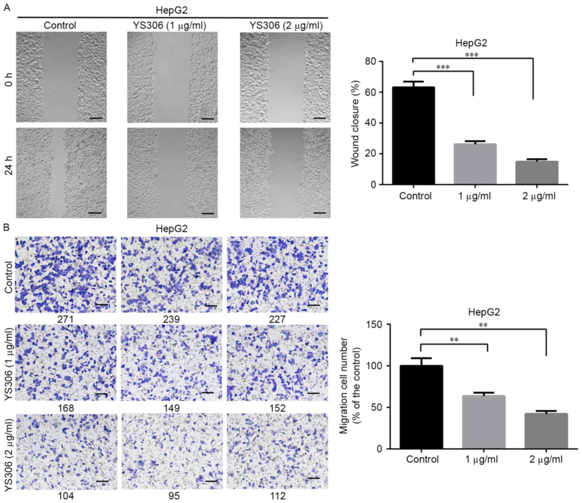
The effects of YS306 and YS206 on cell migration
were also examined. YS306 (1–2 µg/ml) significantly inhibited HepG2
cell migration Fig. 5) and YS206
(1–2 µg/ml) greatly reduced colon cancer migration in vitro
(Figs. 6 and 7). In the scratch wound-healing assay,
the wound closure rate of HepG2 cells was notably decreased when
treated with 1 µg/ml (26.2%) or 2 µg/ml (15.0%) compared with
untreated control HepG2 cells (63.2%; P<0.001; Fig. 5A). Similar reductions in migration
ability were observed using the Transwell assay (Fig. 5B), in which the number of migrating
cells was reduced to 63.6 and 43.1% with 1 or 2 µg/ml YS306,
respectively, compared with untreated control group (n=3;
P<0.01; Fig. 5B).
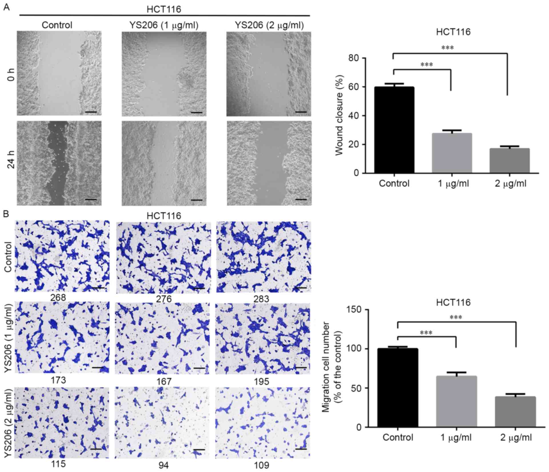
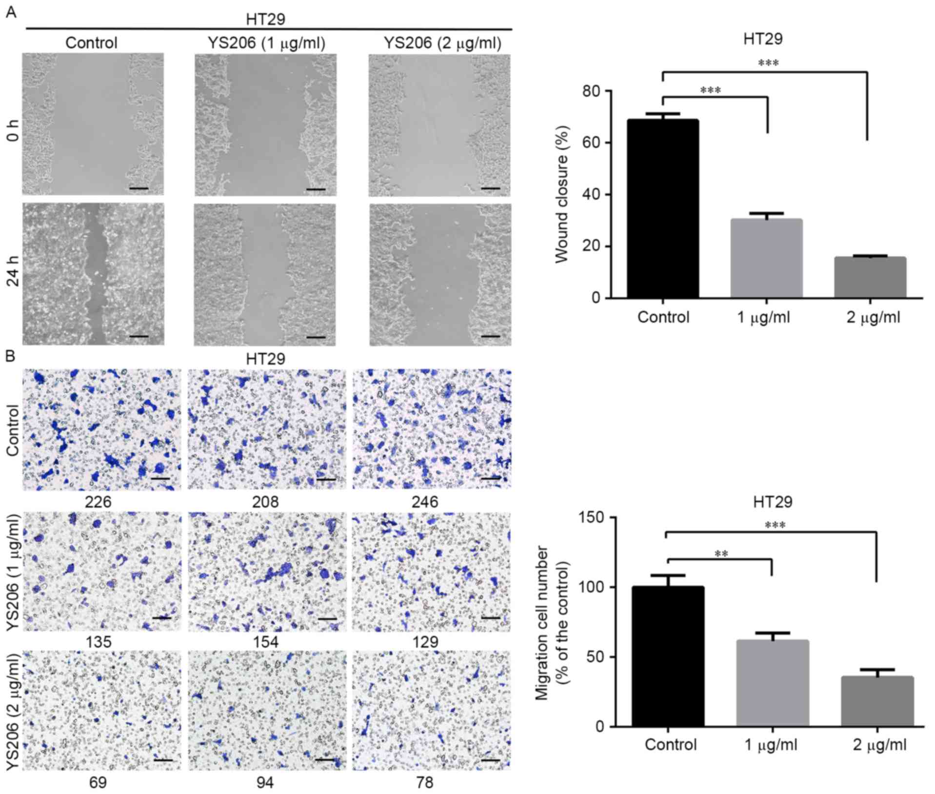
YS206 exhibited similar inhibitory effects on cell
migration of HCT116 and HT29 cells. For HCT116 cells, the wound
closure rate was about 27.5 and 16.9% with 1 or 2 µg/ml YS206
treatment, compared with 59.7% coverage in the control cells
(Fig. 6A). In the Transwell assay,
compared with the untreated control group, cell migration was
decreased to 64.7, 38.4% in response to 1, 2 µg/ml YS206 treatment
(µg/ml) (n=3; P<0.001; Fig.
6B). For HT29 cells, the wound closure rates were 30.2 and
15.6% when treated with 1 or 2 µg/ml YS206, compared with the 68.3%
coverage in the untreated control (Fig. 7A). Transwell assay analysis
revealed that HT29 cells treated with 1 or 2 µg/ml YS206 exhibited
61.5 and 35.4% cell migration compared with cells in the untreated
control group (n=3; P<0.001; Fig.
7B).
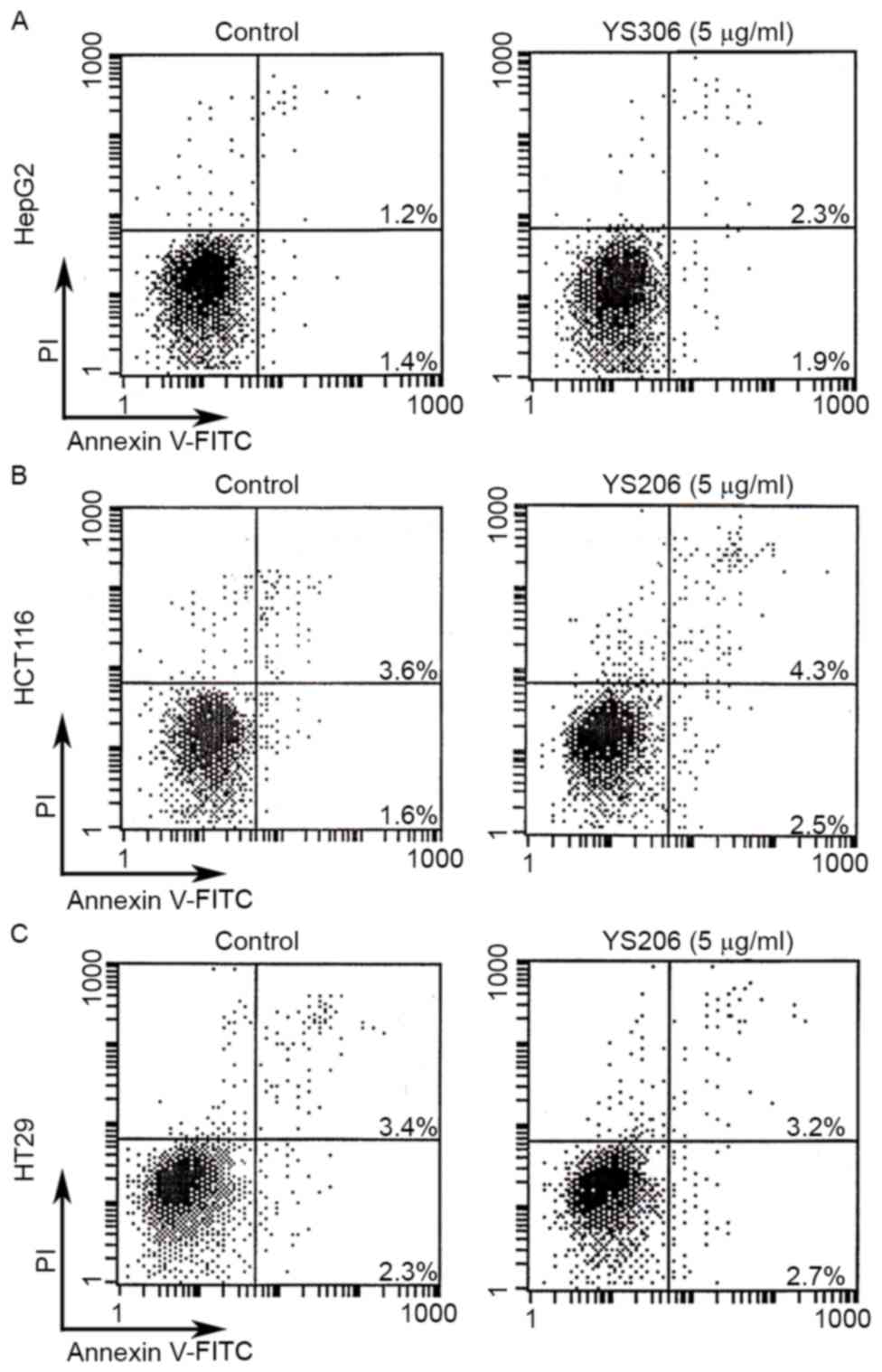
YS306 and YS206 have no effects on
cell apoptosis
The effects of YS306 and YS206 on cell apoptosis
were examined by Annexin V-FITC/PI staining and flow cytometry.
HepG2 cells were incubated with 5 µg/ml YS306, and HCT116 and HT29
cells were incubated with 2 or 5 µg/ml YS206. Cancer cells were
also incubated with 0.01% DMSO as a negative control. However, no
significant differences in the levels of cell apoptosis were
identified for any of the PA treatments (Fig. 8).
Discussion
Over the past ten years, there has been rapid
development of organic synthesis technologies, and subsequently, an
increasing number of alkaloids have been synthesized, which
provides more choices for clinical therapy and experimental
antitumor research (12). These
naturally occurring products and compound derivatives have
exhibited great efficacy as antiviral, antibacterial and antitumor
treatments (13).
The anti-proliferation activity in different cancer
cells is usually the first effect to be examined when evaluating
potential new antitumor agents. The IC50 values of the
compounds synthesized by the present study were mainly at a level
of micromolar concentration, and among them, YS206 and YS306 were
selected for further analysis. Compared with the anticancer drug
paclitaxel, these two compounds exhibited potential growth
inhibition effects on HepG2, HCT116 and HT29 cells with relatively
low toxicity on normal cells. For compound YS306, the average
inhibition rate for HepG2 cells was higher than for LO2 human
normal liver cells, by 1.6-fold, suggesting that YS306 may have
specificity in inhibiting liver cancer cell growth. In addition,
YS306 exhibited greater anti-proliferation effects on HepG2 liver
cancer cells compared with HCT116 and HT29 colon cancer cells. It
was noticed that the only difference between compounds S306 and
S206 is one ethoxyethyl at the 1′ position (Fig. 1), which may contribute to the
difference in cell preference of these two chemical molecules. Two
other, similar compounds within these series, YS307 and YS207 were
slightly inferior in solubility than YS306 and YS206, which leads
to poor cell inhibition activity. In future studies, these PA
compounds may be modified to improve their anticancer activity
in vitro, and nude mouse xenografts may then be performed to
explore their effects in suppressing cancer cell growth and
metastasis in vivo.
Previous studies have indicated that PAs inhibit
cell proliferation through many different mechanisms, including
inhibition of protein and DNA synthesis, induction of cell
apoptosis and suppression of dihydrofolate reductase activity
(14–16). Additional studies have demonstrated
that PAs may also enhance cytotoxic effects and inhibit cell
migration and metastasis of cancer cells (17,18).
To investigate the cell biological effects induced by YS206 and
YS306 treatment, cell cycle distribution, cell migration and cell
apoptosis were analyzed in the three cancer cell lines
aforementioned. HepG2, HCT116 and HT29 cells treated with YS206 or
YS306 exhibited reduced proliferation, significant changes to the
cell cycle and suppressed cell migration. The low concentration of
5 µg/ml YS206 and YS306 was able to induce cell cycle arrest at
G2/M phase. Compared with normal cells, cancer cells are
characteristically sufficient in stimulating their own growth,
which results in abnormal proliferation through the dysregulation
of their cycle progression and fission activities (19). G1/S and G2/M are the most complex
stages of the cell cycle, which are influenced by environmental
conditions (20). Therefore, the
development of new drugs to suppress tumor growth should target
these two cell stages.
Pinosylvin is a naturally occurring trans-stilbenoid
that mainly occurs in Pinus species (21), which has been revealed to regulate
the cell cycle by through the cyclin-dependent kinase complex
(20,22). The novel triterpenoid
25-methoxyhispidol, extracted from Poncirustrifoliata, was
demonstrated to exhibit growth inhibition activity against cancer
cells, with cell cycle arrested at G0/G1 phase (23). Antofine is a naturally occurring PA
found in Cynanchum paniculatum, from which some analogues
and derivatives have also been reported to regulate the cell cycle
at G0-G1 phase (24). In the
present study, 5 µg/ml YS306 and YS206 significantly induced cell
cycle arrest at G2/M phase in HepG2, HCT116 and HT29 cells; cell
distribution in G0/G1 and S phases were notably decreased compared
with Control cells, and the effect was more significant than using
2 µg/ml YS306 and YS206. The present results are similar to a
previous report on human colon cancer Col2 cells that were induced
by another natural antofine that was isolated from Cynanchum
paniculatum (25). Moreover,
vinblastine and vincristine, which are extracts from
Catharanthusroseus, were demonstrated to affect the G2/M
phase to inhibit microtubule assembly (26).
In addition to acting on cell cycle, YS306 and YS206
significantly inhibited cell migration in HepG2, HCT116 and HT29
cells. Cell migration in vitro, or cell metastasis in
vivo, is one of the fundamental features of malignant tumors,
which is one primary cause of mortality in patients with cancer
(27). YS306 and YS206 may be
promising compounds for targeting cancer cell metastasis; however,
neither compound exhibited effects on cell apoptosis, as determined
by flow cytometric analysis following 5 µg/ml YS306 or YS206
treatment in HepG2, HCT116 and HT29 cells. These results were
similar to previous reports on the PA antofine and tylophorine
(24,28), which demonstrated that the
distribution of sub-G1 phase was not notably altered, and that cell
growth inhibition of human lung cancer A549 cells by derivatives of
antofine is not due to cell apoptosis.
Considering the chemical structures of YS306 and
YS206, the biggest difference in their structure is the side chain
compared with the classical PA antofine. In the present study, the
lateral heterocyclic nitrogen was opened and the number of side
element carbons were decreased at the same time, which brought
about a change in polarity. Compounds with increased polarity have
been indicated to possess good anticancer activity, and the
methoxyl group in different positions of the phenanthrene ring may
be another reason for anticancer abilities (29–31).
In addition, a previous report revealed that water solubility may
be a limitation in clinical use (28). In the present study, YS206 and
YS306 had better water solubility than the other 10 PAs, which may
be why the two compounds have more effective anti-cancer activity
than other derivatives.
A number of previous studies have indicated that PAs
may inhibit cancer cell proliferation through different molecular
mechanisms, including inhibition of protein and DNA synthesis,
suppression of dihydrofolate reductase and induction of apoptosis
through the tumor necrosis factor α signaling pathway (14,15,23).
In conclusion, the mechanisms of anti-proliferation and inhibition
of cell migration of YS306 and YS206 may be relative to the
suppression of related protein synthesis and cell signal
transduction pathways, which needs to be analyzed in detail in
future studies.
Acknowledgements
The present study was financially supported by the
National Key Basic Research Program of China (grant nos.
2013CB911303 and 2011CB910703), National 863 High Tech Program
(grant no. 2014AA020608), the National Natural Sciences Foundation
of China (grant no. 31470810), the Science & Technology
Department of Sichuan Province (grant no. 2017JY0232), and the
Health and Family Planning Commission of Sichuan Province (grant
no. 17ZD045).
References
|
1
|
Torre LA, Bray F, Siegel RL, Ferlay J,
Lortet-Tieulent J and Jemal A: Global cancer statistics, 2012. CA
Cancer J Clin. 65:87–108. 2015. View Article : Google Scholar : PubMed/NCBI
|
|
2
|
Chen W, Zheng R, Baade PD, Zhang S, Zeng
H, Bray F, Jemal A, Yu XQ and He J: Cancer statistics in China,
2015. CA Cancer J Clin. 66:115–132. 2016. View Article : Google Scholar : PubMed/NCBI
|
|
3
|
Newman DJ and Cragg GM: Natural products
as sources of new drugs over the 30 years from 1981 to 2010. J Nat
Prod. 75:311–335. 2012. View Article : Google Scholar : PubMed/NCBI
|
|
4
|
Cushnie TT, Cushnie B and Lamb AJ:
Alkaloids: An overview of their antibacterial, antibiotic-enhancing
and antivirulence activities. Int J Antimicrob Agents. 44:377–386.
2014. View Article : Google Scholar : PubMed/NCBI
|
|
5
|
Saraswati S, Kanaujia PK, Kumar S, Kumar R
and Alhaider AA: Tylophorine, a phenanthraindolizidine alkaloid
isolated from Tylophora indica exerts antiangiogenic and
antitumor activity by targeting vascular endothelial growth factor
receptor 2-mediated angiogenesis. Mol Cancer. 12:822013. View Article : Google Scholar : PubMed/NCBI
|
|
6
|
Ibrahim SR and Mohamed GA: Marine
pyridoacridine alkaloids: Biosynthesis and biological activities.
Chem Biodivers. 13:37–47. 2016. View Article : Google Scholar : PubMed/NCBI
|
|
7
|
Ansha C and Mensah K: A review of the
anticancer potential of the antimalarial herbal cryptolepis
sanguinolenta and its major alkaloid cryptolepine. Ghana Med J.
47:137–147. 2014.
|
|
8
|
Staerk D, Lykkeberg AK, Christensen J,
Budnik BA, Abe F and Jaroszewski JW: In vitro cytotoxic activity of
phenanthroindolizidine alkaloids from Cynanchum vincetoxicum
and Tylophora tanakae against drug-sensitive and
multidrug-resistant cancer cells. J Nat Prod. 65:1299–1302. 2002.
View Article : Google Scholar : PubMed/NCBI
|
|
9
|
Su CR, Damu AG, Chiang PC, Bastow KF,
Morris-Natschke SL, Lee KH and Wu TS: Total synthesis of
phenanthroindolizidine alkaloids (±/−)-antofine,
(±/−)-deoxypergularinine, and their dehydro congeners and
evaluation of their cytotoxic activity. Bioorg Med Chem.
16:6233–6241. 2008. View Article : Google Scholar : PubMed/NCBI
|
|
10
|
Jin X, Liu Y, Liu J, Lu W, Liang Z, Zhang
D, Liu G, Zhu H, Xu N and Liang S: The overexpression of IQGAP1 and
β-catenin is associated with tumor progression in hepatocellular
carcinoma in vitro and in vivo. PLoS One. 10:e01337702015.
View Article : Google Scholar : PubMed/NCBI
|
|
11
|
Zhou J, Liang S, Fang L, Chen L, Tang M,
Xu Y, Fu A, Yang J and Wei Y: Quantitative proteomic analysis of
HepG2 cells treated with quercetin suggests IQGAP1 involved in
quercetin-induced regulation of cell proliferation and migration.
OMICS. 13:93–103. 2009. View Article : Google Scholar : PubMed/NCBI
|
|
12
|
Shang S, Monfregola L and Caruthers MH:
Peptide-substituted oligonucleotide synthesis and non-toxic,
passive cell delivery. Sig Trans Target Ther. 16019:2016.
|
|
13
|
Newman DJ, Cragg GM and Snader KM: Natural
products as sources of new drugs over the period 1981–2002. J Nat
Prod. 66:1022–1037. 2003. View Article : Google Scholar : PubMed/NCBI
|
|
14
|
Lv H, Ren J, Ma S, Xu S, Qu J, Liu Z, Zhou
Q, Chen X and Yu S: Synthesis, biological evaluation and mechanism
studies of deoxytylophorinine and its derivatives as potential
anticancer agents. PLoS One. 7:e303422012. View Article : Google Scholar : PubMed/NCBI
|
|
15
|
Rao KN and Venkatachalam S: Inhibition of
dihydrofolate reductase and cell growth activity by the
phenanthroindolizidine alkaloids pergularinine and tylophorinidine:
The in vitro cytotoxicity of these plant alkaloids and their
potential as antimicrobial and anticancer agents. Toxicol In Vitro.
14:53–59. 2000. View Article : Google Scholar : PubMed/NCBI
|
|
16
|
Ueno S, Yamazaki R, Ikeda T, Yaegashi T
and Matsuzaki T: Antitumor effect of a novel phenanthroindolizidine
alkaloid derivative through inhibition of protein synthesis.
Anticancer Res. 34:3391–3397. 2014.PubMed/NCBI
|
|
17
|
Song J, Kwon Y, Kim S and Lee SK:
Antitumor activity of phenanthroindolizidine alkaloids is
associated with negative regulation of met endosomal signaling in
renal cancer cells. Chem Biol. 22:504–515. 2015. View Article : Google Scholar : PubMed/NCBI
|
|
18
|
Wu TS, Su CR and Lee KH: Cytotoxic and
anti-HIV phenanthroindolizidine alkaloids from Cryptocarya
chinensis. Nat Prod Commun. 7:7252012.PubMed/NCBI
|
|
19
|
Hanahan D and Weinberg RA: The hallmarks
of cancer. Cell. 100:57–70. 2000. View Article : Google Scholar : PubMed/NCBI
|
|
20
|
Schwartz GK and Shah MA: Targeting the
cell cycle: A new approach to cancer therapy. J Clin Oncol.
23:9408–9421. 2005. View Article : Google Scholar : PubMed/NCBI
|
|
21
|
Park EJ, Chung HJ, Park HJ, Kim GD, Ahn YH
and Lee SK: Suppression of Src/ERK and GSK-3/β-catenin signaling by
pinosylvin inhibits the growth of human colorectal cancer cells.
Food Chem Toxicol. 55:424–433. 2013. View Article : Google Scholar : PubMed/NCBI
|
|
22
|
Matsushime H, Quelle DE, Shurtleff SA,
Shibuya M, Sherr CJ and Kato JY: D-type cyclin-dependent kinase
activity in mammalian cells. Mol Cell Biol. 14:2066–2076. 1994.
View Article : Google Scholar : PubMed/NCBI
|
|
23
|
Chung HJ, Park EJ, Pyee Y, Xu G Hua, Lee
SH, Kim YS and Lee SK: 25-Methoxyhispidol A, a novel triterpenoid
of Poncirus trifoliata, inhibits cell growth via the
modulation of EGFR/c-Src signaling pathway in MDA-MB-231 human
breast cancer cells. Food Chem Toxicol. 49:2942–2946. 2011.
View Article : Google Scholar : PubMed/NCBI
|
|
24
|
Min HY, Chung HJ, Kim EH, Kim S, Park EJ
and Lee SK: Inhibition of cell growth and potentiation of tumor
necrosis factor-α (TNF-α)-induced apoptosis by a
phenanthroindolizidine alkaloid antofine in human colon cancer
cells. Biochem Pharmacol. 80:1356–1364. 2010. View Article : Google Scholar : PubMed/NCBI
|
|
25
|
Lee SK, Nam KA and Heo YH: Cytotoxic
activity and G2/M cell cycle arrest mediated by antofine, a
phenanthroindolizidine alkaloid isolated from Cynanchum
paniculatum. Planta Med. 69:21–25. 2003. View Article : Google Scholar : PubMed/NCBI
|
|
26
|
Himes RH, Kersey RN, Heller-Bettinger I
and Samson FE: Action of the vinca alkaloids vincristine,
vinblastine, and desacetyl vinblastine amide on microtubules in
vitro. Cancer Res. 36:3798–3802. 1976.PubMed/NCBI
|
|
27
|
Cheng GZ, Chan J, Wang Q, Zhang W, Sun CD
and Wang LH: Twist transcriptionally up-regulates AKT2 in breast
cancer cells leading to increased migration, invasion, and
resistance to paclitaxel. Cancer Res. 67:1979–1987. 2007.
View Article : Google Scholar : PubMed/NCBI
|
|
28
|
Kwon Y, Song J, Lee B, In J, Song H, Chung
HJ, Lee SK and Kim S: Design, synthesis, and evaluation of a
water-soluble antofine analogue with high antiproliferative and
antitumor activity. Bioorg Med Chem. 21:1006–1017. 2013. View Article : Google Scholar : PubMed/NCBI
|
|
29
|
Gao W, Bussom S, Grill SP, Gullen EA, Hu
YC, Huang X, Zhong S, Kaczmarek C, Gutierrez J, Francis S, et al:
Structure-activity studies of phenanthroindolizidine alkaloids as
potential antitumor agents. Bioorg Med Chem Lett. 17:4338–4342.
2007. View Article : Google Scholar : PubMed/NCBI
|
|
30
|
Wang Z, Wu M, Wang Y, Li Z, Wang L, Han G,
Chen F, Liu Y, Wang K, Zhang A, et al: Synthesis and SAR studies of
phenanthroindolizidine and phenanthroquinolizidine alkaloids as
potent anti-tumor agents. Eur J Med Chem. 51:250–258. 2012.
View Article : Google Scholar : PubMed/NCBI
|
|
31
|
Fu Y, Lee SK, Min HY, Lee T, Lee J, Cheng
M and Kim S: Synthesis and structure-activity studies of antofine
analogues as potential anticancer agents. Bioorg Med Chem Lett.
17:97–100. 2007. View Article : Google Scholar : PubMed/NCBI
|