Introduction
Osteoarthritis (OA) is characterized by a gradual
degradation of articular cartilage and pain, resulting in
significant disability in patients, which consumes a significant
amount of healthcare resources (1). Articular cartilage consists of
abundant extracellular matrix that is synthesized by resident
cells, named chondrocytes. Increased apoptotic chondrocyte death
has been detected in OA cartilage, which leads to the matrix
degradation and the loss of the lubricant and swelling properties
to the joint (2). Interleukin
(IL)-1β is considered to be the most important inflammatory
cytokine involved in multiple pathological processes of OA.
Patients with OA have an elevated level of IL-1β in both synovial
fluid and cartilage, and chondrocytes subjected to IL-1β tend to
induce apoptosis (3).
Astragalus membranaceus is a widely used in
traditional Chinese medicine, particularly for autoimmune diseases
(4). The extracts of Astragalus
membranaceus (Fisch) contain various active ingredients,
including saponins, polysacharides and flavonoids. Astragaloside IV
(AST) is a natural saponin purified from Astragalus, which exerts
numerous therapeutic effects. A previous study has indicated that
AST may protect against IL-1β-induced joint inflammation and
cartilage damage in rat adjuvant-induced arthritis (5). However, to date, the role and
mechanism of AST on the IL-1β-induced chondrocyte apoptosis remain
unclear.
Autophagy is a self-degradation process for
balancing sources of energy in response to cell stress (6). Autophagy has been proven to be
important for cartilage homeostasis (7). Autophagy is increased in OA
chondrocytes and cartilage, particularly during the early stage of
the degenerative phase, which acts as an adaptive response to
protect chondrocyte from sublethal conditions (8). Sasaki et al (9) reported that autophagy activation
could inhibit IL-1β-induced apoptosis in chondrocytes. Furthermore,
AST demonstrated beneficial effects against mitochondrial
dysfunction through promotion of autophagy in rat vascular smooth
muscle cells (10). Thus, the
authors hypothesized that AST may serve a protective role in
chondrocytes against IL-1β-induced apoptosis, and its potential
mechanisms may be responsible to autophagy activation.
Materials and methods
Primary chondrocyte isolation and
culture
Primary articular chondrocytes were isolated from 7
patients with OA undergoing total knee replacement surgery. Written
informed consent was obtained from all tissue donors and the
present study approach was approved by Ethics Committee of Beijing
University of Chinese Medicine (Beijing, China) before surgery. The
human degenerative chondrocytes were isolated and cultured
similarly as described previously (11). In brief, articular cartilage was
first separated from harvested joint tissues microscopically
according to their macroscopic morphologies. Following washing by
PBS buffer, cartilage was dissected and digested with by 0.25%
trypsin solution and 0.2% type II collagenase (Sigma-Aldrich; Merck
KGaA, Darmstadt, Germany) at 37°C for 5 h. Chondrocytes were
isolated by a 200 µm filter, and resuspended in Dulbecco's modified
Eagle's medium/Ham's F-12 (DMEM/F12) medium (Gibco; Thermo Fisher
Scientific, Inc., Waltham, MA, USA) containing 10% fetal bovine
serum (FBS; Invitrogen; Thermo Fisher Scientific, Inc.).
Chondrocytes were monolayer cultured at 37°C under 5%
CO2 and 95% oxygen in humidified air; cells between
passages 1 and 3 were used for subsequent experiments.
Cell treatment
AST (Beijing Solarbio Science & Technology Co.,
Ltd., Beijing, China) was dissolved in dimethyl sulfoxide (DMSO) to
produce a stock solution, the final concentration of AST used in
the current study was 50 µg/ml. Differentiated chondrocytes were
subjected to DMEM/F12 containing 1% FBS for 12 h prior to exposure
to various treatment conditions. Subsequently, cells were
pre-incubated with AST (50 µg/ml) for 2 h and then incubated with
10 ng/ml IL-1β for a further 24 h. In addition, chondrocytes were
transfected with adenovirus containing GFP-LC3 to monitor the
autophagic level changes. Finally, 3-methyladenine (3MA; 5 mM) and
rapamycin (100 nM) were used as a chemical inhibitor or activator
for autophagy, which were applied 2 h prior to further treatment.
Chondrocytes treated with DMEM/F12 medium with 10% FBS was
considered as the control.
Cell Counting kit (CCK)-8
measurement
Chondrocyte viability was detected by CCK-8 (Promega
Corporation, Madison, WI, USA). Briefly, chondrocytes were seeded
into a 96-well plate with concentration of 1×10⁴
cells/well overnight prior to treatment. Following exposeure to
IL-1β or AST, 10 µl CCK-8 solution was added to each well and
incubated for 2 h at 37°C. Optical density was determined at 450 nm
using a microplate reader (Thermo Fisher Scientific, Inc.). The
cellular viability was defined as a percentage of the control
group.
Flow cytometry analysis
The apoptotic incidence was evaluated by the Annexin
V-phycoerythrin (PE)/propidium iodide (PI) apoptosis detection kit
(BD Biosciences, Franklin Lakes, NJ, USA) according to the
manufacturer's instructions. Briefly, treated chondrocytes were
collected and washed by PBS buffer, then incubated with 5 µl
Annexin V-PE and 5 µl PI for 30 min at room temperature in the
dark. Finally, Ending buffer was added, and cell apoptotic rate was
analyzed using a flow cytometer (BD Biosciences).
Western blot analysis
Whole cells were lysed using
radioimmunoprecipitation assay lysis buffer (Beyotime Institute of
Biotechnology, Haimen, China) on ice. The protein concentration was
measured using a bicinchoninic acid protein assay kit (Beyotime
Institute of Biotechnology). Equal amounts of protein (40 µg) from
each sample were subjected to 10–15% SDS-PAGE and transferred to
polyvinylidene difluoride membranes. The membranes were blocked
with 5% bovine serum albumin and then incubated with primary
antibody against cleaved caspase-3 (dilution, 1:1,000; Cell
Signaling Technology, Inc., Danvers, MA, USA; cat no. 9661),
cleaved caspase-9 (dilution, 1:1,000; Cell Signaling Technology,
Inc.; cat no. 9501), cleaved poly (ADP-ribose) polymerase (PARP;
dilution, 1:1,000; Cell Signaling Technology, Inc.; cat no. 5625),
LC-3 (dilution, 1:500; Abcam, Cambridge, UK; cat no. ab48394),
P62/SQSTM1 (dilution, 1:500; Abcam; cat no. ab91526) and β-actin
(dilution, 1:5,000; Beyotime Institute of Biotechnology; cat no.
AF0003) at 4°C overnight. The next day, after washing with TBS with
1% Tween-20 (TBST), membranes were incubated with goat anti-mouse
or rabbit HRP-conjugated secondary antibodies (dilution, 1:1,000;
Beyotime Institute of Biotechnology; cat nos. A0216 and A0208) for
1 h at 37°C. Finally, the membranes were visualized by an enhanced
chemiluminescence detection kit (EMD Millipore, Billerica, MA,
USA). The protein expression levels were detected by Quantity One
software (version, 4.62; Bio-Rad Laboratories, Inc., Hercules, CA,
USA).
Caspase activity measurement
Cell apoptosis was also assayed by a caspase-3/9
activity assay kit (Beyotime Institute of Biotechnology; cat no.
C1115). In brief, whole cell protein was lysed and extracted as
aforementioned. Then, 100 µg protein from each sample was incubated
with 50 µl reaction buffer and 5 µl Ac-DEVD-pNA (4 mM) substrate at
37°C for 2 h. The absorbance at 405 nm was determined using a
microplate reader (Thermo Fisher Scientific, Inc.).
Confocal microscopic analysis
To visualize the autophagosome, cells were
transfected with adenovirus containing GFP-LC3 (Hanbio
Biotechnology Co., Ltd., Shanghai, China). Briefly, NP cells were
incubated in complete medium with the adenovirus at a multiplicity
of infection of 50 for 6 h, then replace fresh medium for another
24 h. The transfected cells were used for subsequent experiments.
Autophagy was evaluated by analyzing the number of green
fluorescent puncta of autophagosomes under laser confocal
microscopy.
Transmission electron microscopy
The treated cells were collected and fixed with 2.5%
glutaraldehyde overnight, subsequently fixed with 1% osmium
tetroxide for 1 h. The fixed specimens were dehydrated and embedded
in tetraoctadecylammonium bromide (TAAB) Epon resin (TAAB
Laboratories Equipment Ltd., Aldermaston, UK). The samples were cut
into 60 nm ultrathin sections and stained with uranyl acetate and
lead citrate. A transmission electron micoroscope (Hitachi-7500,
Hitachi, Ltd., Tokyo, Japan) was used for observing the cellular
ultrastructure at an accelerating voltage of 100 kV.
Statistical analysis
Quantitative data are presented as the mean ±
standard deviation. Statistical analyses were performed using
Student's t test or one-way analysis of variance by SPSS software
(version, 19.0; IBM SPSS, Armonk, NY, USA). P<0.05 was
considered to indicate a statistically significant difference.
Results
AST maintains chondrocyte viability
under IL-1β treatment
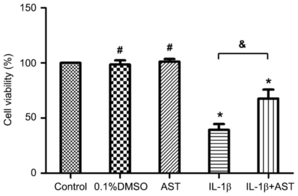
CCK-8 analysis was performed to evaluate the effect
of AST on chondrocyte viability under IL-1β treatment. Results
demonstrated that the cell viability was significantly decreased to
(39.3±5.3%) following stimulation with 10 ng/ml IL-1β. However,
pretreatment with AST (50 µg/ml) dramatically increased the cell
viability to (67.7±8.1%). Otherwise, the viabilities of the
chondrocytes were not altered significantly by AST or 0.1% DMSO
alone (Fig. 1). Therefore, AST
demonstrated a protective effect on maintaining chondrocyte
viability under IL-1β treatment.
AST protects against IL-1β-induced
chondrocyte apoptosis
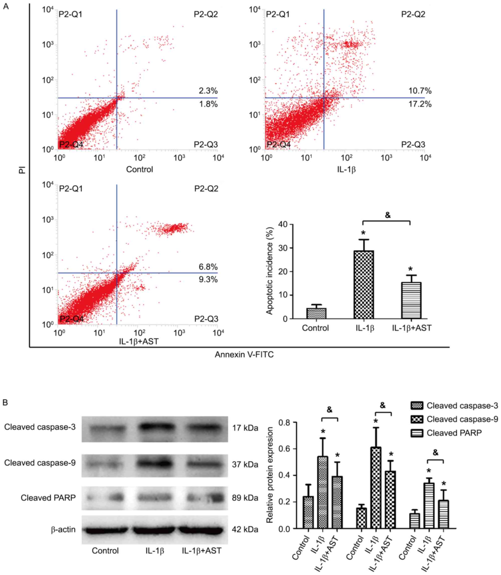
Annexin V-fluorescein isothiocyanate/propidium
iodide double staining by flow cytometry analysis was performed to
test the effect of AST on IL-1β-induced cell apoptosis. Results
indicated that the apoptotic ratio in the control group was
(4.3±1.6%), and IL-1β treatment led to a significant increase in
cell apoptosis to (28.7±4.8%). However, co-treatment of AST and
IL-1β presented an anti-apoptotic effect that decreased the
apoptotic ratio to (15.3±3.1%; Fig.
2A). Western blotting further confirmed that IL-1β alone
strongly increased the protein levels of cleaved caspase-3/9 and
cleaved PARP, while AST under treatment with IL-1β inhibited the
increase in these three pro-apoptotic protein expressions (Fig. 2B). These results indicated that
IL-1β-induced apoptosis of chondrocytes may be suppressed by
AST.
AST stimulates autophagic flux in
chondrocytes
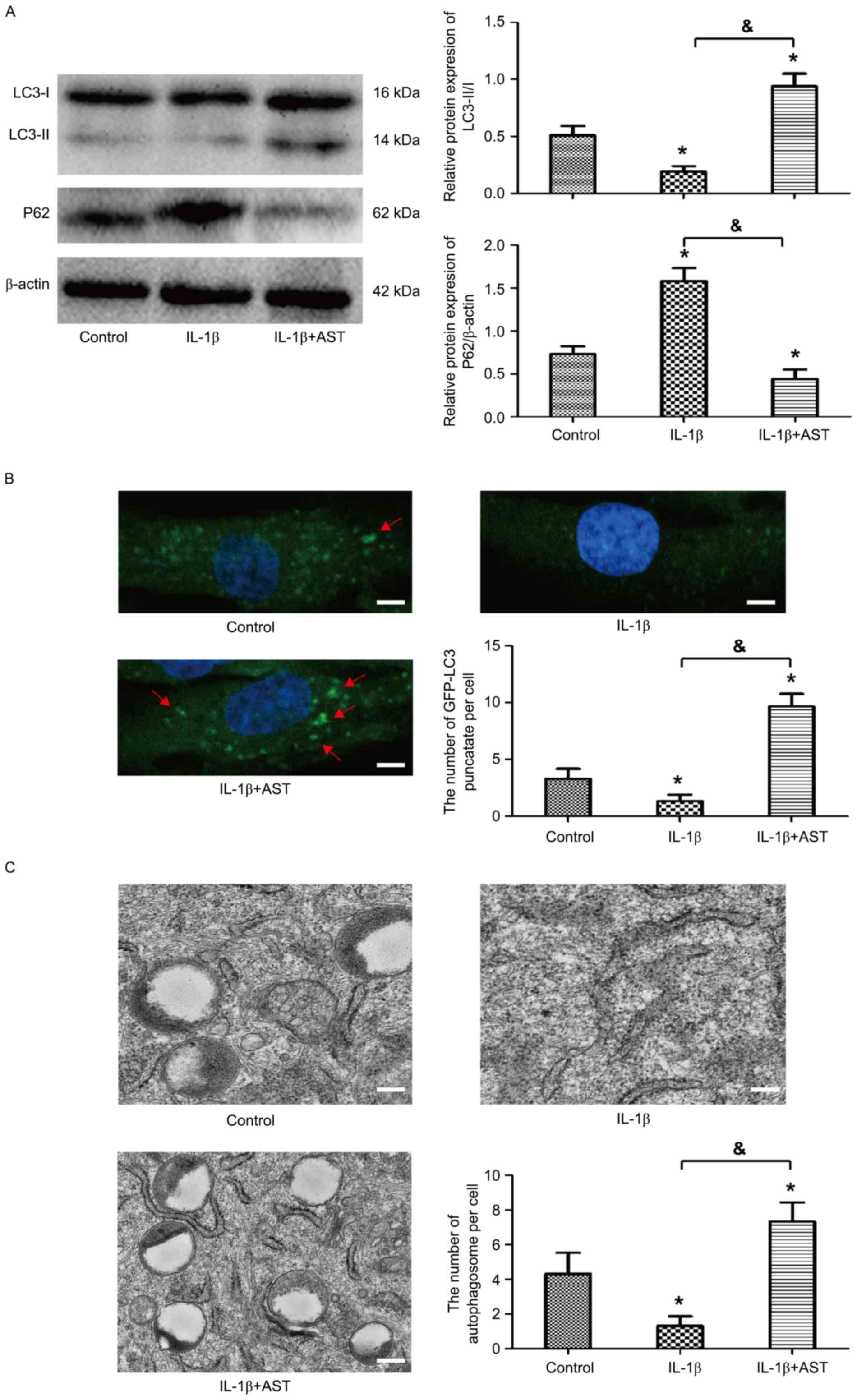
To investigate whether autophagy was activated in
chondrocytes stimulated by AST, western blot analysis was performed
to detect the protein levels of LC3 and P62/SQSTM1, which are
markers of autophagy. Results demonstrated that the expression of
LC3-II/I was downregulated significantly in chondrocytes with
treatment of IL-1β, in contrast, expression of P62/SQSTM1 was
obviously upregulated. However, AST presented promoting effect on
autophagic flux in chondrocytes, which reversed the expression of
LC-II/I and P62/SQSTM1 (Fig. 3A).
The autophagy activation was further confirmed by monitoring the
subcellular localization of fluorescent GFP-LC3 puncta. Confocal
microscopic analysis indicated that AST significantly increased the
fluorescent puncta in cytoplasm, and punctate green fluorescence
could hardly be observed with IL-1β treatment (Fig. 3B). Furthermore, TEM observation
demonstrated that the number of double-membrane autophagosomes was
much greater under AST treatment (Fig.
3C). All the evidence suggests that AST stimulated autophagy
response in the chondrocyte.
AST inhibits chondrocyte apoptosis by
promoting autophagy
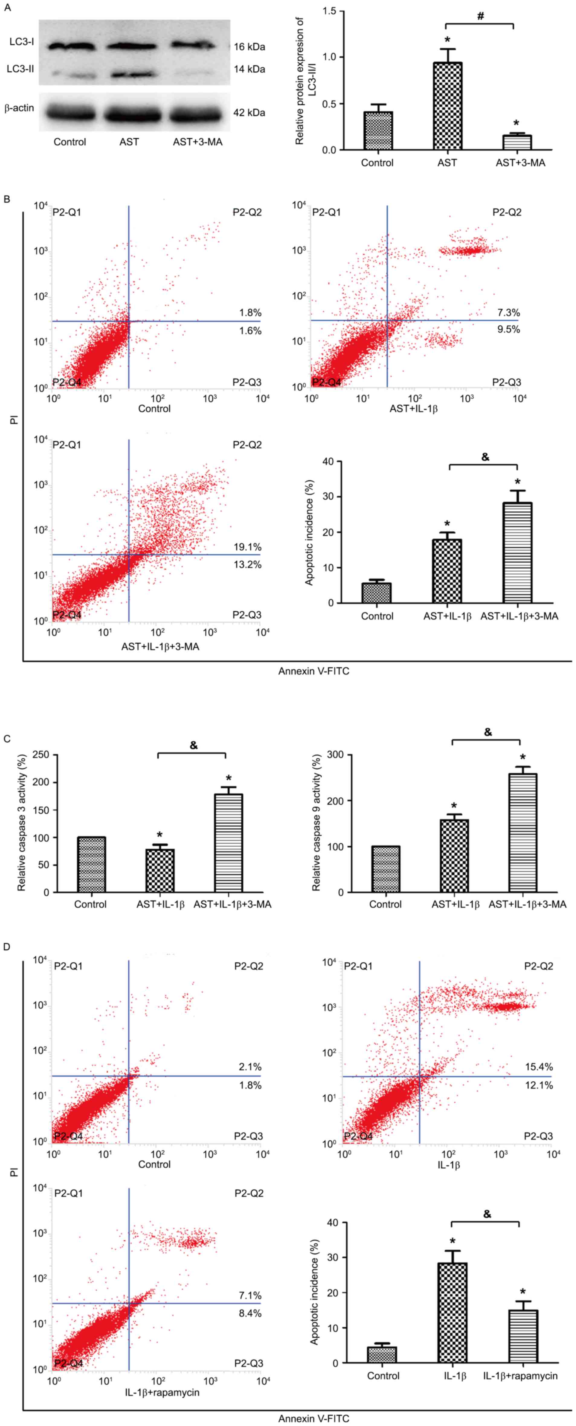
To investigate the interaction between autophagy and
apoptosis in chondrocytes under AST treatment, 3MA was used as a
chemical inhibitor of autophagy. 3MA treatment significantly
attenuated the LC3-II expression that indicated 3MA decreased the
autophagy incidence by AST treatment (Fig. 4A). Flow cytometric analysis
revealed that the suppressed apoptosis ratio by AST was obvious
increased under 3MA treatment (Fig.
4B). Colorimetric assay revealed that, under 3MA treatment, the
activities of caspase-3 and caspase-9 increased to 178 and 258%,
respectively, compared to the control group (Fig. 4C). Ultimately, to verify the exact
mechanism of autophagy in chondrocytes, rapamycin was used to
activate autophagy. The flow analysis presented similar results
with those treated by AST. Rapamycin also reversed the increased
apoptotic rate induced by IL-1β (Fig.
4D). These results suggested that activation of autophagy could
downregulate the IL-1β-induced apoptosis in chondrocytes under AST
treatment.
Discussion
To the best of the authors' knowledge, the present
study was the first to demonstrate that AST prevents against
IL-1β-induced apoptosis of chondrocytes via autophagy activation.
IL-1β was proven to reduce cell viability and increase apoptosis in
chondrocytes, however, AST significantly attenuated the
IL-1β-induced apoptosis and maintain cell viability. Furthermore,
AST treatment induced autophagic flux in chondrocytes, and, after
inhibiting this autophagy, the suppressed apoptosis ratio by AST
was reversed.
Increased expression of proinflammatory cytokines in
cartilage, synovial membrane and subchodral bone is related to the
pathological process in the OA joint (12). IL-1β is one of the most important
cytokines responsible for chondrocyte apoptosis and cartilage
matrix degradation. Therefore, IL-1β is considered as the potential
target in OA therapy. AST is a characteristic active saponin
compound present in Astragalus, which presents an immunomodulating
effect on lymphocyte apoptosis (13) and inflammatory cytokine production
(14). IL-1β was used to induce
chondrocyte apoptosis in the presence or absence of AST in the
present study. Results demonstrated that AST possessed the ability
to reverse the inhibition of cell proliferation and enhancement of
cell apoptosis induced by IL-1β. These findings suggested that AST
could be treated as a candidate for the antiarthritic treatment.
However, the underlying mechanism of anti-apoptotic effect by AST
needs to be further investigated.
The role of autophagy on the pathogenesis of OA is a
research hotspot during recent years. During the early stage of OA,
autophagy activation has been indicated to protect from limiting
catabolic degradation (9); while
in late OA, the level of autophagy decreases in chondrocytes, which
is associated with the increase of apoptosis of chondrocytes
(15). Previous studies have
suggested that autophagy serves a prominent influence on
chondrocyte survival. In aged mice, the number of autophagic
vesicles decreased, accompanied by reduced expression of LC3 and
increased expression of PARP, a marker of apoptosis (16). In addition, in neural cells, AST
can concomitantly inhibit multiple cell death pathways following
oxygen and glucose deprivation (17). Therefore, the authors speculated
that the anti-apoptotic role of AST in chondrocytes is also based
on autophagy activation. The present results indicated that IL-1β
decreased the expression ratio of LC-3II/I and increased the
expression of P62, suggesting IL-1β inhibited autophagy in
chondrocytes. However, AST treatment may be activating autophagy,
which reverses the expression of LC-3II/I and P62. This was further
confirmed by fluorescent microscopy and transmission electron
microscopy observation. All the evidence indicated that AST had the
effect of promoting autophagy in chondrocytes.
The effect of autophagy on apoptosis is varying
depending on the cell type and different stimulations (18). To further investigate the role of
autophagy by AST on the apoptosis under IL-1β stimulation,
chondrocytes were pre-treated with MA to inhibit autophagy. Results
showed that 3MA significantly reduced the level of LC3-II/I that
was originally boosted by AST. On the contrary, downregulation of
autophagy via 3MA inhibited the anti-apoptotic effect of AST in
chondrocytes. This result was in accordance with Huang et al
(19) who demonstrated that leptin
promotes apoptosis via inhibition of autophagy in chondrocytes
during osteoarthritis pathogenesis. Ultimately, rapamycin was used
as a positive control to investigate the potency of autophagy in
chondrocytes. Results demonstrated that rapamycin reversed the
increased apoptotic rate induced by IL-1β, which has a similar
effect to AST. Thus, these findings indicated that regulation of
autophagy activation may be the underlying mechanism of AST to
reduce the apoptosis of chondrocytes under IL-1β stimulation.
The current study also has some limitations. First,
chondrocytes were monolayer cultured; it is easy to lose cell
phenotype during cell propagation (20). Thus, the authors used the passage
1–3 chondrocytes to avoid and decrease this phenomenon. Secondly,
though sufficient results were acquired to elucidate the role of
autophagy in the regulation of apoptosis in chondrocytes, the
underlying intracellular signaling pathway for regulation of
autophagy needs to be further investigated.
In conclusion, the present study demonstrated that
inhibition of autophagy enhanced IL-1β-induced chondrocyte
apoptosis, while induction of autophagy by AST prevented
chondrocyte survival. The findings suggested that autophagy serves
an important role in human chondrocytes to protect them from
inflammatory stress, and its natural agonist, AST may be a novel
candidate for the treatment of OA. In a future investigation, the
potential mechanism of AST to regulate autophagy in chondrocytes is
required.
Acknowledgements
This study was supported by a grant from the
National Natural Science Foundation of China (grant no.
81473800).
References
|
1
|
Xie F, Kovic B, Jin X, He X, Wang M and
Silvestre C: Economic and humanistic burden of osteoarthritis: A
systematic review of large sample studies. Pharmacoeconomics.
34:1087–1100. 2016. View Article : Google Scholar : PubMed/NCBI
|
|
2
|
Zamli Z and Sharif M: Chondrocyte
apoptosis: A cause or consequence of osteoarthritis? Int J Rheum
Dis. 14:159–166. 2011. View Article : Google Scholar : PubMed/NCBI
|
|
3
|
Wojdasiewicz P, Poniatowski ŁA and
Szukiewicz D: The role of inflammatory and anti-inflammatory
cytokines in the pathogenesis of osteoarthritis. Mediators Inflamm.
2014:5614592014. View Article : Google Scholar : PubMed/NCBI
|
|
4
|
He Y, Du M, Gao Y, Liu H, Wang H, Wu X and
Wang Z: Astragaloside IV attenuates experimental autoimmune
encephalomyelitis of mice by counteracting oxidative stress at
multiple levels. PLoS One. 8:e764952013. View Article : Google Scholar : PubMed/NCBI
|
|
5
|
Wang B and Chen MZ: Astragaloside IV
possesses antiarthritic effect by preventing interleukin 1β-induced
joint inflammation and cartilage damage. Arch Pharm Res.
37:793–802. 2014. View Article : Google Scholar : PubMed/NCBI
|
|
6
|
Rockel JS and Kapoor M: Autophagy:
Controlling cell fate in rheumatic diseases. Nat Rev Rheumatol.
12:517–531. 2016. View Article : Google Scholar : PubMed/NCBI
|
|
7
|
Li YS, Zhang FJ, Zeng C, Luo W, Xiao WF,
Gao SG and Lei GH: Autophagy in osteoarthritis. Joint Bone Spine.
83:143–148. 2016. View Article : Google Scholar : PubMed/NCBI
|
|
8
|
Almonte-Becerril M, Navarro-Garcia F,
Gonzalez-Robles A, Vega-Lopez MA, Lavalle C and Kouri JB: Cell
death of chondrocytes is a combination between apoptosis and
autophagy during the pathogenesis of Osteoarthritis within an
experimental model. Apoptosis. 15:631–638. 2010. View Article : Google Scholar : PubMed/NCBI
|
|
9
|
Sasaki H, Takayama K, Matsushita T, Ishida
K, Kubo S, Matsumoto T, Fujita N, Oka S, Kurosaka M and Kuroda R:
Autophagy modulates osteoarthritis-related gene expression in human
chondrocytes. Arthritis Rheum. 64:1920–1928. 2012. View Article : Google Scholar : PubMed/NCBI
|
|
10
|
Lu Y, Li S, Wu H, Bian Z, Xu J, Gu C, Chen
X and Yang D: Beneficial effects of astragaloside IV against
angiotensin II-induced mitochondrial dysfunction in rat vascular
smooth muscle cells. Int J Mol Med. 36:1223–1232. 2015.PubMed/NCBI
|
|
11
|
Xu J and Zhang C: In vitro isolation and
cultivation of human chondrocytes for osteoarthritis renovation. In
Vitro Cell Dev Biol Anim. 50:623–629. 2014. View Article : Google Scholar : PubMed/NCBI
|
|
12
|
Philp AM, Davis ET and Jones SW:
Developing anti-inflammatory therapeutics for patients with
osteoarthritis. Rheumatology (Oxford). 56:869–881. 2017.PubMed/NCBI
|
|
13
|
Liu R, Jiang H, Tian Y, Zhao W and Wu X:
Astragaloside IV protects against polymicrobial sepsis through
inhibiting inflammatory response and apoptosis of lymphocytes. J
Surg Res. 200:315–323. 2016. View Article : Google Scholar : PubMed/NCBI
|
|
14
|
Cho WC and Leung KN: In vitro and in vivo
immunomodulating and immunorestorative effects of Astragalus
membranaceus. J Ethnopharmacol. 113:132–141. 2007. View Article : Google Scholar : PubMed/NCBI
|
|
15
|
Zhang Y, Vasheghani F, Li YH, Blati M,
Simeone K, Fahmi H, Lussier B, Roughley P, Lagares D, Pelletier JP,
et al: Cartilage-specific deletion of mTOR upregulates autophagy
and protects mice from osteoarthritis. Ann Rheum Dis. 74:1432–1440.
2015. View Article : Google Scholar : PubMed/NCBI
|
|
16
|
Caramés B, Olmer M, Kiosses WB and Lotz
MK: The relationship of autophagy defects to cartilage damage
during joint aging in a mouse model. Arthritis Rheumatol.
67:1568–1576. 2015. View Article : Google Scholar : PubMed/NCBI
|
|
17
|
Chiu BY, Chang CP, Lin JW, Yu JS, Liu WP,
Hsu YC and Lin MT: Beneficial effect of astragalosides on stroke
condition using PC12 cells under oxygen glucose deprivation and
reperfusion. Cell Mol Neurobiol. 34:825–837. 2014. View Article : Google Scholar : PubMed/NCBI
|
|
18
|
Levine B and Yuan J: Autophagy in cell
death: An innocent convict? J Clin Invest. 115:2679–2688. 2005.
View Article : Google Scholar : PubMed/NCBI
|
|
19
|
Huang ZM, Du SH, Huang LG, Li JH, Xiao L
and Tong P: Leptin promotes apoptosis and inhibits autophagy of
chondrocytes through upregulating lysyl oxidase-like 3 during
osteoarthritis pathogenesis. Osteoarthritis Cartilage.
24:1246–1253. 2016. View Article : Google Scholar : PubMed/NCBI
|
|
20
|
Ma B, Leijten JC, Wu L, Kip M, van
Blitterswijk CA, Post JN and Karperien M: Gene expression profiling
of dedifferentiated human articular chondrocytes in monolayer
culture. Osteoarthritis Cartilage. 21:599–603. 2013. View Article : Google Scholar : PubMed/NCBI
|