Introduction
Tongue squamous cell carcinoma (TSCC) is one of the
most frequent types of oral carcinoma, and is characterized by a
high growth and metastatic potential (1,2).
TSCC usually causes dysfunctions in speech, mastication and
deglutition (2). Currently, the
predominant treatment options available to patients with TSCC are
surgery, radiotherapy and chemotherapy (3). Despite the improvements in its
diagnosis and treatment, the prognosis of TSCC has not improved in
recent decades, probably due to its late diagnosis, as ~50% of
patients are diagnosed at stages III and IV (4,5).
Previous studies have demonstrated that oncogene activation and
tumor suppressor gene inactivation may be implicated in the
pathogenesis of TSCC (6,7). However, the detailed molecular
mechanisms underlying TSCC development and progression have yet to
be elucidated (8). Therefore, it
is essential to investigate the molecular mechanisms underlying the
pathogenesis of TSCC and to develop novel therapeutic approaches,
in order to improve the diagnosis, treatment and prognosis of
patients with TSCC.
MicroRNAs (miRNAs/miRs) are a large group of
single-stranded, highly conserved, non-coding, short RNAs, 19–24
nucleotides in length, which are expressed in mammalian cells
(9,10). miRNAs suppress the expression of
their target genes through the action of the RNA-induced silencing
complex, following binding of the miRNA molecule to the 3′
untranslated region (UTR) of its target mRNA; this results in the
degradation of the target mRNA or the repression of mRNA
translation (11). Current
predictions suggest the existence of 800–1,000 miRNAs, which can
regulate the expression of >30% of all human genes (12). Previous studies have demonstrated
that miRNAs act as essential regulators of numerous biological
processes, including cell growth, development, differentiation,
apoptosis and endocrine homeostasis (13–15).
The aberrant expression of cancer-associated miRNAs has been
reported to participate in tumorigenesis and tumor development, via
promoting the uncontrolled proliferation, enhancing the survival,
inhibiting the differentiation and promoting the metastasis of
cancer cells (16,17). Cancer-associated miRNAs have been
suggested to function as oncogenes or tumor suppressors, depending
on the type of tumor and their target genes (18). These findings suggested that miRNAs
may be critical regulators of tumorigenesis and may have potential
as novel therapeutic targets for the treatment of various types of
human cancer.
Previous studies have reported that miR-509 is
aberrantly expressed and may serve important roles in numerous
types of human cancer (19–22).
However, the expression levels and the biological roles of miR-509
in TSCC, as well as the molecular mechanisms underlying the effects
of miR-509 on TSCC development and progression, have yet to be
elucidated. Therefore, the present study aimed to assess the
expression of miR-509 in TSCC tissues and cell lines, to explore
the effects of miR-509 on TSCC cells, and to investigate the
underlying molecular mechanisms that may be involved in the actions
of miR-509.
Materials and methods
Clinical sample collection
Primary TSCC tissue samples and paired adjacent
normal tissue samples were collected from 28 patients with TSCC
(mean age, 57±9 years old; male, n=18; female, n=10; I–II stage,
n=6; III–IV stage, n=22) undergoing surgery at the Department of
Stomatology, Zaozhuang Municipal Hospital (Zaozhuang, China)
between March 2012 and February 2014. None of the patients had
received chemotherapy or radiotherapy prior to surgery. Tissue
samples were trimmed, snap-frozen in liquid nitrogen and then
stored at −80°C until RNA isolation. The present study was approved
by the Ethics Committee of Zaozhuang Municipal Hospital, and
written informed consent was obtained from all patients prior to
enrollment in the present study.
Cell lines and culture conditions
Human Tca8113, SCC-15 and CAL-27 TSCC cells, and
normal gingival epithelial cells (ATCC® PCS-200-014™)
were purchased from the American Type Culture Collection (ATCC;
Manassas, VA, USA). TSCC cells were maintained in Dulbecco's
modified Eagle's medium supplemented with 10% heat-inactivated
fetal bovine serum (FBS) (both from Gibco; Thermo Fisher
Scientific, Inc., Waltham, MA, USA), 100 U/ml penicillin and 100
mg/ml streptomycin. Normal gingival epithelial cells were cultured
in minimum essential media (Gibco; Thermo Fisher Scientific, Inc.)
supplemented with 10% FBS, 100 U/ml penicillin and 100 mg/ml
streptomycin. Cells were maintained at 37°C in a humidified
atmosphere containing 5% CO2 and 95% air.
Oligonucleotide transfection
miR-509 mimics and non-targeting negative control
miRNA (miR-NC) were purchased from Shanghai GenePharma Co., Ltd.
(Shanghai, China). The miR-509 mimics sequence was
5′-UGAUUGGUACGUCUGUGGGUAG-3′ and the miR-NC sequence was
5′-UUCUCCGAACGUGUCACGUTT-3′. Small interfering (si)RNA targeting
EGFR (si-EGFR) and the corresponding negative control siRNA (si-NC)
were obtained from Shanghai Integrated Biotech Solutions Co., Ltd.
(Ibsbio, Shanghai, China). The si-EGFR sequences were
5′-CCUUAGCAGUCUUAUCUAATT-3′ (forward) and
5′-AAGAGGACAAACCAGCCdTdT-3′ (reverse). The si-NC sequences were
5′-UUCUCCGAACGUGUCACGU-3′ (forward) and
5′-ACGUGACACGUUCGGAGAAdTdT-3′ (reverse). Tca8113 and CAL-27 cells,
grown in DMEM containing 10% FBS as aforementioned, were seeded
into 6-well plates at a density of 8×10⁵ cells/well.
miR-509 mimics (100 pmol) or miR-NC (100 pmol), and si-EGFR (100
pmol) or si-NC (100 pmol) were transfected into Tca8113 and CAL-27
cells using Lipofectamine 2000 transfection reagent (Invitrogen;
Thermo Fisher Scientific, Inc.) according to the manufacturer's
protocol.
Reverse transcription-quantitative
polymerase chain reaction (RT-qPCR)
Total RNA was isolated from tissue samples and cells
(Tca8113, SCC-15, CAL-27 and normal gingival epithelial cells)
using TRIzol reagent (Invitrogen; Thermo Fisher Scientific, Inc.)
according to the manufacturer's protocol. For miRNA expression, RT
was performed using TaqMan MicroRNA Reverse Transcription kit,
followed by qPCR using TaqMan PCR kit (both from Applied
Biosystems; Thermo Fisher Scientific, Inc.). The temperature
protocol for reverse transcription was as follows: 16°C for 30 min,
42°C for 30 min and 85°C for 5 min. The thermocycling conditions
for qPCR were as follows: 50°C for 2 min, 95°C for 10 min, then 40
cycles of denaturation at 95°C for 15 sec and annealing/extension
at 60°C for 60 sec. For mRNA expression, cDNA was synthesized using
PrimeScript RT Reagent kit and qPCR was performed using SYBR Premix
Ex Taq II (both from Takara Biotechnology Co., Ltd., Dalian,
China). The temperature protocol for reverse transcription was as
follows: 37°C for 15 min and 85°C for 5 sec. The thermocycling
conditions for qPCR were as follows: 5 min at 95°C, followed by 40
cycles of 95°C for 30 sec and 65°C for 45 sec. U6 and GAPDH were
used as housekeeping genes for miR-509 and EGFR mRNA expression,
respectively. The primers were designed as follows: miR-509,
5′-TGCGGTACTGCAGACAGTGGCAA-3′ (forward) and
5′-CCAGTGCAGGGTCCGAGGT-3′ (reverse); U6,
5′-GCTTCGGCAGCACATATACTAAAAT-3′ (forward) and
5′-CGCTTCACGAATTTGCGTGTCAT-3′ (reverse); EGFR,
5′-GTGGCGGGACATAGTCAGCA-3′ (forward) and 5′-CCCATTGGGACAGCTTGGA-3′
(reverse); and GAPDH, 5′-CATGAGAAGTATGACAACAGCCT-3′ (forward) and
5′-AGTCCTTCCACGATACCAAAGT-3′ (reverse). Each sample was analyzed in
triplicate and experiments were performed three times. Relative
gene expression was quantified according to the comparative Cq
method (23).
MTT assay
The effects of miR-509 overexpression and EGFR
knockdown on TSCC cell proliferation were evaluated using an MTT
assay (Sigma-Aldrich; Merck KGaA, Darmstadt, Germany). Briefly,
3×10³ cells/well were seeded into 96-well plates and
transfected with miR-509 mimics, miR-NC, si-EGFR or si-NC, at 37°C
in a humidified atmosphere containing 5% CO2 and 95% air
for 0, 24, 48 and 72 h. At the indicated time points, an MTT assay
was performed. MTT solution (20 µl; 5 mg/ml) was added to each well
and cells were incubated at 37°C for 4 h. Subsequently, the culture
medium was carefully removed and 150 µl dimethyl sulfoxide
(Sigma-Aldrich; Merck KGaA) was added to each well. Following
agitation for 10 min, the absorbance of each well at 490 nm was
detected using a microplate spectrophotometer (BioTek Instruments,
Inc., Winooski, VT, USA). Each sample was analyzed in triplicate
and experiments were performed three times.
Invasion assay
Transwell chambers (8-µm pore size; Corning
Incorporated, Corning, NY, USA) coated with Matrigel (BD
Biosciences, San Jose, CA, USA) were used for the invasion assay.
Following incubation at 37°C for 48 h, transfected cells were
harvested using trypsinization and seeded into the upper chambers
of the Transwell inserts at a density of 5×10⁴ cells in
200 µl FBS-free DMEM, whereas the lower chambers were filled with
500 µl culture medium containing 20% FBS as a chemoattractant.
Following incubation at 37°C for 48 h, non-invaded cells were
removed using cotton swabs. Cells that had invaded into the lower
membrane were fixed with 100% methanol at room temperature for 10
min, stained with 0.5% crystal violet at room temperature for 10
min, washed with PBS, air-dried and observed under an inverted
microscope (Olympus Corporation, Tokyo, Japan). Photomicrographs
were captured and invaded cells were counted by eye using an
inverted microscope. Each sample was analyzed in triplicate.
miR-509 target prediction
The potential target genes of miR-509 were
predicated using the online software miRanda (http://www.microrna.org) and TargetScan (http://www.targetscan.org).
Luciferase reporter assay
Luciferase reporter plasmids,
pmirGLO-EGFR-3′UTR-wild type (Wt) or pmirGLO-EGFR-3′UTR-mutant
(Mut), were synthesized by Shanghai GenePharma Co., Ltd. Human
embryonic kidney (HEK)293T cells (ATCC) were seeded in 48-well
plates at room temperature and grown overnight in DMEM with 10%
FBS. Upon reaching 80–90% confluence, HEK293T cells were
cotransfected with pmirGLO-EGFR-3′UTR-Wt (0.4 µg) or
pmirGLO-EGFR-3′UTR-Mut (0.4 µg), and miR-509 mimics (10 pmol) or
miR-NC (10 pmol) using Lipofectamine 2000 (Invitrogen; Thermo
Fisher Scientific, Inc.) at room temperature. A total of 48 h
post-transfection, the cotransfected cells were harvested and
luciferase activity was detected using a Dual-Luciferase Reporter
assay system (Promega Corporation, Madison, WI, USA) according to
the manufacturer's protocol. Experiments were performed in
triplicate.
Western blot analysis
Western blot analysis was performed using specific
antibodies against EGFR (cat no. sc-71033; 1:1,000 dilution), Akt
(cat no. sc-56878; 1:1,000 dilution), phosphorylated (p)-AKT (cat
no. sc-514,032; 1:1,000 dilution), extracellular signal-regulated
kinase (ERK; cat no. sc-514302; 1:1,000 dilution), p-ERK (cat no.
sc-81492; 1:1,000 dilution) and GAPDH (cat no. sc-166574; 1:1,000
dilution) purchased from Santa Cruz Biotechnology, Inc. (Dallas,
TX, USA). GAPDH was used as the loading control. Total protein was
extracted from transfected cells using Pierce cell lysis buffer
(Pierce; Thermo Fisher Scientific, Inc.) and quantified using a
bicinchoninic acid protein assay (Beyotime Institute of
Biotechnology, Haimen, China). Equal amounts of extracted protein
samples (20 µg) were separated by 10% SDS-PAGE and transferred onto
polyvinylidene difluoride membranes (EMD Millipore, Billerica, MA,
USA), which were blocked overnight in 5% skim milk in TBS
containing 0.05% Tween-20 (TBST) at room temperature. Subsequently,
the membranes were incubated overnight at 4°C with the primary
antibodies. Following washing with TBST, the membranes were
incubated for 2 h at room temperature with a secondary antibody
conjugated to horseradish peroxidase (1:5,000 dilution; cat no.
sc-2005; Santa Cruz Biotechnology, Inc.), washed three times in
TBST, and protein bands were visualized using enhanced
chemiluminescence reagents (Pierce; Thermo Fisher Scientific,
Inc.). Blots were semi-quantified by densitometry using Quantity
One software version 4.62 (Bio-Rad Laboratories, Inc., Hercules,
CA, USA).
Statistical analysis
Data were expressed as the mean ± standard deviation
of at least three repeated experiments. SPSS software version 17.0
(SPSS, Inc., Chicago, IL, USA) was used for statistical analysis.
The statistical significance of the differences between groups was
assessed using Student's t-test or one-way analysis of variance
followed by a Student-Newman-Keuls post hoc multiple comparisons
test. Spearman's correlation analysis was used to evaluate the
correlation between miR-509 and EGFR mRNA expression in TSCC
tissues. P<0.05 was considered to indicate a statistically
significant difference.
Results
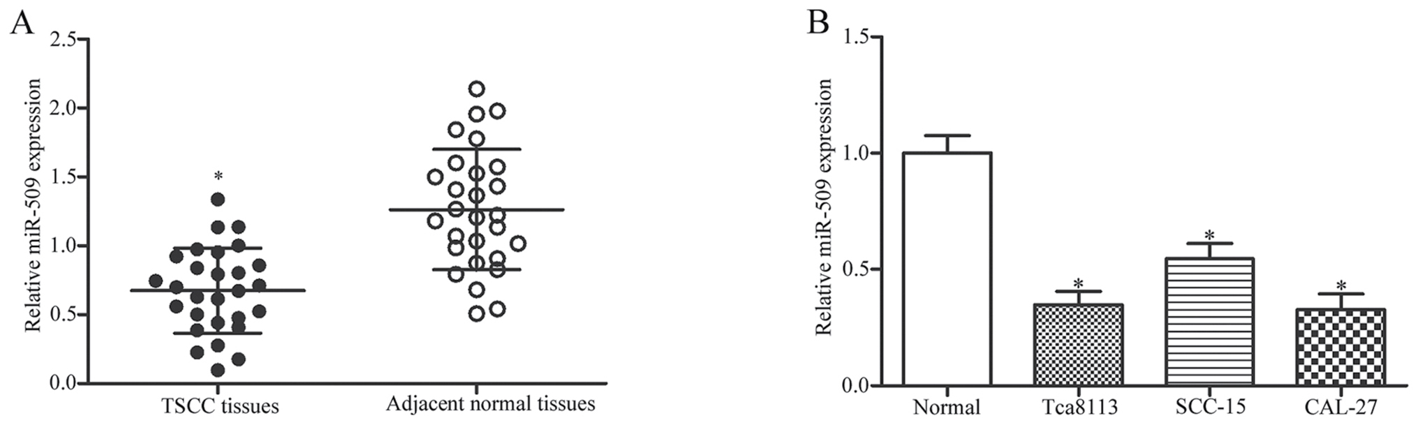
miR-509 is downregulated in TSCC
tissues and cell lines
The expression of miR-509 was detected in 28 TSCC
tissues and paired adjacent normal tissue samples using RT-qPCR. As
presented in Fig. 1A, miR-509 was
significantly downregulated in TSCC tissues compared with in
adjacent normal tissues (P<0.05). In addition, miR-509
expression was assessed in human TSCC cell lines and in normal
gingival epithelial cells. As demonstrated in Fig. 1B, the expression levels of miR-509
were significantly decreased in the TSCC cell lines compared with
in normal epithelial cells (P<0.05).
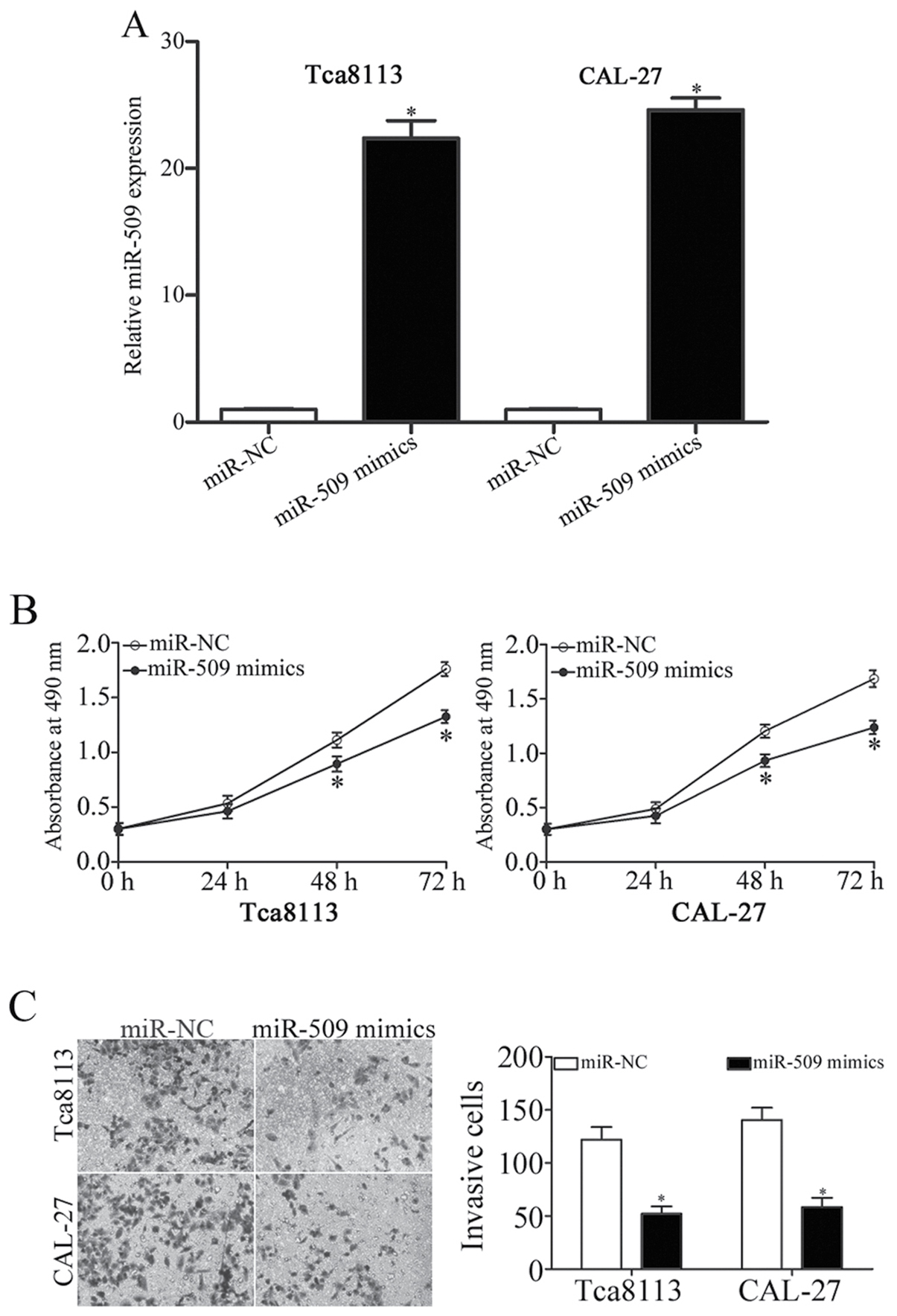
miR-509 inhibits TSCC cell
proliferation and invasion
To evaluate the effects of miR-509 on TSCC cell
function, Tca8113 and CAL-27 cells, as they expressed relatively
lower levels of miR-509, were transfected with miR-509 mimics or
miR-NC. A total of 48 h post-transfection, RT-qPCR demonstrated
that miR-509 was significantly upregulated in Tca8113 and CAL-27
cells transfected with miR-509 mimics compared with in
miR-NC-transfected cells (P<0.05; Fig. 2A). An MTT assay revealed that the
ectopic expression of miR-509 resulted in the inhibition of Tca8113
and CAL-27 cell proliferation compared with NC-transfected cells
(P<0.05; Fig. 2B). In addition,
an invasion assay revealed that the invasive capabilities of
Tca8113 and CAL-27 cells were significantly suppressed following
miR-509 overexpression (P<0.05; Fig. 2C). These findings suggested that
miR-509 may act as a tumor suppressor in TSCC cells and inhibit
their growth and metastasis.
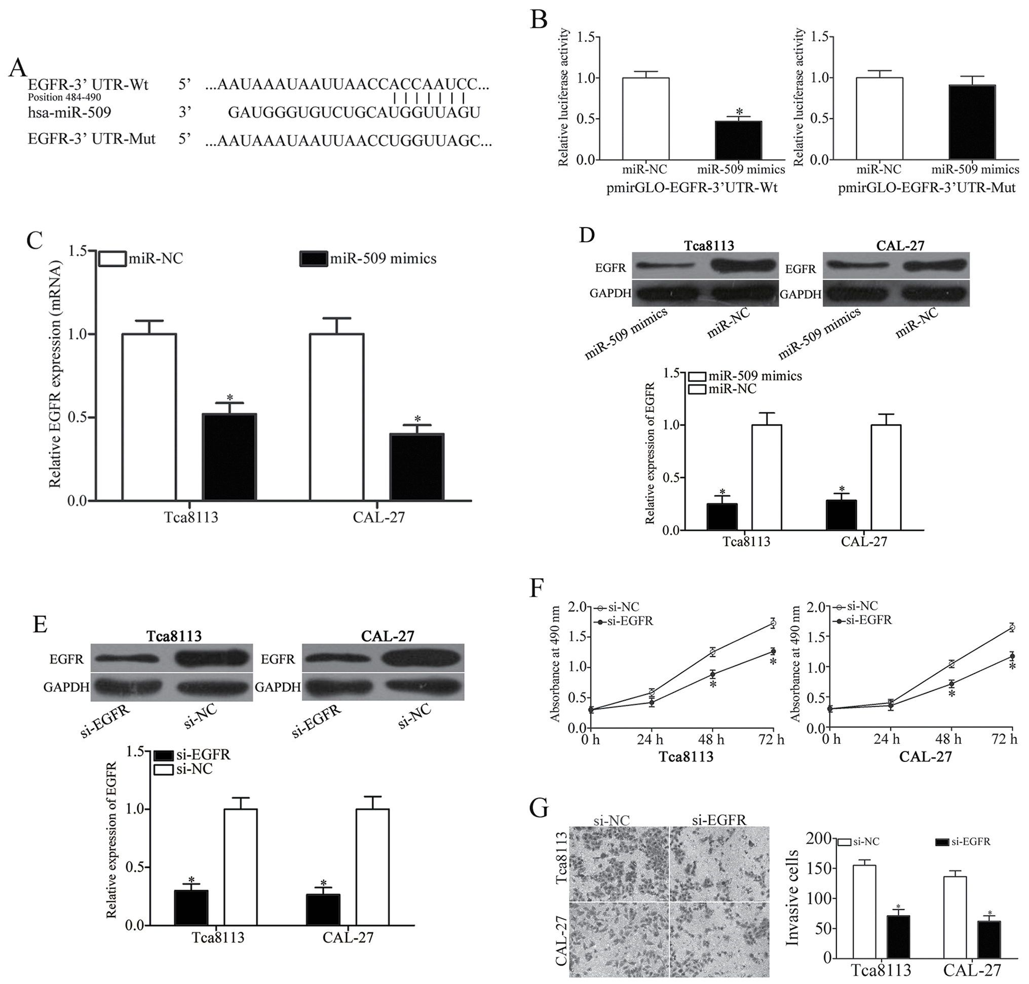
miR-509 directly targets EGFR in TSCC
cells
To investigate the molecular mechanisms underlying
the effects of miR-509 on TSCC cell proliferation and invasion, the
putative targets of miR-509 were predicted using miRNA target
prediction software. Among the potential target genes of miR-509,
EGFR (Fig. 3A) has been reported
to be overexpressed in TSCC cells, and has been correlated with
TSCC development and progression (24,25).
Therefore, a luciferase reporter assay was conducted; the results
indicated that luciferase activity was significantly decreased in
pmirGLO-EGFR-3′UTR-Wt-transfected cells that were cotransfected
with miR-509 mimics (P<0.05; Fig.
3B). Conversely, no inhibition in luciferase activity was
detected among cells transfected with the Mut EGFR 3′UTR sequence.
In addition, RT-qPCR and western blot analysis demonstrated that
transfection with the miR-509 mimic markedly decreased the mRNA and
protein expression levels of EGFR in Tca8113 and CAL-27 cells
(Fig. 3C and D). The present
findings suggested that EGFR may be a direct target gene of miR-509
in TSCC cells.
To investigate the roles of EGFR on TSCC cell
biological activity, si-EGFR was used to knock down the expression
of EGFR in Tca8113 and CAL-27 cells. Successful silencing was
confirmed by western blot analysis (P<0.05; Fig. 3E). An MTT assay demonstrated that
EGFR knockdown significantly inhibited the proliferation of Tca8113
and CAL-27 cells compared with si-NC-transfected cells (P<0.05;
Fig. 3F). Furthermore, a Transwell
invasion assay revealed that the downregulation of EGFR expression
significantly suppressed the invasive capabilities of Tca8113 and
CAL-27 cells (P<0.05; Fig. 3G).
These findings suggested that miR-509 may regulate the progression
of TSCC, through the direct regulation of EGFR expression.
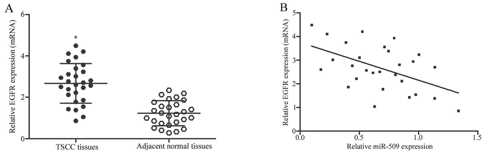
EGFR mRNA expression is negatively
correlated with miR-509 expression in TSCC tissues
In the present study, EGFR was identified as a
direct target gene of miR-509 in TSCC cells; therefore, the
expression of EGFR was assessed in TSCC tissues and in paired
adjacent normal tissues, and the correlation between EGFR and
miR-509 expression was investigated. As presented in Fig. 4A, EGFR mRNA expression was
significantly upregulated in TSCC tissues compared with in adjacent
normal tissue samples (P<0.05). Furthermore, an inverse
correlation was detected between EGFR mRNA and miR-509 expression
in TSCC tissues (r=−0.5109, P=0.0055; Fig. 4B). The present findings suggested
that miR-509 may be involved in the development and progression of
TSCC, through the negative regulation of EGFR.
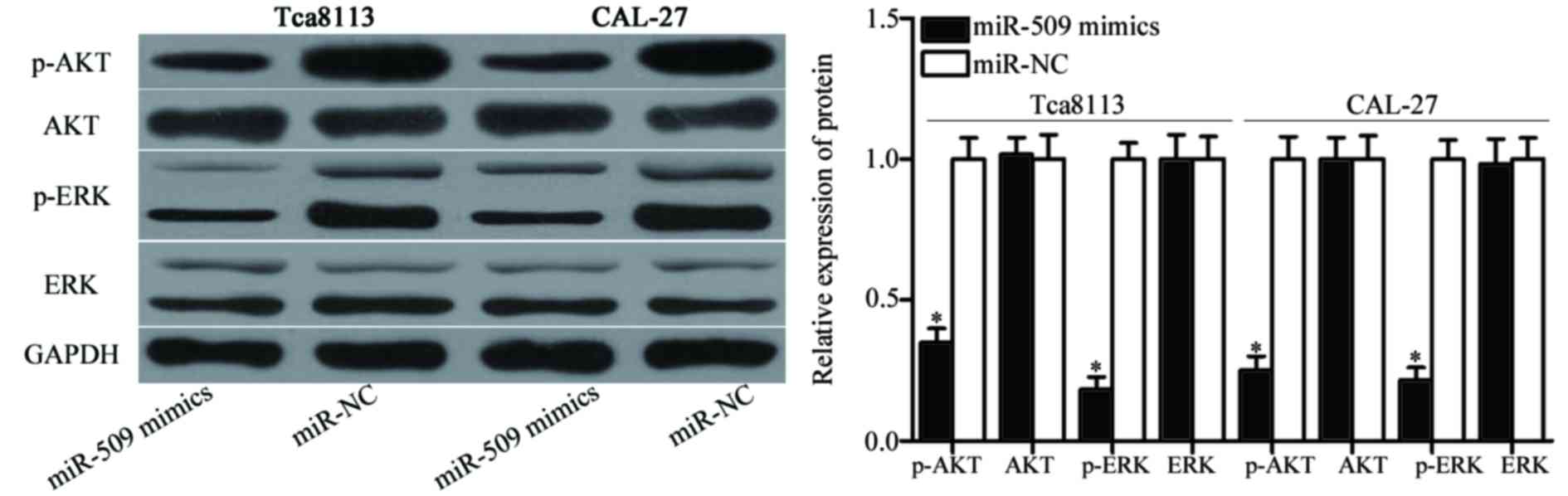
miR-509 suppresses EGFR-associated
signaling in TSCC cells
Since EGFR was identified as a direct target gene of
miR-509, the effects of miR-509 upregulation were examined on the
signaling pathways downstream of EGFR. As demonstrated in Fig. 5, miR-509 overexpression
downregulated the protein expression levels of p-Akt (P<0.05)
and p-ERK (P<0.05) in Tca8113 and CAL-27 cells, whereas it
exerted no significant effect on the expression of Akt and ERK
protein expression. The present results suggested that miR-509 may
inhibit EGFR-associated signaling pathways in TSCC cells.
Discussion
miRNA-based therapeutic approaches may have
potential as effective anticancer treatment strategies, through the
regulation of target genes that are implicated in numerous
physiological and pathological processes (26,27).
Therefore, it is essential to investigate the expression, roles and
underlying molecular mechanisms of cancer-associated miRNAs in
humans, and develop novel therapeutic targets for miRNA-based
cancer treatment strategies. In the present study, miR-509 was
revealed to be significantly downregulated in TSCC tissues and cell
lines compared with in normal tissues and normal epithelial cells,
respectively. Functional assays demonstrated that overexpression of
miR-509 inhibited the proliferation and invasion of TSCC cells
in vitro. Furthermore, EGFR was identified as a direct
target gene of miR-509 in TSCC cells. miR-509 overexpression also
resulted in the downregulation of p-AKT and p-ERK expression in
TSCC cells, thus suggesting that EGFR-associated signaling were
inhibited. The present findings indicated that miR-509 may exert
tumor-suppressive roles in TSCC cells, through the regulation of
EGFR-mediated signaling pathways.
Previous studies have identified miR-509 as a
critical regulator of tumorigenesis and tumor development, via
acting as a tumor suppressor gene in numerous types of human
cancer: Zhai et al (28)
reported that miR-509 was downregulated in renal carcinoma tissue
samples, whereas the restoration of miR-509 expression attenuated
the proliferation and migration, and induced the apoptosis of renal
carcinoma cells. Su et al (19) also demonstrated that miR-509
expression was decreased in renal carcinoma cells, whereas its
upregulation resulted in the significant suppression of cancer cell
proliferation and migration. A study by Chen et al (20) revealed that miR-509 expression
levels were decreased in chemoresistant epithelial ovarian cancer
tissues. Conversely, the overexpression of miR-509 suppressed the
proliferation and migration of ovarian cancer epithelial cells,
disrupted multi-cellular spheroids and improved their sensitivity
to cisplatin-induced apoptosis (20,29).
In addition, miR-509 expression has been revealed to be
downregulated in gastric cancer tissues, and low miR-509 expression
has been associated with decreased overall survival of patients
with gastric cancer; conversely, the restoration of miR-509
expression suppressed gastric cancer cell motility (21). Zhang et al (22) reported that miR-509 served
tumor-suppressive roles in triple-negative breast cancer cell
proliferation, invasion and apoptosis. These studies suggested that
miR-509 may have potential as a novel therapeutic target for the
development of cancer treatments.
The molecular mechanisms underlying the
anti-proliferative and anti-invasive effects of miR-509 in TSCC
cells were investigated in the present study. Several targets of
miR-509 have been identified, including mitogen-activated protein
kinase kinase kinase 8 (19),
X-linked inhibitor of apoptosis protein (20,21),
tumor necrosis factor-α (22),
cyclin-dependent kinase 2 (30),
Ras-related C3 botulinum toxin substrate 1 (30), and phosphatidylinositol-4-phosphate
3-kinase C2 domain-containing α polypeptide (30). In the present study, bioinformatics
analysis identified numerous candidate target genes for miR-509.
Among them, EGFR contained a putative binding site for miR-509 in
its 3′UTR, and has been reported to be upregulated in TSCC cells,
where it contributes to the development and progression of TSCC
(24,25). Therefore, the present study
investigated whether the tumor-suppressive roles of miR-509 in TSCC
cells may be attributed to the negative regulation of EGFR.
Luciferase reporter assays demonstrated that miR-509 directly
targeted the 3′UTR of EGFR. In addition, the mRNA and protein
expression levels of EGFR were revealed to be downregulated in TSCC
cells following transfection with miR-509 mimics, whereas EGFR
knockdown suppressed the proliferation and invasion of TSCC cells,
similar to miR-509 overexpression. Furthermore, an inverse
correlation was revealed between miR-509 and EGFR mRNA expression
in TSCC tissues, whereas the upregulation of miR-509 expression was
revealed to inhibit EGFR-associated signaling pathways in TSCC
cells. Taken together, these findings suggested that the
downregulation of EGFR expression and the inhibition of its
downstream signaling pathways may be important mechanisms
implicated in the miR-509 induced suppression of TSCC development
and progression.
EGFR is a cell-surface receptor for members of the
EGF family and transduces critical growth factor signals from the
extracellular to intracellular environment (31). EGFR has been reported to be
activated by various ligands, including EGF, transforming growth
factor-α, amphiregulin, heparin-binding EGF, betacellulin and
epiregulin (32). In addition,
EGFR has been revealed to be abnormally upregulated in numerous
tumor types, including colorectal (33), gastric (34) and bladder cancer (35), and osteosarcoma (36). Increasing evidence has indicated
that the upregulation of EGFR is strongly correlated with tumor
development and progression, and poor disease prognosis (37–40).
In TSCC, Ulanovski et al (24) reported that EGFR was highly
expressed in tumor specimens and its expression was obviously
correlated with tumor differentiation. Nakata et al
(41) revealed that the expression
levels of EGFR were significantly associated with reduced
disease-free survival and overall survival of patients with TSCC.
Previous functional assays also indicated an oncogenic role for
EGFR during TSCC cell growth, metastasis and apoptosis (42,43).
These findings suggested that targeting EGFR may have potential as
a novel and efficient therapeutic strategy for the treatment of
patients with TSCC.
In conclusion, the present study was, to the best of
our knowledge, the first to demonstrate that miR-509 acted as a
tumor suppressor in TSCC, via negatively regulating EGFR and
inhibiting its downstream signaling pathways. Further studies are
required to fully elucidate the roles of miR-509 and EGFR in TSCC
and reveal novel strategies for the treatment of this
malignancy.
References
|
1
|
Tsushima N, Sakashita T, Homma A,
Hatakeyama H, Kano S, Mizumachi T, Kakizaki T, Suzuki T and Fukuda
S: The role of prophylactic neck dissection and tumor thickness
evaluation for patients with cN0 tongue squamous cell carcinoma.
Eur Arch Otorhinolaryngol. 273:3987–3992. 2016. View Article : Google Scholar : PubMed/NCBI
|
|
2
|
Lao XM, Liang YJ, Su YX, Zhang SE, Zhou XI
and Liao GQ: Distribution and significance of interstitial fibrosis
and stroma-infiltrating B cells in tongue squamous cell carcinoma.
Oncol Lett. 11:2027–2034. 2016.PubMed/NCBI
|
|
3
|
Rao SV Krishna, Mejia G, Roberts-Thomson K
and Logan R: Epidemiology of oral cancer in Asia in the past
decade-an update (2000–2012). Asian Pac J Cancer Prev.
14:5567–5577. 2013. View Article : Google Scholar : PubMed/NCBI
|
|
4
|
Rusthoven K, Ballonoff A, Raben D and Chen
C: Poor prognosis in patients with stage I and II oral tongue
squamous cell carcinoma. Cancer. 112:345–351. 2008. View Article : Google Scholar : PubMed/NCBI
|
|
5
|
Yuen PW, Lam KY, Chan AC, Wei WI and Lam
LK: Clinicopathological analysis of local spread of carcinoma of
the tongue. Am J Surg. 175:242–244. 1998. View Article : Google Scholar : PubMed/NCBI
|
|
6
|
Regezi JA, Dekker NP, McMillan A,
Ramirez-Amador V, Meneses-Garcia A, Rivera Ruiz-Godoy LM,
Chrysomali E and Ng IO: p53, p21, Rb, and MDM2 proteins in tongue
carcinoma from patients <35 versus >75 years. Oral Oncol.
35:379–383. 1999. View Article : Google Scholar : PubMed/NCBI
|
|
7
|
Knopf A, Lempart J, Bas M,
Slotta-Huspenina J, Mansour N and Fritsche MK: Oncogenes and tumor
suppressor genes in squamous cell carcinoma of the tongue in young
patients. Oncotarget. 6:3443–3451. 2015. View Article : Google Scholar : PubMed/NCBI
|
|
8
|
Squarize CH, Castilho RM, Abrahao AC,
Molinolo A, Lingen MW and Gutkind JS: PTEN deficiency contributes
to the development and progression of head and neck cancer.
Neoplasia. 15:461–471. 2013. View Article : Google Scholar : PubMed/NCBI
|
|
9
|
Voorhoeve PM and Agami R: Classifying
microRNAs in cancer: The good, the bad and the ugly. Biochim
Biophys Acta. 1775:274–282. 2007.PubMed/NCBI
|
|
10
|
Seven M, Karatas OF, Duz MB and Ozen M:
The role of miRNAs in cancer: From pathogenesis to therapeutic
implications. Future Oncol. 10:1027–1048. 2014. View Article : Google Scholar : PubMed/NCBI
|
|
11
|
Bartel DP: MicroRNAs: Genomics,
biogenesis, mechanism, and function. Cell. 116:281–297. 2004.
View Article : Google Scholar : PubMed/NCBI
|
|
12
|
Sand M, Gambichler T, Sand D, Skrygan M,
Altmeyer P and Bechara FG: MicroRNAs and the skin: Tiny players in
the body's largest organ. J Dermatol Sci. 53:169–175. 2009.
View Article : Google Scholar : PubMed/NCBI
|
|
13
|
Yan B, Fu Q, Lai L, Tao X, Fei Y, Shen J,
Chen Z and Wang Q: Downregulation of microRNA 99a in oral squamous
cell carcinomas contributes to the growth and survival of oral
cancer cells. Mol Med Rep. 6:675–681. 2012.PubMed/NCBI
|
|
14
|
Xu P, Guo M and Hay BA: MicroRNAs and the
regulation of cell death. Trends Genet. 20:617–624. 2004.
View Article : Google Scholar : PubMed/NCBI
|
|
15
|
Karp X and Ambros V: Developmental
biology. Encountering microRNAs in cell fate signaling. Science.
310:1288–1289. 2005. View Article : Google Scholar : PubMed/NCBI
|
|
16
|
Jin Y, Chen Z, Liu X and Zhou X:
Evaluating the microRNA targeting sites by luciferase reporter gene
assay. Methods Mol Biol. 936:117–127. 2013. View Article : Google Scholar : PubMed/NCBI
|
|
17
|
Sun Z, Li S, Kaufmann AM and Albers AE:
miR-21 increases the programmed cell death 4 gene-regulated cell
proliferation in head and neck squamous carcinoma cell lines. Oncol
Rep. 32:2283–2289. 2014. View Article : Google Scholar : PubMed/NCBI
|
|
18
|
Zhang B, Pan X, Cobb GP and Anderson TA:
microRNAs as oncogenes and tumor suppressors. Dev Biol. 302:1–12.
2007. View Article : Google Scholar : PubMed/NCBI
|
|
19
|
Su Z, Chen D, Zhang E, Li Y, Yu Z, Shi M,
Jiang Z, Ni L, Yang S, Gui Y, et al: MicroRNA-509-3p inhibits
cancer cell proliferation and migration by targeting the
mitogen-activated protein kinase kinase kinase 8 oncogene in renal
cell carcinoma. Mol Med Rep. 12:1535–1543. 2015.PubMed/NCBI
|
|
20
|
Chen W, Zeng W, Li X, Xiong W, Zhang M,
Huang Y, Zhou L and Jiang S: MicroRNA-509-3p increases the
sensitivity of epithelial ovarian cancer cells to cisplatin-induced
apoptosis. Pharmacogenomics. 17:187–197. 2016. View Article : Google Scholar : PubMed/NCBI
|
|
21
|
Sun J, Li J, Zhang W, Zhang J, Sun S, Li
G, Song H and Wan D: MicroRNA-509-3p inhibits cancer cell
proliferation and migration via upregulation of XIAP in gastric
cancer cells. Oncol Res. 25:455–461. 2017. View Article : Google Scholar : PubMed/NCBI
|
|
22
|
Zhang G, Liu Z, Han Y, Wang X and Yang Z:
Overexpression of miR-509 increases apoptosis and inhibits invasion
via suppression of tumor necrosis factor-a in triple-negative
breast cancer Hs578T cells. Oncol Res. 24:233–238. 2016. View Article : Google Scholar : PubMed/NCBI
|
|
23
|
Livak KJ and Schmittgen TD: Analysis of
relative gene expression data using real-time quantitative PCR and
the 2(-Delta Delta C(T)) method. Methods. 25:402–408. 2001.
View Article : Google Scholar : PubMed/NCBI
|
|
24
|
Ulanovski D, Stern Y, Roizman P, Shpitzer
T, Popovtzer A and Feinmesser R: Expression of EGFR and Cerb-B2 as
prognostic factors in cancer of the tongue. Oral Oncol. 40:532–537.
2004. View Article : Google Scholar : PubMed/NCBI
|
|
25
|
Ekberg T, Nestor M, Engström M, Nordgren
H, Wester K, Carlsson J and Anniko M: Expression of EGFR, HER2,
HER3, and HER4 in metastatic squamous cell carcinomas of the oral
cavity and base of tongue. Int J Oncol. 26:1177–1185.
2005.PubMed/NCBI
|
|
26
|
Thai TH, Calado DP, Casola S, Ansel KM,
Xiao C, Xue Y, Murphy A, Frendewey D, Valenzuela D, Kutok JL, et
al: Regulation of the germinal center response by microRNA-155.
Science. 316:604–608. 2007. View Article : Google Scholar : PubMed/NCBI
|
|
27
|
Poy MN, Eliasson L, Krutzfeldt J, Kuwajima
S, Ma X, Macdonald PE, Pfeffer S, Tuschl T, Rajewsky N, Rorsman P
and Stoffel M: A pancreatic islet-specific microRNA regulates
insulin secretion. Nature. 432:226–230. 2004. View Article : Google Scholar : PubMed/NCBI
|
|
28
|
Zhai Q, Zhou L, Zhao C, Wan J, Yu Z, Guo
X, Qin J, Chen J and Lu R: Identification of miR-508-3p and
miR-509-3p that are associated with cell invasion and migration and
involved in the apoptosis of renal cell carcinoma. Biochem Biophys
Res Commun. 419:621–626. 2012. View Article : Google Scholar : PubMed/NCBI
|
|
29
|
Pan Y, Robertson G, Pedersen L, Lim E,
Hernandez-Herrera A, Rowat AC, Patil SL, Chan CK, Wen Y, Zhang X,
et al: miR-509-3p is clinically significant and strongly attenuates
cellular migration and multi-cellular spheroids in ovarian cancer.
Oncotarget. 7:25930–25948. 2016. View Article : Google Scholar : PubMed/NCBI
|
|
30
|
Yoon S, Han E, Choi YC, Kee H, Jeong Y,
Yoon J and Baek K: Inhibition of cell proliferation and migration
by miR-509-3p that targets CDK2, Rac1, and PIK3C2A. Mol Cells.
37:314–321. 2014. View Article : Google Scholar : PubMed/NCBI
|
|
31
|
Herbst RS: Review of epidermal growth
factor receptor biology. Int J Radiat Oncol Biol Phys. 59 2
Suppl:S21–S26. 2004. View Article : Google Scholar
|
|
32
|
Harris RC, Chung E and Coffey RJ: EGF
receptor ligands. Exp Cell Res. 284:2–13. 2003. View Article : Google Scholar : PubMed/NCBI
|
|
33
|
Spano JP, Lagorce C, Atlan D, Milano G,
Domont J, Benamouzig R, Attar A, Benichou J, Martin A, Morere JF,
et al: Impact of EGFR expression on colorectal cancer patient
prognosis and survival. Ann Oncol. 16:102–108. 2005. View Article : Google Scholar : PubMed/NCBI
|
|
34
|
Zhen Y, Guanghui L and Xiefu Z: Knockdown
of EGFR inhibits growth and invasion of gastric cancer cells.
Cancer Gene Ther. 21:491–497. 2014. View Article : Google Scholar : PubMed/NCBI
|
|
35
|
Naik DS, Sharma S, Ray A and Hedau S:
Epidermal growth factor receptor expression in urinary bladder
cancer. Indian J Urol. 27:208–214. 2011. View Article : Google Scholar : PubMed/NCBI
|
|
36
|
Boulytcheva IV, Soloviev YN, Kushlinskii
NE and Mahson AN: Expression of molecular markers in the tumor and
survival prognosis in osteosarcoma. Bull Exp Biol Med. 150:237–242.
2010.(In English, Russian). View Article : Google Scholar : PubMed/NCBI
|
|
37
|
Galizia G, Lieto E, Orditura M, Castellano
P, Mura AL, Imperatore V, Pinto M, Zamboli A, De Vita F and
Ferraraccio F: Epidermal growth factor receptor (EGFR) expression
is associated with a worse prognosis in gastric cancer patients
undergoing curative surgery. World J Surg. 31:1458–1468. 2007.
View Article : Google Scholar : PubMed/NCBI
|
|
38
|
Traynor AM, Weigel TL, Oettel KR, Yang DT,
Zhang C, Kim K, Salgia R, Iida M, Brand TM, Hoang T, et al: Nuclear
EGFR protein expression predicts poor survival in early stage
non-small cell lung cancer. Lung Cancer. 81:138–141. 2013.
View Article : Google Scholar : PubMed/NCBI
|
|
39
|
de Melo Maia B, Fontes AM, Lavorato-Rocha
AM, Rodrigues IS, de Brot L, Baiocchi G, Stiepcich MM, Soares FA
and Rocha RM: EGFR expression in vulvar cancer: Clinical
implications and tumor heterogeneity. Hum Pathol. 45:917–925. 2014.
View Article : Google Scholar : PubMed/NCBI
|
|
40
|
Ema A, Waraya M, Yamashita K, Kokubo K,
Kobayashi H, Hoshi K, Shinkai Y, Kawamata H, Nakamura K, Nishimiya
H, et al: Identification of EGFR expression status association with
metastatic lymph node density (ND) by expression microarray
analysis of advanced gastric cancer. Cancer Med. 4:90–100. 2015.
View Article : Google Scholar : PubMed/NCBI
|
|
41
|
Nakata Y, Uzawa N, Takahashi K, Sumino J,
Michikawa C, Sato H, Sonoda I, Ohyama Y, Okada N and Amagasa T:
EGFR gene copy number alteration is a better prognostic indicator
than protein overexpression in oral tongue squamous cell
carcinomas. Eur J Cancer. 47:2364–2372. 2011. View Article : Google Scholar : PubMed/NCBI
|
|
42
|
Dai W, Li Y, Zhou Q, Xu Z, Sun C, Tan X
and Lu L: Cetuximab inhibits oral squamous cell carcinoma invasion
and metastasis via degradation of epidermal growth factor receptor.
J Oral Pathol Med. 43:250–257. 2014. View Article : Google Scholar : PubMed/NCBI
|
|
43
|
Huang HJ, Ping FY, Hu JA and Zhao SF:
Effects of epidermal growth factor receptor gene silencing mediated
by short hairpin RNA on proliferation and apoptosis of human tongue
carcinoma cells. Zhonghua Kou Qiang Yi Xue Za Zhi. 44:365–369.
2009.(In Chinese). PubMed/NCBI
|