Introduction
Glioma is the most common malignant brain tumor in
adults and represents one of the most aggressive and
life-threatening types of human cancer (1). The morbidity rate of glioma is
3–8,000,000/10,000,000 individuals each year worldwide (2). Similar to other tumors, glioma
primarily results from multiple genetic risk factors, poor
lifestyle choices, infection and carcinogenic environmental
factors, including biochemicals in the environment, ionizing
radiation, nitrous compounds and air pollution (3). According to the 2007 World Health
Organization classification, glioma can be classified into three
groups: Astrocytomas, which are well-differentiated, anaplastic
astrocytomas, and glioblastoma multiforme, which is the most
aggressive form (4). There has
been substantial progress in early diagnosis and multimodal
treatments for glioma, including surgery, local irradiation and
conventional chemotherapy; however, there has been no marked
improvement in the survival rates for patients with this disease
(5). The high rates of recurrence
and ineffective therapeutic strategies contribute to the poor
prognosis of patients with glioma (6). Therefore, it is imperative to
investigate the molecular mechanism involved in the occurrence and
development of glioma, and identify novel effective therapeutic
targets for the treatment of glioma.
MicroRNAs (miRNAs) are a group of conserved,
endogenous, non-protein-coding, single stranded, short RNA
molecules, which have a length of 17–27 nucleotides. miRNAs can
negatively regulate gene expression through binding to
complementary sequences in the 3′untranslated regions (3′UTRs) of
their target genes, resulting in their translational regulation
and/or direct cleavage (7,8). miRNAs are widely expressed in animal
and plant cells, and are involved in several biological processes,
including cell proliferation, cell cycle, apoptosis,
differentiation, metastasis, angiogenesis and metabolism (9–11).
Increasing studies have revealed that miRNAs are dysregulated in
various types of human cancer, and exert oncogenic or tumor
suppressive roles, which primarily depend on the type of cancer
(12–14). For example, miRNA (miR)-211 is
upregulated in non-small cell lung cancer, and acts as an oncogene
through enhanced cell proliferation, colony formation and invasion
(15). By contrast, the expression
of miR-211 is lower in gastric cancer tissues. The ectopic
expression of miR-211 has also been shown to suppress the
progression of gastric cancer (16). Therefore, miRNAs offer potential
for investigation as specific diagnostic and prognostic markers,
and as therapeutic targets in oncology.
The aim of the present study was to examine the
expression, functional roles and underlying mechanism of miR-186 in
glioma. This may provide a novel basis for development of the
mechanism of glioma.
Materials and methods
Tissue specimens and cell lines
The present study was approved by the Ethical
Committee of the Affiliated Huai'an First Hospital of Nanjing
Medical University (Huai'an, China). Fresh glioma tissues (n=16)
and adjacent normal tissues (n=16) were collected from patients
with glioma during surgery at the Department of Neurosurgery, the
Affiliated Huai'an First Hospital of Nanjing Medical University,
between June 2014 and January 2016. None of these patients had been
treated with chemotherapy or radiotherapy prior to surgery. All
fresh tissues were frozen in liquid nitrogen and stored at −80°C
until use.
A total of five glioma cell lines (U251, U87, U373,
H4 and A172) were obtained from the Institute of Biochemistry and
Cell Biology of the Chinese Academy of Sciences (Shanghai, China).
The normal human astrocyte cell line (NHA) was purchased from
American Type Culture Collection (Manassas, VA, USA). All cell
lines were maintained in Dulbecco's modified Eagle's medium (DMEM;
Invitrogen; Thermo Fisher Scientific, Inc., Waltham, MA, USA)
containing 10% fetal bovine serum (FBS; Invitrogen; Thermo Fisher
Scientific, Inc.) and 1% penicillin/streptomycin (Invitrogen;
Thermo Fisher Scientific, Inc.) in a humid atmosphere containing 5%
CO2 at 37°C.
Reverse transcription-quantitative
polymerase chain reaction (RT-qPCR) analysis
Total RNA was isolated from tissues and cells using
TRIzol (Invitrogen; Thermo Fisher Scientific, Inc.) according to
the manufacturer's protocol. The concentration and purity of total
RNA was determined using a NanoDrop® ND-1000
spectrophotometer (Thermo Fisher Scientific, Inc.). The RT process
was performed using a TaqMan microRNA Reverse Transcription kit
(Applied Biosystems; Thermo Fisher Scientific, Inc.). qPCR was
performed using a TaqMan microRNA assay kit (Applied Biosystems;
Thermo Fisher Scientific, Inc.) with U6 as an internal control for
determining the expression of miR-186. The reaction system for qPCR
contained 1 µl TaqMan® Small RNA assay (20X), 1.33 µl
cDNA, 10 µl TaqMan® Universal PCR Master mix II (2X) and
7.67 µl nuclease-free water. The thermocycling conditions for qPCR
were as follows: 50°C for 2 min, 95°C for 10 min, followed by 40
cycles of denaturation at 95°C for 15 sec and annealing/extension
at 60°C for 60 sec. To determine the mRNA expression of IGF-1R, RT
was performed using the M-MLV Reverse Transcription system (Promega
Corporation, Madison, WI, USA). SYBR-Green I mix (Takara
Biotechnology Co., Ltd., Dalian, China) was then used to detect the
mRNA expression of IGF-1R with GAPDH as the internal control. This
reaction for qPCR included 2 µl cDNA (100 ng), 2 µl forward primer,
2 µl reverse primer, 10 µl SYBR-Green PCR Master mix and 4 µl
ddH2O. The thermocycling conditions for qPCR were as
follows: 95°C for 10 min, followed by 40 cycles of 95°C for 15 sec
and 60°C for 1 min. The RT-qPCR analysis was performed using an
Applied Biosystems 7900HT Fast Real-Time PCR system (Applied
Biosystems; Thermo Fisher Scientific, Inc.). The relative mRNA
expression of miR-186 and IGF-1R were quantified using the
2−ΔΔCq method (17).
Cell transfection
To examine the functions of miR-186 in glioma,
miR-186 mimics and miRNA negative control (miR-NC) were purchased
from GenePharma (Shanghai, China). The miR-186 mimics sequence was
5′-AUUUCUUAGGUCUCAUAUAGCGU-3′ and the miR-NC sequence was
5′-UUCUCCGAACGUGUCACGUTT-3′. Two small interfering RNAs (siRNAs),
targeting IGF-1R (si-IGF-1R) and negative control (si-NC), were
obtained from Guangzhou RiboBio Co., Ltd. (Guangzhou, China). The
si-IGF-1R sequence was 5′-CCACGTCGAAGAATCGCAT-3′ and the si-NC
sequence was 5′-UUCUCCGAACGUGUCACGUTT-3′. Lipofectamine 2000
reagent (Invitrogen; Thermo Fisher Scientific, Inc.) was used to
transfect oligonucleotides.
MTT assay
The cells were seeded into 96-well plates at a
density of 3×10³ per well. Following incubation for 6–8
h, the cells were transfected with the miRNA mimics or siRNAs. The
transfected cells were then incubated at 37°C in a humid atmosphere
containing 5% CO2 for 24, 48, 72 and 96 h. At each time
point, 10 µl MTT solution (5 mg/ml; Sigma-Aldrich; Merck Millipore,
Darmstadt, Germany) was added to each well and incubated at 37°C
for 4 h. Following incubation, the medium containing MTT solution
was removed and 150 µl DMSO (Sigma-Aldrich; Merck Millipore) was
added to resolve MTT formazan. The absorbance of solution was then
measured at 490 nm on a microplate reader.
Cell invasion assay
Transwell chambers (8-µm; Corning Costar, Cambridge,
MA, USA) and pre-coated Matrigel (BD Biosciences, San Jose, CA,
USA) were used to perform cell invasion assays. Briefly,
4×10⁴ of the transfected cells were resuspended in 100
µl FBS-free culture medium and seeded into the upper chambers, and
500 µl culture medium containing 20% FBS was added to the lower
chambers. Following incubation for 48 h, any cells that had not
invaded through the pores on the polycarbonate membranes, were
carefully removed using a cotton swab. The invaded cells were fixed
with 100% methanol, stained with 0.5% crystal violet and then
counted under a light microscope (CKX41; Olympus Corporation,
Tokyo, Japan; magnification, ×200).
Target prediction
The potential target genes of miR-186 were analyzed
using bioinformatics analysis with multiple target prediction
algorithms, including TargetScan 7.1 (http://www.targetscan.org/vert_71/), miRanda
(http://www.microrna.org/microrna/home.do) and PicTar
(http://www.pictar.mdc-berlin.de/).
Luciferase reporter assay
The luciferase reporter plasmids,
pGL3-Wt-IGF-1R-3′UTR and pGL3-Mut-IGF-1R-3′UTR, were synthesized
and confirmed by GenePharma. The HEK293T cell line was obtained
from the Institute of Biochemistry and Cell Biology of the Chinese
Academy of Sciences. For the luciferase reporter assay, the HEK293T
cells were plated into 48-well plates at a density of
5×10⁴ cells/well and transfected with
pGL3-Wt-IGF-1R-3′UTR or pGL3-Mut-IGF-1R-3′UTR, and miR-186 mimics
or miR-NC using Lipofectamine 2000 reagent. At 48 h
post-co-transfection, luciferase activities were detected using
Dual-Luciferase reporter assay system (Promega Corporation)
according to the manufacturer's protocol. All experiments were
performed with three independent replicates.
Western blot analysis
The transfected cells were harvested at 48 h
post-transfection and lysed by RIPA buffer, containing 10 mM
Tris-HCl (pH 7.4), 1% Triton X-100, 0.1% SDS, 1% NP-40, 1 mM
MgCl2 and protease inhibitors. The protein concentration
was measured using a BCA protein assay kit (Beyotime Institute of
Biotechnology, Haimen, China). Equal quantities of protein (20 µg)
were separated by 10% SDS-PAGE and then transferred onto
polyvinylidene fluoride membranes (EMD Millipore). Following
blocking with 5% nonfat milk in TBS containing 0.1% Tween-20
(TBST), the membranes were incubated with primary antibodies,
including mouse anti-human monoclonal IGF-1R (1:1,000 dilution;
cat. no. sc-81464; Santa Cruz Biotechnology, Inc., Santa Cruz, CA,
USA) and mouse anti-human monoclonal GAPDH (1:1,000 dilution; cat.
no. sc-59540; Santa Cruz Biotechnology, Inc.), at 4°C overnight.
The following day, the membranes were washed with TBST three times.
Goat anti-mouse horseradish peroxidase-conjugated secondary
antibody (1:5,000 dilution; cat. no. sc-2005; Santa Cruz
Biotechnology, Inc.) was used to detect primary antibodies at room
temperature. Finally, ECL solution (Pierce; Thermo Fisher
Scientific, Inc.) was used to visualize protein bands. GAPDH was
used as an internal control. Band densities were quantified using
ImageJ v1.49 software (National Institutes of Health, Bethesda, MD,
USA).
Statistical analysis
Data are expressed as the mean ± standard deviation.
Statistical analyses were performed with Student's t-test using
SPSS 19.0 software (IBM SPSS, Armonk, NY, USA). P<0.05 was
considered to indicate a statistically significant difference.
Results
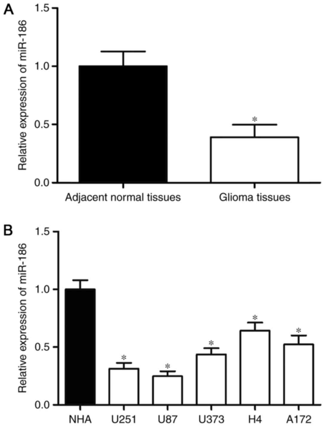
miR-186 is downregulated in glioma
tissues and cell lines
To investigate the association between miR-186 and
glioma, RT-qPCR analysis was performed to examine the expression of
miR-186 in glioma tissues. As shown in Fig. 1A, the expression of miR-186 was
downregulated in the glioma tissues, compared with that in the
adjacent normal tissues. The expression levels of miR-186 in glioma
cell lines were also detected, and the results of the RT-qPCR
analysis revealed that the expression levels of miR-186 were lower
in the glioma cell lines, compared with that in the NHA cell line
(P<0.05; Fig. 1B). As the
expression of miR-186 was lowest in the U251 and U87 cells, these
two cell lines were selected for use in the subsequent
experiments.
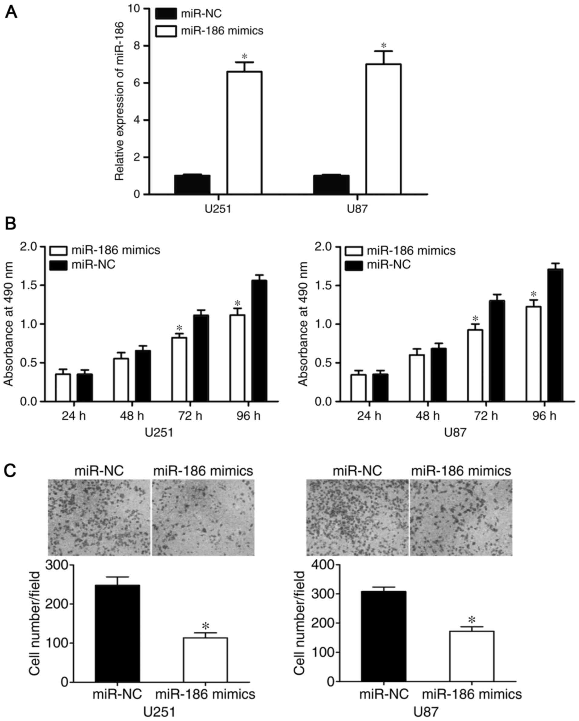
miR-186 suppresses the proliferation
and invasion of U251 and U87 cells
To assess the functional roles of miR-186 in glioma,
miR-186 mimics or miR-NC were injected into U251 and U87 cells. The
transfection efficiency was evaluated using RT-qPCR analysis, which
revealed that the expression of miR-186 was significantly higher in
the U251 and U87 cells following transfection with miR-186 mimics
(P<0.05; Fig. 2A). The possible
effects of the overexpression of miR-186 on the proliferation and
invasion of glioma cells were determined using MTT and cell
invasion assays, respectively. The results of the MTT assay showed
that miR-186 reduced the proliferation of U251 and U87 cells,
compared with proliferation in the miR-NC groups (P<0.0 5;
Fig. 2B). Furthermore, the
overexpression of miR-186 significantly reduced the invasive
ability of the U251 and U87 cells (P<0.05; Fig. 2C). These results suggested that
miR-186 acted as a tumor suppressor in the tumorigenesis and
progression of glioma.
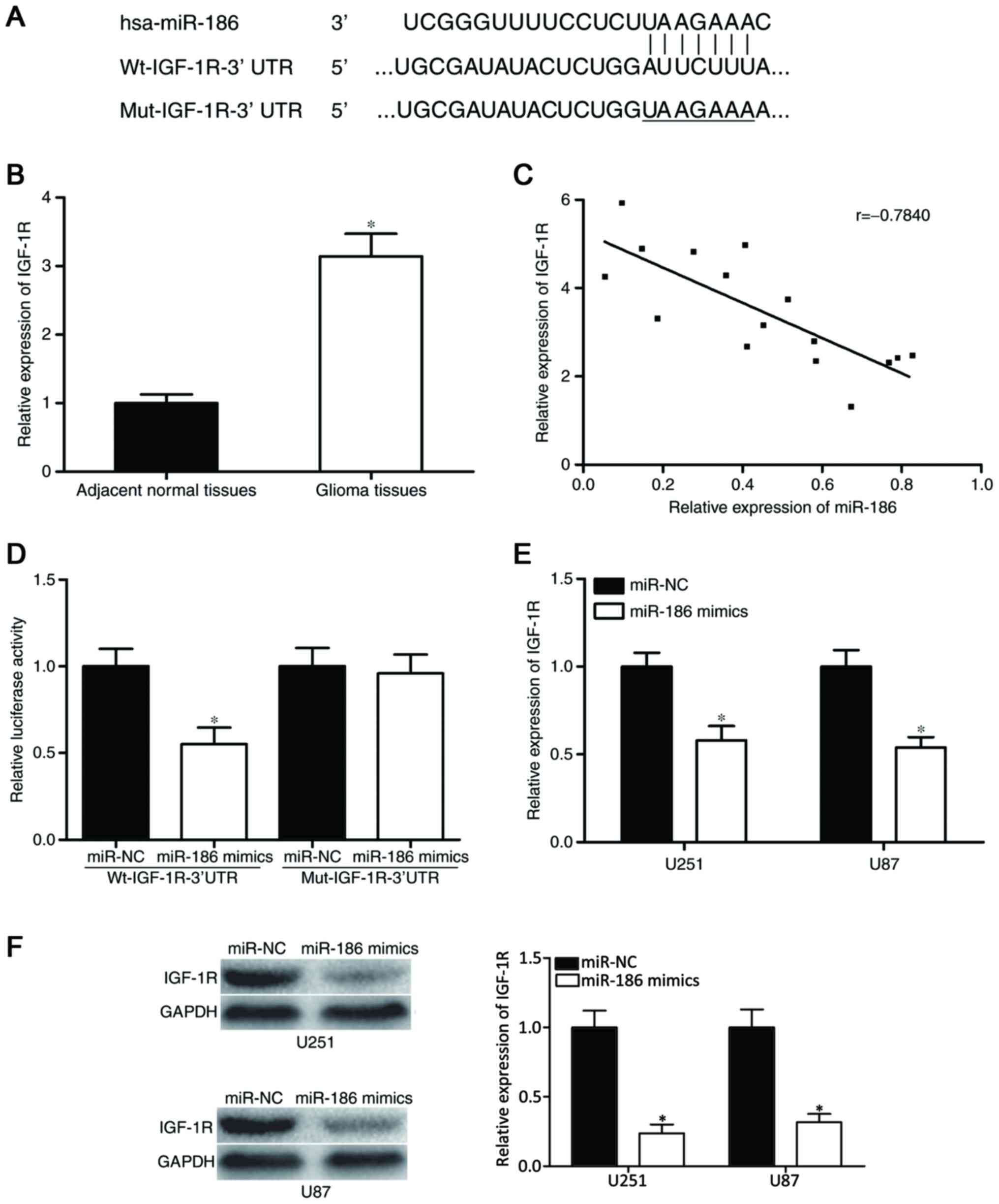
IGF-1R is a direct target of
miR-186
To investigate the molecular mechanism underlying
the action of miR-186 in glioma, bioinformatics analysis was
performed to determine the potential target genes of miR-186. The
analysis found that the ‘seed sequence’ of miR-186 matched the
3′UTR of the IGF-1R (Fig. 3A).
The correlation between the expression of miR-186
and the IGF-1R in glioma tissues was then examined. As shown in
Fig. 3B, the mRNA expression of
IGF-1R was high in glioma tissues, compared with adjacent normal
tissues (P<0.05). Spearman's correlation analysis was then
performed to analyze the association between miR-186 and IGF-1,
which revealed that the expression of miR-186 was inversely
correlated with that of IGF-1R in glioma (r=−0.7840; P=0.0003;
Fig. 3C).
To investigate the direct interaction between
miR-186 and its binding site in the 3′UTR of IGF-1R, a luciferase
reporter assay was used. The pGL3-Wt-IGF-1R-3′UTR or
pGL3-Mut-IGF-1R-3′UTR construct, with miR-186 mimics or miR-NC were
transfected into HEK293T cells. The results indicated that the
overexpression ofmiR-186 decreased the luciferase activity of
pGL3-Wt-IGF-1R-3′UTR (P<0.05; Fig.
3D); however, the luciferase activity of pGL3-Mut-IGF-1R-3′UTR
was not affected by simultaneous transfection with miR-186
mimics.
Western blot and RT-qPCR analyses were performed to
evaluate the regulatory effects of the overexpression ofmiR-186 on
the mRNA and protein expression of IGF-1R, respectively. The
results showed that the overexpression of miR-186 significantly
reduced the mRNA (P<0.05; Fig.
3E) and protein (P<0.05; Fig.
3F) expression levels of IGF-1R in the U251 and U87 cells.
Collectively, these results demonstrated that miR-186 negatively
regulated the expression of IGF-1R via binding to the 3′UTR of
IGF-1R.
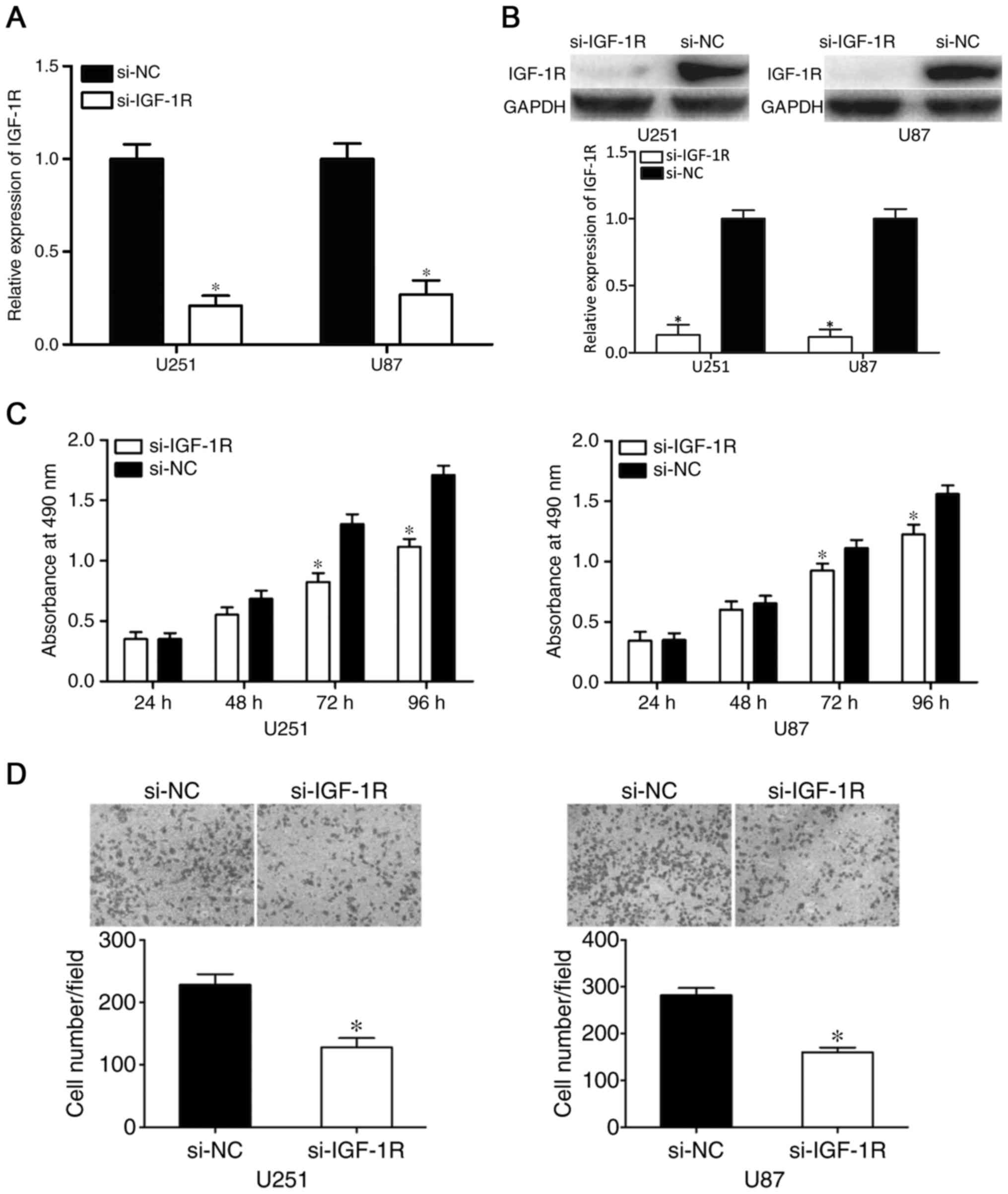
Knockdown of IGF-1R exerts similar
effects as the overexpression of miR-186 in glioma
IGF-1R was identified as a direct target gene of
miR-186. Therefore, it was hypothesized that IGF-1R contributes to
the suppressive effects of miR-186 on glioma cells. To assess this,
si-IGF-1R was transfected into U251 and U87 cells to knockdown the
expression of IGF-1R. After 48 h, RT-qPCR and western blot analyses
were performed to measure the mRNA and protein levels of IGF-1R. As
shown in Fig. 4A and B, the
expression levels of IGF-1R were reduced in the
si-IGF-1R-transfected U251 and U87 cells (P<0.05). The results
of the MTT and cell invasion assays revealed that the effects of
si-IGF-1R on U251 and U87 cell proliferation (P<0.05; Fig. 4C) and invasion (P<0.05; Fig. 4D) were similar to those induced by
the overexpression of miR-186. These results indicated that IGF-1R
contributed to the tumor suppressive effects of miR-186 in
glioma.
Discussion
Increasing studies have reported that the aberrant
expression of miR-186 is a characteristic of malignancies. For
example, miR-186 has been found to be downregulated in non-small
cell lung cancer clinical tissues and cell lines, and the
expression level of miR-186 was correlated with patient survival
rates (18). Chen et al
(19) demonstrated that the
relative expression of miR-186 was lower in colon carcinoma tissues
and cell lines. A study by Zhang et al (20) demonstrated that miR-186 was reduced
in acute myeloid leukemia (AML). Patients with AML with low
expression levels ofmiR-186 had significantly lower rates of
complete remission and shorter overall survival rates, compared
with patients with a high level of miR-186 (20). He et al (21) reported that miR-186 was commonly
downregulated in esophageal squamous cell carcinoma (ESCC), and was
correlated with the level of differentiation, tumor-node-metastasis
stage and lymph node metastasis in patients with ESCC. Liu et
al (22) found that miR-186
was expressed at low levels in multiple myeloma tissues and cell
lines, and miR-186 has been shown to be downregulated in oral
squamous cell carcinoma (23),
bladder cancer (24), colorectal
neuroendocrine tumors (25),
gastric cancer (26) and
hepatocellular carcinoma (27).
These studies suggest that the downregulation of miR-186 is a
frequent event in malignancies.
There is increasing evidence that miR-186 is
important in tumorigenesis and tumor development. In non-small cell
lung cancer, the upregulation of miR-186 targets CCND1, CDK2 and
CDK6 to decrease proliferation by inducing G(1)-S checkpoint arrest (18). Cui et al (28) found that the enforced expression of
miR-186 suppressed cell proliferation, migration and invasion
through the downregulation of Rho-associated protein kinase 1 in
non-small cell lung cancer. In colon carcinoma, the ectopic
expression of miR-186 was found to significantly inhibit growth and
metastasis via directly targeting Yin Yang 1 (19). In ESCC, the re-expression of
miR-186 was shown to suppress cell proliferation, invasion and
enhance apoptosis through the negative regulation of S-phase
kinase-associated protein 2 (21).
Yao et al (24)
demonstrated that the enforced expression of miR-186 reduces cell
proliferation and invasion via the inhibition of nucleosome-binding
protein 1 in bladder cancer. In multiple myeloma, the
overexpression of miR-186 decreases cell growth in vitro and
in vivo, and improves cell cycle G0/G1 arrest through the
downregulation of Jagged 1 (22).
A previous study also showed that the upregulation of miR-186
downregulates the mRNA and protein expression levels of YAP1,
resulting in downregulation of the Hippo signaling pathway, and
resulting in the repression of cell growth and metastasis in
hepatocellular carcinoma (27).
These findings implicate miR-186 as an attractive candidate
therapeutic target in antitumor therapy.
In the present study, it was found that miR-186 was
downregulated in glioma tissues, compared with that in adjacent
normal tissues. The expression of miR-186 was also measured in
glioma cell lines, and it was found that the expression levels of
miR-186 were lower in glioma cell lines, compared with that in the
normal HAC line. Subsequently, the functions of the overexpression
of miR-186 in glioma cells were evaluated. The results of the MTT
and cell invasion assays revealed that the upregulation of miR-186
suppressed the proliferation and invasion of glioma cells,
respectively. This finding indicated that miR-186 may be a
tumor-inhibiting factor.
To investigate the molecular mechanism underlying
the involvement of miR-186 in the progression of glioma, the
present study aimed to confirm the direct target genes of miR-186.
According to bioinformatics analysis with several target prediction
algorithms, IGF-1R was identified as a potential target gene of
miR-186. A luciferase reporter assay was then performed to confirm
this hypothesis. The results showed that miR-186 directly targeted
the 3′UTR of IGF-1R. Subsequent experiments revealed that the mRNA
expression of IGF-1R was high in glioma tissues and inversely
correlated with the expression of miR-186. Subsequently, the
induced expression of miR-186 decreased the mRNA and protein
expression of IGF-1R in glioma cells. Finally, IGF-1R knockdown was
found to exhibit similar tumor suppressive effects as that observed
by miR-186 on glioma cell proliferation and invasion. These results
indicated thatmiR-186 acted as a tumor-suppressive molecule in
glioma genesis and progression, at least partially through
regulating the expression of IGF-1R.
IGF-1R, a tyrosine kinase receptor, contains two
extracellular α subunits with a ligand-binding site, two
transmembrane β subunits and intracellular tyrosine kinase activity
(29). It has previously been
demonstrated that IGF-1R is involved in IGF-I and IGF-II signaling,
and therefore regulates cell proliferation, cell cycle, apoptosis,
metastasis, survival and differentiation (30–33).
IGF-1R has been investigated widely in glioma. Harrington et
al (34) found that IGF-1R was
important in the tumorigenesis of glioma. In addition, studies have
shown that apoptosis (35), growth
(36,37), invasion (38), migration (39) and glucose metabolism (40) are regulated by IGF-1R in glioma.
These findings suggest that selecting IGF-1R as a therapeutic
approach is practicable for patients with glioma.
In conclusion, the present study showed that the
miR-186/IGF-1R pathway regulated cell proliferation and invasion in
glioma. These findings suggested that miR-186 may be a potential
therapeutic target for the treatment of glioma in the future.
References
|
1
|
Taylor LP: Diagnosis, treatment and
prognosis of glioma: Five new things. Neurology. 75 18 Suppl
1:S28–S32. 2010. View Article : Google Scholar : PubMed/NCBI
|
|
2
|
Karsy M, Albert L, Tobias ME, Murali R and
Jhanwar-Uniyal M: All-trans retinoic acid modulates cancer stem
cells of glioblastoma multiforme in an MAPK-dependent manner.
Anticancer Res. 30:4915–4920. 2010.PubMed/NCBI
|
|
3
|
Zhu GY, Shi BZ and Li Y: FoxM1 regulates
Sirt1 expression in glioma cells. Eur Rev Med Pharmacol Sci.
18:205–211. 2014.PubMed/NCBI
|
|
4
|
Louis DN, Ohgaki H, Wiestler OD, Cavenee
WK, Burger PC, Jouvet A, Scheithauer BW and Kleihues P: The 2007
WHO classification of tumours of the central nervous system. Acta
Neuropathol. 114:97–109. 2007. View Article : Google Scholar : PubMed/NCBI
|
|
5
|
Xu Z, Zeng X, Tian D, Xu H, Cai Q, Wang J
and Chen Q: MicroRNA-383 inhibits anchorage-independent growth and
induces cell cycle arrest of glioma cells by targeting CCND1.
Biochem Biophys Res Commun. 453:833–838. 2014. View Article : Google Scholar : PubMed/NCBI
|
|
6
|
Omuro A and DeAngelis LM: Glioblastoma and
other malignant gliomas: A clinical review. JAMA. 310:1842–1850.
2013. View Article : Google Scholar : PubMed/NCBI
|
|
7
|
Bartel DP: MicroRNAs: Target recognition
and regulatory functions. Cell. 136:215–233. 2009. View Article : Google Scholar : PubMed/NCBI
|
|
8
|
Shukla GC, Singh J and Barik S: MicroRNAs:
Processing, maturation, target recognition and regulatory
functions. Mol Cell Pharmacol. 3:83–92. 2011.PubMed/NCBI
|
|
9
|
Hwang HW and Mendell JT: MicroRNAs in cell
proliferation, cell death, and tumorigenesis. Br J Cancer. 96
Suppl:R40–R44. 2007.PubMed/NCBI
|
|
10
|
Bueno MJ and Malumbres M: MicroRNAs and
the cell cycle. Biochim Biophys Acta. 1812:592–601. 2011.
View Article : Google Scholar : PubMed/NCBI
|
|
11
|
Zhang J, Gong X, Tian K, Chen D, Sun J,
Wang G and Guo M: miR-25 promotes glioma cell proliferation by
targeting CDKN1C. Biomed Pharmacother. 71:7–14. 2015. View Article : Google Scholar : PubMed/NCBI
|
|
12
|
Lu J, Getz G, Miska EA, Alvarez-Saavedra
E, Lamb J, Peck D, Sweet-Cordero A, Ebert BL, Mak RH, Ferrando AA,
et al: MicroRNA expression profiles classify human cancers. Nature.
435:834–838. 2005. View Article : Google Scholar : PubMed/NCBI
|
|
13
|
Li T, Pan H and Li R: The dual regulatory
role of miR-204 in cancer. Tumour Biol. 37:11667–11677. 2016.
View Article : Google Scholar : PubMed/NCBI
|
|
14
|
Bertoli G, Cava C and Castiglioni I: The
potential of miRNAs for diagnosis, treatment and monitoring of
breast cancer. Scand J Clin Lab Invest Suppl. 245:34–39. 2016.
View Article : Google Scholar
|
|
15
|
Ye L, Wang H and Liu B: miR-211 promotes
non-small cell lung cancer proliferation by targeting SRCIN1.
Tumour Biol. 37:1151–1157. 2016. View Article : Google Scholar : PubMed/NCBI
|
|
16
|
Wang CY, Hua L, Sun J, Yao KH, Chen JT,
Zhang JJ and Hu JH: MiR-211 inhibits cell proliferation and
invasion of gastric cancer by down-regulating SOX4. Int J Clin Exp
Pathol. 8:14013–14020. 2015.PubMed/NCBI
|
|
17
|
Livak KJ and Schmittgen TD: Analysis of
relative gene expression data using real-time quantitative PCR and
the 2(-Delta Delta C(T)) method. Methods. 25:402–408. 2001.
View Article : Google Scholar : PubMed/NCBI
|
|
18
|
Cai J, Wu J, Zhang H, Fang L, Huang Y,
Yang Y, Zhu X, Li R and Li M: miR-186 downregulation correlates
with poor survival in lung adenocarcinoma, where it interferes with
cell-cycle regulation. Cancer Res. 73:756–766. 2013. View Article : Google Scholar : PubMed/NCBI
|
|
19
|
Chen F, Zhou C, Lu Y, Yuan L, Peng F,
Zheng L and Li X: Expression of hsa-miR-186 and its role in human
colon carcinoma cells. Nan Fang Yi Ke Da Xue Xue Bao. 33:654–660.
2013.(In Chinese). PubMed/NCBI
|
|
20
|
Zhang TJ, Wang YX, Yang DQ, Yao DM, Yang
L, Zhou JD, Deng ZQ, Wen XM, Guo H, Ma JC, et al: Down-regulation
of miR-186 correlates with poor survival in de novo acute myeloid
leukemia. Clin Lab. 62:113–120. 2016. View Article : Google Scholar : PubMed/NCBI
|
|
21
|
He W, Feng J, Zhang Y, Wang Y, Zang W and
Zhao G: microRNA-186 inhibits cell proliferation and induces
apoptosis in human esophageal squamous cell carcinoma by targeting
SKP2. Lab Invest. 96:317–324. 2016. View Article : Google Scholar : PubMed/NCBI
|
|
22
|
Liu Z, Zhang G, Yu W, Gao N and Peng J:
miR-186 inhibits cell proliferation in multiple myeloma by
repressing Jagged1. Biochem Biophys Res Commun. 469:692–697. 2016.
View Article : Google Scholar : PubMed/NCBI
|
|
23
|
Ries J, Vairaktaris E, Agaimy A, Kintopp
R, Baran C, Neukam FW and Nkenke E: miR-186, miR-3651 and miR-494:
Potential biomarkers for oral squamous cell carcinoma extracted
from whole blood. Oncol Rep. 31:1429–1436. 2014. View Article : Google Scholar : PubMed/NCBI
|
|
24
|
Yao K, He L, Gan Y, Zeng Q, Dai Y and Tan
J: MiR-186 suppresses the growth and metastasis of bladder cancer
by targeting NSBP1. Diagn Pathol. 10:1462015. View Article : Google Scholar : PubMed/NCBI
|
|
25
|
Wang M, Xia X, Chu W, Xia L, Meng T, Liu L
and Liu Y: Roles of miR-186 and PTTG1 in colorectal neuroendocrine
tumors. Int J Clin Exp Med. 8:22149–22157. 2015.PubMed/NCBI
|
|
26
|
Liu L, Wang Y, Bai R, Yang K and Tian Z:
MiR-186 inhibited aerobic glycolysis in gastric cancer via HIF-1α
regulation. Oncogenesis. 5:e2242016. View Article : Google Scholar : PubMed/NCBI
|
|
27
|
Ruan T, He X, Yu J and Hang Z:
MicroRNA-186 targets Yes-associated protein 1 to inhibit Hippo
signaling and tumorigenesis in hepatocellular carcinoma. Oncol
Lett. 11:2941–2945. 2016.PubMed/NCBI
|
|
28
|
Cui G, Cui M, Li Y, Liang Y, Li W, Guo H
and Zhao S: MiR-186 targets ROCK1 to suppress the growth and
metastasis of NSCLC cells. Tumour Biol. 35:8933–8937. 2014.
View Article : Google Scholar : PubMed/NCBI
|
|
29
|
Liu Q, Wang H, Singh A and Shou F:
Expression and function of microRNA-497 in human osteosarcoma. Mol
Med Rep. 14:439–445. 2016. View Article : Google Scholar : PubMed/NCBI
|
|
30
|
Singh RK, Gaikwad SM, Jinager A, Chaudhury
S, Maheshwari A and Ray P: IGF-1R inhibition potentiates cytotoxic
effects of chemotherapeutic agents in early stages of
chemoresistant ovarian cancer cells. Cancer Lett. 354:254–262.
2014. View Article : Google Scholar : PubMed/NCBI
|
|
31
|
Chen HX and Sharon E: IGF-1R as an
anti-cancer target-trials and tribulations. Chin J Cancer.
32:242–252. 2013. View Article : Google Scholar : PubMed/NCBI
|
|
32
|
Singh I, Amin H, Rah B and Goswami A:
Targeting EGFR and IGF 1R: A promising combination therapy for
metastatic cancer. Front Biosci (Schol Ed). 5:231–246. 2013.
View Article : Google Scholar : PubMed/NCBI
|
|
33
|
Ma Y, Cheng Q, Ren Z, Xu L, Zhao Y, Sun J,
Hu S and Xiao W: Induction of IGF-1R expression by EGR-1
facilitates the growth of prostate cancer cells. Cancer Lett.
317:150–156. 2012. View Article : Google Scholar : PubMed/NCBI
|
|
34
|
Harrington EA, Bennett MR, Fanidi A and
Evan GI: c-Myc-induced apoptosis in fibroblasts is inhibited by
specific cytokines. EMBO J. 13:3286–3295. 1994.PubMed/NCBI
|
|
35
|
Resnicoff M, Abraham D, Yutanawiboonchai
W, Rotman HL, Kajstura J, Rubin R, Zoltick P and Baserga R: The
insulin-like growth factor I receptor protects tumor cells from
apoptosis in vivo. Cancer Res. 55:2463–2469. 1995.PubMed/NCBI
|
|
36
|
Ambrose D, Resnicoff M, Coppola D, Sell C,
Miura M, Jameson S, Baserga R and Rubin R: Growth regulation of
human glioblastoma T98G cells by insulin-like growth factor-1 and
its receptor. J Cell Physiol. 159:92–100. 1994. View Article : Google Scholar : PubMed/NCBI
|
|
37
|
Naidu KA, Tang JL, Naidu KA, Prockop LD,
Nicosia SV and Coppola D: Antiproliferative and apoptotic effect of
ascorbyl stearate in human glioblastoma multiforme cells:
Modulation of insulin-like growth factor-I receptor (IGF-IR)
expression. J Neurooncol. 54:15–22. 2001. View Article : Google Scholar : PubMed/NCBI
|
|
38
|
He Z, Cen D, Luo X, Li D, Li P, Liang L
and Meng Z: Downregulation of miR-383 promotes glioma cell invasion
by targeting insulin-like growth factor 1 receptor. Med Oncol.
30:5572013. View Article : Google Scholar : PubMed/NCBI
|
|
39
|
Lian HW, Zhou Y, Jian ZH and Liu RZ:
MiR-323-5p acts as a tumor suppressor by targeting the insulin-like
growth factor 1 receptor in human glioma cells. Asian Pac J Cancer
Prev. 15:10181–10185. 2014. View Article : Google Scholar : PubMed/NCBI
|
|
40
|
Wang B, Sun F, Dong N, Sun Z, Diao Y,
Zheng C, Sun J, Yang Y and Jiang D: MicroRNA-7 directly targets
insulin-like growth factor 1 receptor to inhibit cellular growth
and glucose metabolism in gliomas. Diagn Pathol. 9:2112014.
View Article : Google Scholar : PubMed/NCBI
|