Introduction
Several surgical options are available for the
repair of adipose tissues following trauma or resection, and these
options generally involve autologous tissue transplantation.
Although these methods are generally sufficient for tissue repair,
they are associated with a number of limitations regarding the
long-term survival and functionality of the transplanted tissue
(1). A tissue-engineered adipose
substitute that promotes tissue regeneration rather than repair
would be of significant value to plastic surgeons in reconstructive
and cosmetic applications. Such a substitute would provide a
biocompatible scaffold that defines the appropriate
three-dimensional tissue architecture and promotes host integration
and implant vascularization. Ultimately, the scaffold should
degrade as it is replaced by healthy host soft tissue. A number of
different synthetic scaffolds have been investigated for adipose
tissue engineering applications, including polyethylene glycol
diacrylate (2), polyglycolic acid
(3), polyethylene terephthalate
(4), poly(lactic-co-glycolic acid)
(5) and polytetrafluoroethylene
(6). In addition,
naturally-derived materials, such as collagen (7) and hyaluronic acid derivatives
(8), Matrigel (9) and silk fibrin (10) have been studied for use in adipose
tissue engineering. However, a number of problems have been
observed with their application in soft tissue filling, such as
absorption, poor cellular compatibility and foreign body reactions,
which affect the widespread clinical application of these
materials.
The extracellular matrix (ECM) is a complex network
of macromolecules that provide an appropriate local
microenvironment for the survival and activity of cells in
vivo, and affect cellular shape, metabolism, function,
migration, proliferation and differentiation (11,12).
ECM scaffolds have been prepared from the small intestine
submucosalmatrix (13), blood
vessels, skin (14–16), nerves (17), skeletal muscles (18) and bladder tissues (19), and ECM scaffolds derived from
different tissues and organs have different components and
ultrastructural properties (11).
Adipose tissue represents a potentially abundant source of ECM, and
previous studies have indicated that adipose tissue may represent
an ideal scaffold material for the growth, differentiation and
phenotypic maintenance of cells that have been harvested from
adipose tissues (20,21). However, the methods used to prepare
adipose tissue ECM scaffolds have distinct effects upon the
structural and functional components of the resultant scaffold
material, and thus determine the functional effectiveness of the
material in tissue engineering and regenerative medicine
applications (22,23). Flynn et al (21) prepared decellularized adipose
tissues using physical and chemical treatments, including enzyme
preparations; however, the prolonged time-periods required for
chemical and enzyme treatments may affect the biocompatibility of
materials. Choi et al (20)
used high-speed centrifugation combined with chemical reagents,
such as SDS, to achieve decellularization; however, this process
was revealed to be cumbersome and lengthy.
The aim of the present study was to characterize the
adipose ECM material derived using a distinct decellularization
method, involving a series of successive physical (freezing,
pressure and agitation) and chemical treatments (enzymatic
digestion and immersion in polar solvents), that are commonly
customized according to the tissue type. The authors of the present
study focused on the removal of potential immunogenic components
with the aim of using the resultant scaffold material for the
purposes of allograft tissue engineering, and specifically
evaluated the efficiency of cellular content removal, the effect on
the scaffold ultrastructure, the retention of key ECM components
and the ability of the resulting scaffold material to support the
in vitro growth and differentiation of human adipose-derived
stem cells (hASCs) toward an adipogenic lineage.
Materials and methods
Procurement of adipose tissues
Human adipose tissue was obtained from 8 healthy
female donors (20–40 years old) who had undergone routine
abdominoplasty at the Burns and Plastic Surgery Center of General
Hospital of Lanzhou Military Command of the People's Liberation
Army (Lanzhou, China) between October 2013 and January 2014.
Written informed consent was obtained from all patients, and
experiments were performed according to the Human Research
Guidelines by the Institutional Review Board of General Hospital of
Lanzhou Military Command of China. The samples were transported to
the Clinical Laboratories of General Hospital of Lanzhou Military
Command at room temperature (RT) within 30 min.
Decellularization of adipose
tissue
Conical tubes (50 ml) containing the adipose tissue
samples were submersed in liquid nitrogen for 10 min and then
immediately placed in a 37°C water bath for 30 min. This
freeze-thaw cycle was repeated a further five times, before tissues
were centrifuged at 1,800 × g for 10 min at RT. Following removal
of the fatty liquid portion, the tissues were subjected to a 12-h
polar solvent extraction using 99.9% isopropanol. The processed
tissues were then rinsed in phosphate-buffered saline (PBS) three
times for 30 min each time, and incubated in a solution of 0.05%
trypsin, 0.05% EDTA, 20 ng/ml DNAse I (Sigma-Aldrich; Merck KGaA,
Darmstadt, Germany), and 20 ng/ml RNAse (Sigma-Aldrich; Merck KGaA)
for 4 h with slow rotation at RT. Following four washes with PBS
for 30 min each time, the tissues were incubated in 1% penicillin
(Sigma-Aldrich; Merck KGaA) and streptomycin (Sigma-Aldrich; Merck
KGaA) for 12 h at 4°C and stored in sterile distilled water at 4°C
until required.
Scanning electron microscopy
(SEM)
Decellularized adipose tissue samples were fixed in
2.5% glutaraldehyde for 1 h at RT. Following extensive rinsing with
PBS, each sample was mounted onto a cover glass slide and air-dried
at RT. The surface morphology of the ECM was observed by SEM
(Hitachi S-4800 FE-SEM; Hitachi, Ltd., Tokyo, Japan) following
coating with platinum at an accelerating voltage of 15 kV.
Evaluation of decellularization and
delipidization
To determine the extent of decellularization of the
adipose tissue, fresh and decellularized tissue samples were fixed
in 10% neutral buffered formalin for 24 h at RT, rinsed with 70%
ethanol, embedded in paraffin, and sectioned into 6-mm sections.
Hematoxylin and eosin (H&E) staining and Masson's trichrome
staining were used to observe residual cells or cell fragments, and
to characterize the collagen structure of the decellularized ECM.
For H&E staining, the tissue slices were deparaffinized and
rinsed in anhydrous alcohol twice for 2 min and immersed in 95, 80
and 75% alcohol for 1 min each before rinsing in PBS for 1 min.
After staining with hematoxylin for 5 min at RT, the slices were
rinsed in PBS and differentiated with 10 ml/l hydrochloric acid
alcohol. Then they were incubated in eosin staining for 1 min at
RT, rinsed with PBS for 30 sec, dehydrated in alcohol and observed
under a CX31-12C04 light microscope (Olympus Corporation, Tokyo,
Japan).
To elucidate changes in tissue components, a Masson
trichrome staining kit (cat. no. 87019; Richard-Allan Scientific;
Thermo Fisher Scientific, Inc., Waltham, MA, USA) was used
according to the manufacturer's protocol. Stained tissue sections
were imaged using bright field microscopy (Nikon EC600; Nikon
Corporation, Tokyo, Japan).
Immunohistochemical staining was conducted to
confirm the effectiveness of the cellular extraction process, as
well as to characterize the distribution of the basement membrane
components, including collagen type IV and laminin (LN). Sections
of both fresh adipose tissue and decellularized adipose tissue were
fixed with acetone and then treated with 3%
H2O2 in methanol for 30 min to block
endogenous peroxidase activity. Samples were then stained with
primary antibodies against collagen IV (ab21295; Abcam, Cambridge,
MA, USA; 1:100 dilution) and laminin (ab11575; Abcam; 1:100
dilution) at 4°C overnight, and then washed four times with PBS (5
min/wash). HRP-labeled anti-rabbit secondary antibody (goat
anti-rabbit IgG; A6154; Sigma-Aldrich; Merck KGaA) was added and
the tissue was incubated at room temperature for 35 min before the
secondary antibody was discarded. Tissue was washed four times with
PBS (5 min/wash). DAB was added to detect the positive signal.
Hematoxylin was added to stain the nucleus at room temperature for
1 min, then after washing five times with PBS, neutral gum was used
to close the slice. Finally the sections were observed under a
light microscope.
Both native and decellularized tissue sections were
deparaffinized, dehydrated using a graded ethanol series, and
stained with DAPI at RT for 20 min (Thermo Fisher Scientific, Inc.)
to identify nucleic acids. The stained sections were examined using
a fluorescence microscope (IX81, Olympus Corporation). A light blue
color indicated residual nucleic acids.
DNA content was assessed using the DNeasy kit
(Qiagen, Inc., Valencia, CA, USA). To achieve this, samples of
lyophilized adipose matrix were weighed, and DNA was extracted
according to the manufacturer's specifications (Qiagen, Inc.). The
DNA content (µg/mg dry weight ECM) was estimated from absorbance
readings at 260 nm using a Synergy H4 microplate reader (BioTek
Instruments, Inc., Winooski, VT, USA) and normalized to the initial
dry weight of the sample.
Lipid removal from the tissue was assessed by
staining with Oil red O dye (Sigma-Aldrich; Merck KGaA). The
samples were fixed with 4% paraformaldehyde for 24 h and then
dehydrated with ethanol and embedded in Leica JUNG tissue-freezing
medium. The embedded tissue sections were then cut into 8 µm
sections. Oil Red O solution was added with a pipette and reacted
for 7 min at RT. 1% hydrochloric acid was added to differentiate
for 3 sec, then the sections were rinsed with distilled water for 5
sec to terminate the differentiation. Sections were stained with
hematoxylin for 1 min, then rinsed in tap water for 2 min, with gum
arabic to seal and finally observed under a light microscope. Red
colour indicated the presence of lipids.
Glycosaminoglycan (GAG) content of
prepared scaffolds
The concentration of GAG in decellularized adipose
tissue samples was measured using the Blyscan Sulfated
Glycosaminoglycan assay kit (Biocolor Ltd., Carrickfergus, UK).
Samples were prepared by digestion of 50 mg/ml dry weight ECM in a
solution containing 0.1 mg/ml proteinase K, 10 mM Tris buffer (pH
8.0), 50 mM NaCl and 1 mM EDTA for 24 h at 50°C. GAG content was
then assayed following the manufacturer's protocol (Biocolor,
Ltd.). The absorbance was measured in a 96-well plate at 656 nm
using a Synergy H4 microplate reader (BioTek Instruments, Inc.).
Each assay was performed in duplicate, and one-way analysis of
variance with Tukey's post hoc test was used to determine
statistical significance.
Evaluation of mechanical
properties
Tensile tests were conducted using a universal
tensile machine (UTM) equipped with a 50-N load cell (H100K-S;
Tinius Olsen Inc., Horsham, PA, USA). Tissue samples (15 mm in
lengthx10 mm in widthx0.15 mm in thickness) were pulled at a rate
of 10 mm/min. Strain values were calculated by crosshead
displacement, and stress values were calculated by dividing the
measured force by the cross-sectional area of the sample. A total
of 5 specimens were measured, and the data was averaged. True
stress (σ) and true strain (ε) were calculated using the following
formulas: σ=F/A, where F is the force and A is the cross-sectional
area of the tissue strip. Because the tissue undergoes pronounced
deformation during testing, the cross-sectional area decreases as
the strip is stretched. Thus, the cross-sectional area can be
calculated as A=LWH/(L+ΔL), where W is the width of the tissue, H
is the thickness, and ΔL is the elongation. Strain (ε) is defined
as ε=ΔL/L. Because the Young's modulus (E) at any point of the
curve is equal to the value of the slope, it can be obtained by
E=dσ/dε.
In order to assess swelling behavior, each dried
sample was incubated in 5 ml PBS at 37°C for up to 14 days. At
different time points (1, 24 h and 3, 7, 10 and 14 days), samples
were removed, and the wet weight (Ww) was determined.
Scaffolds were then dried in a vacuum oven at 37°C until a constant
mass was reached (Wd), and sample swelling ratios were
calculated using the following formula: Swelling
ratio=(Ww-Wd)/Wd.
The porosity of the prepared scaffolds was estimated
through liquid displacement. Each sample was immersed in 5 ml
deionized water for 4 h at RT, and the change in the fluid volume
was recorded prior to (DV1) and following (DV2) immersion of the
scaffold samples. The porosity was estimated using the following
formula: Porosity (%)=(DV1-DV2)/DV1 × 100.
In order to determine the biodegradability of
scaffolds, acellular ECM samples were weighed and incubated in PBS
containing 0.05 and 0.2% collagenase I at 37°C. At predetermined
time intervals (4, 12, 24, 36, 48 and 72 h), the samples were
removed, rinsed with distilled water, dried under vacuum and
weighed. The biodegradability was calculated using the following
equation: Degradation (%)=100 ×
(W0-Wt)/W0, where W0
represents the initial weight and Wt represents the
weight following a specific time-interval, t. The results
are presented as the mean ± standard deviation (n=5).
Human adipose-derived stem cell (hASC)
culture on adipose-derived ECM material
hASCs were isolated from the subcutaneous adipose
tissue of 3 healthy female donors (20–40 years old) that had
undergone liposuction at Lanzhou General Hospital (Lanzhou, China)
between October 2013 and January 2014. According to the modified
procedure for the isolation of hASCs (24), the adipose tissues were washed with
PBS containing 5% penicillin/streptomycin (Gibco; Thermo Fisher
Scientific, Inc.). The tissues were then digested in PBS
supplemented with 0.01% (w/v) collagenase type II (Gibco; Thermo
Fisher Scientific, Inc.) for 1 h at 37°C. The digested tissues were
filtered through a 100-µm mesh to remove aggregated tissue and
debris. The filtered suspension was centrifuged at 200 × g for 7
min, and the pellet was washed several times with PBS. The isolated
cells were incubated in Dulbecco's modified Eagle's medium (DMEM;
Gibco; Thermo Fisher Scientific, Inc.) supplemented with 10% fetal
bovine serum (Gibco; Thermo Fisher Scientific, Inc.) and 1%
penicillin-streptomycin at 37°C in 5% CO2.
The ECM scaffolds were sterilized by ethylene oxide
gas (800 mg/l), washed with PBS and incubated at 37°C and 5%
CO2 overnight in growth medium prior to seeding. Each
scaffold was then transferred to a 24-well cell culture dish and
seeded with 1×10⁶ hASCs (at passage 2)/100
mm2 in complete growth medium. The medium was refreshed
every 2–3 days. Following 24 h in culture, an MTT assay was used to
measure the viability of hASCs in triplicate samples for 14
consecutive days. hASCs that were not incubated with scaffolds were
used as controls. Briefly, at predetermined time intervals (every
day), 0.4 ml MTT solution (5 mg/ml) was injected into the hydrogel
and incubated for 4 h. Thereafter, 4 ml DMSO was added into each
well and allowed to react until dissolution of the formazan
pigment. Afterwards, 200 µl of the pigment solution of each sample
was transferred to a new 96-well plate, and the absorbance was
measured at 570 nm. In addition, cell viability analysis was
performed using a commercially available Live/Dead®
Viability/Cytotoxicity kit (Invitrogen; Thermo Fisher Scientific,
Inc.). The cell-seeded adipose ECM scaffolds were washed twice with
PBS and then incubated for 10 min in a 1:1 mixture of 0.01 mg/ml
propidium-iodide (PI) and 5 mg/ml fluorescein diacetate (FDA).
Seeded materials were then imaged using a confocal microscope
(Olympus Fluoview 1000MP, Olympus Corporation). Images were taken
at sequential planes and flattened into one single plane using
Metamorph software 7.7 (Molecular Devices, LLC, Sunnyvale, CA,
USA). Five different representative low power fields were taken
from each sample, and signal densities from PI and FDA were
measured with Metamorph software and used to determine the
percentages of live and dead cells within the materials. Cell
morphology was visualized using SEM using the aforementioned
methods. Collagen type I scaffolds (Sigma-Aldrich; Merck KGaA) were
used according to the manufacturer's instructions as a control
substrate for hASC growth.
Induction of adipogenic
differentiation
At 48 h following the seeding of hASCs on ECM
scaffolds, serum-free adipogenic differentiation medium (serum-free
DMEM, Ham's F-12 supplemented with 15 mM NaHCO3, 15 mM
HEPES, 33 mM biotin, 17 mM pantothenate, 10 mg/ml transferrin, 100
nM cortisol, 66 nM insulin, 1 nM tri-iodothyronine, 100 U/ml
penicillin and 0.1 mg/ml streptomycin) was added to hASCs to induce
adipogenic differentiation. Oil Red O staining was performed to
characterize intracellular lipid accumulation in the hASCs
following 14 days. To quantitatively assess the adipogenic
response, glycerol-3-phosphate dehydrogenase (GPDH) enzyme activity
levels were measured following 72 h, 7 and 14 days, using a GPDH
Activity Measurement kit (cat. no. KT-010; Kamiya Biomedical
Company, Seattle, WA, USA; n=3 per group) according to the
manufacturer's protocol. Differentiated (positive control) and
undifferentiated (negative control) hASCs were seeded in 24-well
plates at 1×10⁶ cells/well in 5 ml of complete growth
medium. Within each sample, the absorbance (340 nm) was measured
over a 10-min period at 25°C using a microplate reader. The results
were normalized to the total intracellular protein content as
determined using the Bio-Rad Protein assay kit (Bio-Rad
Laboratories, Inc., Hercules, CA, USA). For GPDH activity, 1 unit
(U) was defined as the level of activity required to oxidize 1 mmol
NADH in 1 min.
Statistical analysis
Data are expressed as the mean ± standard deviation,
and statistical analyses were performed using one-way analysis of
variance with Tukey's post hoc comparison of the means. The SPSS
13.0 software program (SPSS, Inc., Chicago, IL, USA) was used to
perform statistical tests. P<0.05 was considered to indicate a
statistically significant difference.
Results
Generation and characterization of
adipose tissue-derived ECM scaffolds
The hydrated mass of decellularized ECM exhibited a
white, shiny and stretchable appearance, and typically represented
between 30–40% of the original tissue mass (Fig. 1A). Following freeze-drying, the
material appeared as a soft porous sponge with a well-defined
three-dimensional architecture, as observed in SEM images (Fig. 1B). The network structure of these
scaffolds was composed primarily of collagen fibers of different
diameters. The scaffold pore sizes ranged from 20–200 µm, and
sections of the fibers were not continuous (Fig. 1B). The scaffolds displayed uneven
and smooth surfaces with an extensive surface area, which may be
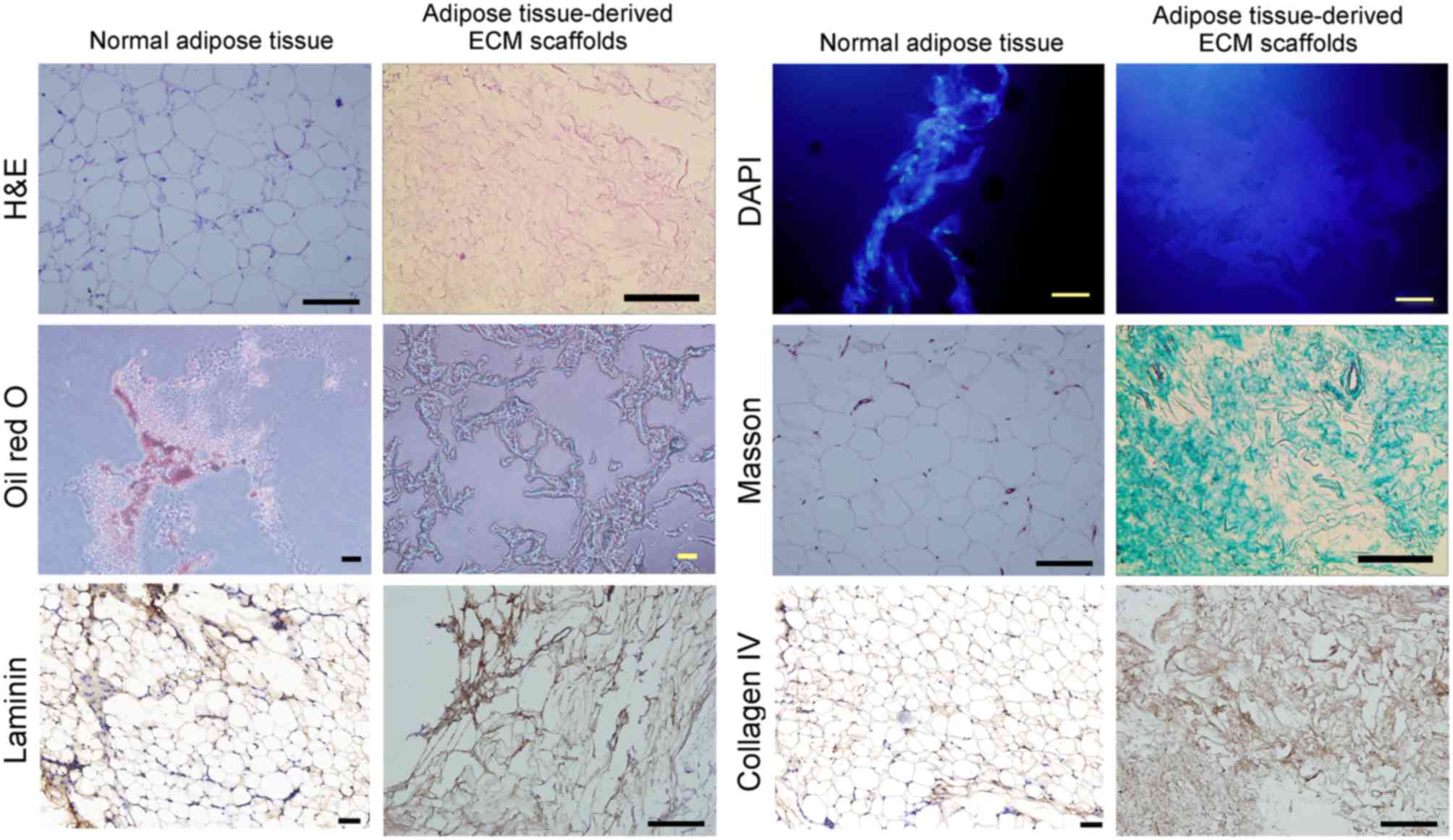
favorable for cell adhesion and proliferation. No intact nuclei
were observed in tissue sections stained with H&E or labeled
with DAPI (Fig. 2). Oil red O
staining demonstrated a complete absence of lipids in adipose
tissue-derived ECM scaffolds (Fig.
2).
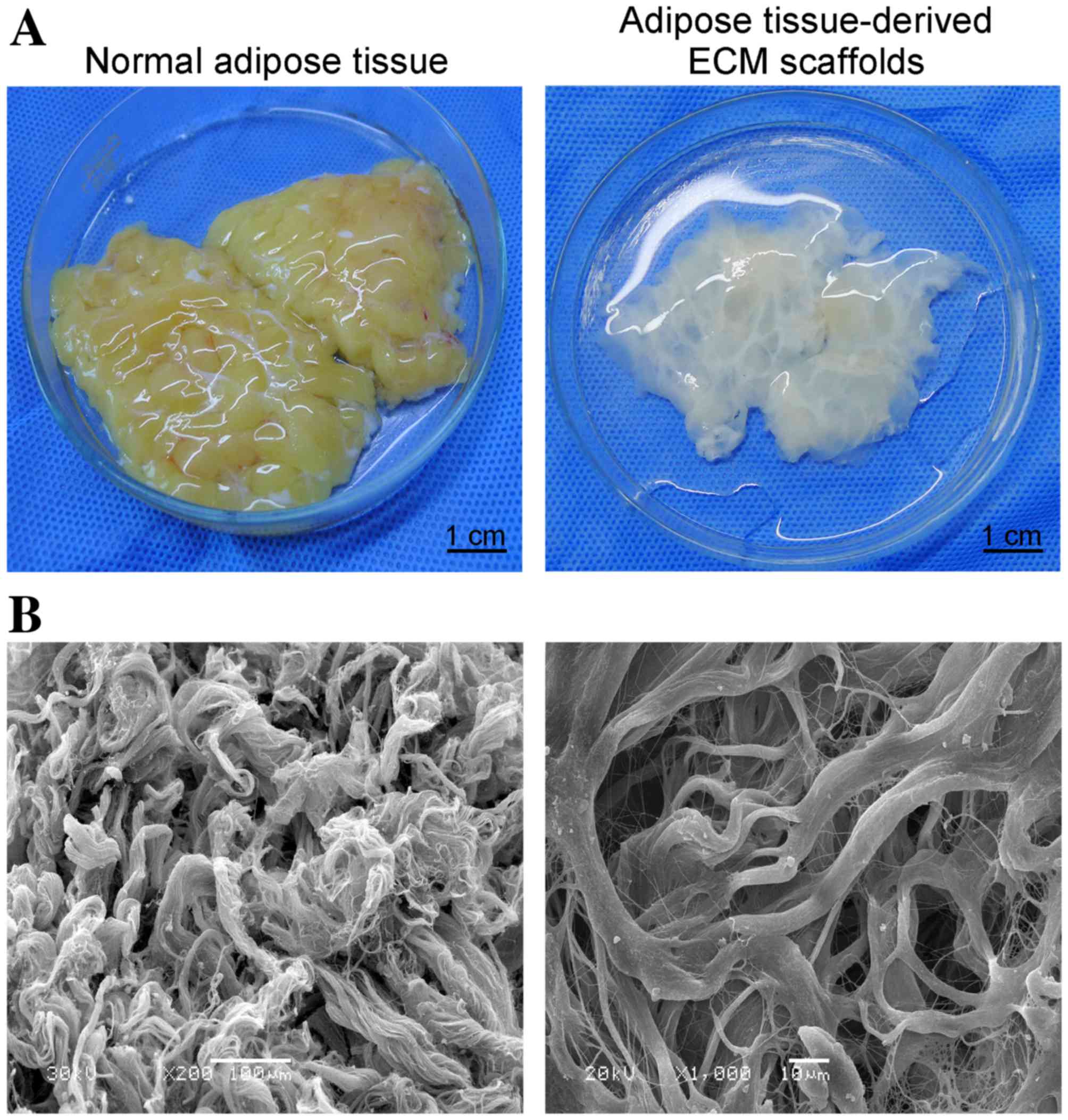 | Figure 1.(A) Macroscopic images of a human
fresh adipose tissue and human adipose tissue-derived ECM scaffolds
(scale bar, 1 cm). (B) Scanning electron microscopy images of the
ECM scaffolds demonstrating the network structure of collagen
fibers and the range of pore sizes (left panel, magnification,
×200, scale bar, 100 µm; right panel, magnification, ×1,000, scale
bar, 10 µm). ECM, extracellular matrix. |
Composition of adipose tissue-derived
ECM scaffolds
Masson's trichrome staining demonstrated that the
primary component of the ECM scaffolds following decellularization
was collagen fibers, and no additional cellular structures were
identified (Fig. 2).
Immunohistochemical staining demonstrated that LN and collagen type
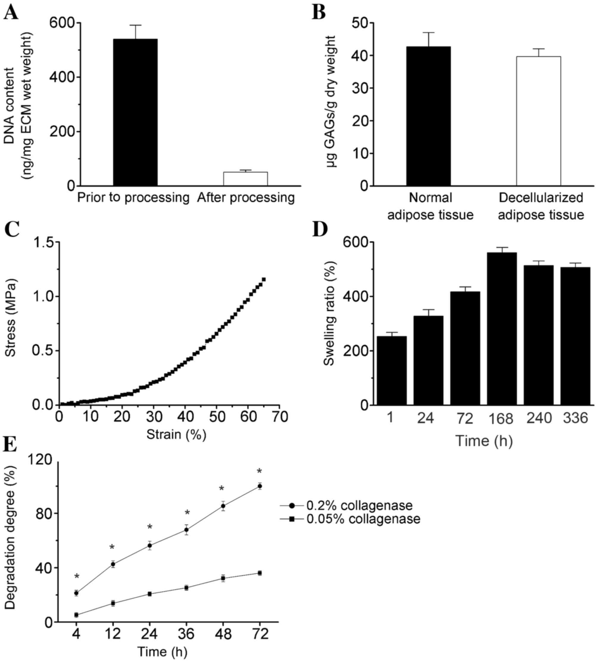
IV remained abundant in the decellularized matrix (Fig. 2). The total DNA content was <50
ng double-stranded DNA/mg of scaffold dry weight (Fig. 3A). GAG analysis estimated an
average of 39.67±2.31 µg sulfated GAG/g dry adipose ECM, and
42.15±4.36 µg sulfated GAG/g normal adipose tissue (Fig. 3B).
Mechanical properties of adipose
tissue-derived ECM scaffolds
The mechanical properties of the decellularized
samples were investigated using an UTM, and a typical stress-strain
curve is shown in Fig. 3C. The
initial tensile strength and Young's modulus were 0.22±0.05 and
65.70±5.97 MPa (at 0.1% of initial strain), respectively (Fig. 3C). The maximum elongation at the
point of breakage was 65.02±8.26% of the initial length (data not
shown). The ECM scaffolds swelled in time-dependent manner until
168 h, and the maximum PBS uptake ratio was 561±19.3% at this time
point (Fig. 3D), which indicates
that the scaffold absorbed ~6 times its own weight of medium. By
determining liquid displacement measurements, the ECM scaffolds
exhibited a porosity of 90.43±6.5% (mean ± standard error, n=5;
data not shown), which may facilitate cellular penetration and
proliferation.
The ECM scaffolds demonstrated sustained degradation
in the presence of collagenase, and the degradation rate
significantly increased with increasing collagenase concentration
(P<0.05; Fig. 3E). The
scaffolds exhibited rapid degradation (>50%) within the first 24
h, and complete degradation within 3 days of incubation in PBS
containing 0.2% collagenase at 37°C (Fig. 3E).
In vitro cell viability and adipogenic
differentiation
The levels of hASC adhesion and viability on ECM
scaffolds were compared to those observed on collagen type I
scaffolds by MTT assay analysis. As demonstrated in Fig. 4A, the mean optical density value
for hASCs on the ECM scaffolds at day 1 was slightly lower than
that observed on the collagen type I scaffolds. However, the
viability of hASCs on ECM scaffolds increased at a slightly faster
rate when compared with collagen type I scaffolds following 1 week
(Fig. 4A). The mean optical
density value for cells cultured on the ECM scaffolds increased
with time in culture, and was significantly higher when compared
with cells cultured on the collagen type I scaffolds at day 14
(P<0.05; Fig. 4A). These
results suggest that the adipose tissue-derived ECM scaffold
promoted the adhesion and growth of hASCs.
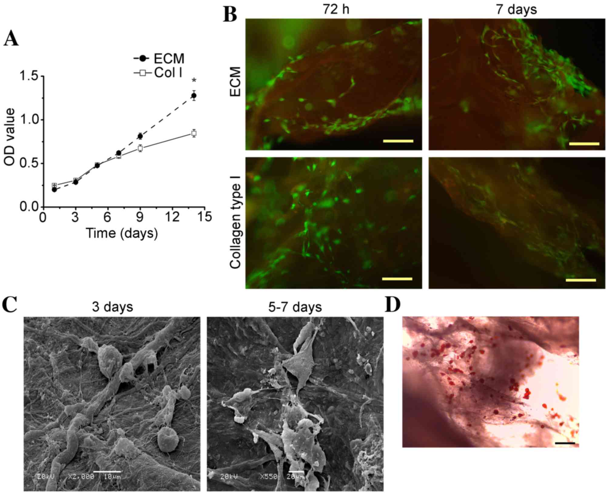 | Figure 4.hASC culture on adipose
tissue-derived ECM scaffolds in vitro. (A) The growth of
hASCs on ECM or Col I scaffolds, as determined using MTT assays.
Data are presented as the mean ± standard deviation. *P<0.05 vs.
Col I. (B) Analysis of hASC viability at 72 h and 7 days following
culture on ECM scaffolds and collagen type I scaffolds, as
determined using a Live/Dead® Viability/Cytotoxicity
kit. Green fluorescence indicates live cells, and red fluorescence
indicates dead cells (magnification, ×100; scale bar, 50 µm). (C)
Scanning electron microscopy images showing the adherence of hASCs
to ECM scaffolds during the first 3 days in culture (magnification,
×2,000; scale bar, 10 µm) and hASC dispersal and extension of
pseudopodia following 5–7 days (magnification, ×550; scale bar, 20
µm). (D) Representative optical microscopy image of Oil Red O
staining for intracellular lipid accumulation within hASCs cultured
on ECM scaffolds at 14 days following induction of adipogenic
differentiation (magnification, ×100; scale bar, 50 µm). hASC,
human adipose-derived stem cell; ECM, extracellular matrix; OD,
optical density; Col I, collagen type 1 scaffolds. |
Representative images of cellular organization at 72
h and 7 days post-cell seeding are shown in Fig. 4B. Staining of cells with a
Live/Dead® Viability/Cytotoxicity kit revealed that the
majority of cells were viable when cultured on ECM and collagen
type I scaffolds for 72 h (Fig.
4B). However, following 7 days, a greater number of dead cells
were observed on the collagen type I scaffolds when compared with
the adipose tissue-derived ECM scaffolds, and a higher number of
viable cells were attached to the ECM scaffolds (Fig. 4B). These results indicated that the
ECM scaffolds promoted hASC adhesion and growth. SEM analysis
revealed that hASCs cultured on the ECM scaffolds exhibited good
dispersal and distribution patterns on the scaffold surface area
(Fig. 4C). During the first 1–3
days, hASCs exhibited a spherical morphology, whereas they
progressively dispersed and displayed extended pseudopodia by 5–7
days, potentially due to cell proliferation (Fig. 4C). A representative Oil Red O
staining image obtained following 14 days of hASC differentiation
on adipose tissue-derived ECM scaffolds is presented in Fig. 4D, demonstrating that lipid loading
was detected in hASCs.
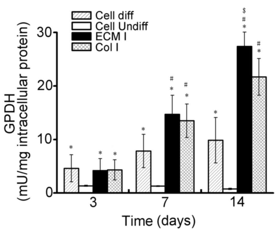
The average GPDH activity levels in hASCs cultured
on the two scaffold types are presented in Fig. 5. At 3 days following induction of
differentiation, GPDH activity levels in hASCs cultured on the
adipose tissue-derived ECM scaffolds (4.18±2.25 mU/mg;
α=0.018, P<0.05) and collagen type I scaffolds (4.32±1.89
mU/mg; α=0.014, P<0.05), as well as in the induced
positive control cells (4.61±2.53 mU/mg; α=0.009, P<0.05)
were significantly higher when compared with the non-induced
negative control cells (1.33±0.11 mU/mg). However, no statistically
significant differences were observed among the GPDH activity
levels in the former three groups following 3 days. GPDH activity
levels in the ECM scaffold, collagen type I scaffold and induced
positive control groups were greater at 7 days following induction
of differentiation when compared with 3 days (Fig. 5). However, at this time point, the
GPDH activity levels of hASCs on the ECM scaffolds (14.7±3.56
mU/mg; α=0.001, P<0.05) and collagen type I scaffolds
(13.5±3.14 mU/mg; α=0.004, P<0.05) were significantly
greater than those of the induced positive control cells (7.83±3.12
mU/mg) and non-induced negative control cells (1.28±0.09 mU/mg). No
significant difference in GPDH activity in hASCs cultured on the
ECM scaffolds and collagen type I scaffolds was observed at 7 days.
At 14 days following induction of differentiation, although the
GPDH activity of hASCs cultured on the collagen type I scaffolds
(21.7±9.45 mU/mg) was significantly higher than that of the induced
positive control cells (9.86±4.26 mU/mg; α=0, P<0.05) and
the non-induced negative control cells (0.75±0.13 mU/mg;
α=0.001, P<0.05), GPDH activity in hASCs cultured on the
ECM scaffolds (27.4±7.82 mU/mg; α=0.001, P<0.05) was
significantly greater compared with hASCs cultured on collagen type
I scaffolds (Fig. 5). As expected,
low GPDH activity levels were observed for the non-induced control
samples at all time points. Notably, GPDH activity in hASCs
increased by >6-fold on the ECM scaffolds from 3 (4.18±2.25
mU/mg) to 14 days (27.4±7.82 mU/mg).
Discussion
Decellularized tissues derived from the aorta, heart
valve, pericardium and tendons of human and animal cadavers have
been developed and investigated for clinical application in humans
(25–28). Adipose tissue contains multiple
cell types, including adipocytes, fibroblasts, smooth muscle cells,
endothelial cells, immune cells and ASCs (29). Therefore, adipose tissue is a rich
source of ECM components, such as type I, II and IV collagens. In
addition, adipose tissue is a major endocrine and secretory organ
that releases a wide variety of cytokines (20). Substantial quantities of adipose
tissue obtained during clinical liposuction and abdominoplasty
procedures are discarded as medical waste. The ability to obtain
cells and scaffold materials from human adipose tissue was a key
factor for the authors' approach to employ this tissue type as a
source for a decellularized ECM scaffold in the present study, and
this approach may be considered for autologous tissue
engineering.
The aim of decellularization is to remove
immunogenic cellular components from the tissue structure, while
maintaining the biological activity and mechanical integrity of the
ECM network. Different methods for decellularization, involving
physical, chemical and enzymatic treatments, have been widely
studied (30). Generally, the use
of any of these treatment methods alone is insufficient to achieve
complete decellularization, and combinations of these treatments
have been demonstrated to increase the decellularization efficiency
and optimize the tissue texture (31,32).
Previous studies involving the decellularization of tissues or
organs have used SDS more extensively than any other chemical
reagent. However, the decellularization protocol presented by Flynn
et al (21) was designed to
avoid the use SDS, as this reagent has been demonstrated to alter
the matrix architecture and affect cellular repopulation of the
matrix. Although 99.9% isopropanol has been demonstrated to be
effective for the decellularization of adipose tissues within 48 h
(21), the results of the present
study indicated that complete decellularization of adipose tissue
was possible following 12 h of 99.9% isopropanol extraction and
enzymatic digestion in combination with repeated freeze-thaw
treatments. Based on previous experience, human adipose tissue is a
loose connective tissue that is easily disrupted by repeated
freeze-thaw cycles, and the addition of strong chemical and
enzymatic agents is necessary to facilitate the removal of cellular
components. A shorter processing time, as demonstrated in the
current study, may be very beneficial in terms of the cost and
scalability of this approach for clinical use. Histologic and SEM
analyses indicated that the finalized protocol applied in the
current study was highly effective at removing the cellular
components from adipose tissue, while minimizing macroscopic
alterations to the ECM network in the shortest processing
time-frame.
Biochemical studies performed in the present study
indicated that the decellularized adipose tissue retained a complex
composition of proteins, peptides and GAGs. Sulfated GAGs are known
for their ability to sequester growth factors and subsequently
present them to cells (33,34).
Therefore, their presence within the matrix provides a possible
avenue for bioactive molecule delivery in vitro and in
vivo. Immunochemical staining revealed the preservation of
collagen fibers, a major component of native adipose ECM. Collagen
type IV, which is a fibrillar collagen, contains various binding
sites for bioactive molecules that modulate cellular behavior and
therefore not only functions as a structural protein (35). In the present study, LN was
detected in decellularized adipose ECM tissues, despite exhibiting
a slight reduction when compared with native tissues. LN promotes
the attachment of epithelial cells to the basal lamina, and is
involved in the migration and growth of these cells (16). Therefore, retention of these
adhesion molecules within ECM scaffolds following
chemically-induced cell extraction may promote the migration and
growth of cells in these scaffolds during repopulation in
vivo or in vitro.
In the present study, mechanical testing of the ECM
scaffolds indicated that they maintained a mechanical strength
comparable to that of the native tissue. This suggests that the
applied decellularization process preserved the ECM scaffold
structure. The average porosity of the acellular ECM scaffolds was
90.43%, which is sufficient for cell infiltration, oxygen
transport, and nutrient and waste exchange (36). Finally, the median pore sizes
within the ECM scaffolds ranged from 20–200 µm, which are
appropriate for the adhesion of many types of cells (37).
The results of the present study demonstrated that
hASCs cultured on the human adipose tissue-derived ECM scaffolds
exhibited satisfactory adhesion and viability in vitro. In
addition, the surface features and chemical and biological
compositions of human adipose tissue-derived ECM scaffolds were
highly supportive of three-dimensional stem cell culture. SEM
analysis indicated that the cells exhibited a star-shaped
morphology upon adhesion to the ECM scaffolds, which was distinct
from their original spherical morphology. The results suggest that
the prepared ECM scaffolds provided sufficient space to support the
growth and proliferation of hASCs.
In subsequent in vitro experiments
investigating hASC adipogenesis, the adipose tissue-derived ECM
scaffolds were observed to strongly support adipogenesis of these
cells. Oil red O staining revealed that the hASCs differentiated
into adipose cells on the ECM scaffolds, as they contained a large
quantity of lipid droplets. In addition, the observed levels of
GPDH enzyme activity provided additional evidence that the ECM
scaffolds were adipo-inductive (21,38).
At 14 days following induction of differentiation, the GPDH
activity in cells cultured on the ECM scaffolds was significantly
higher when compared with cells cultured on collagen type I
scaffolds. These results clearly demonstrated that the ECM
composition and scaffold architecture serve critical roles in
directing the cellular response, which supports the tissue-specific
approach for adipose regeneration.
In conclusion, the results of the present study
demonstrated that human adipose tissue may be successfully
decellularized through treatment with repeated freeze-thaw cycles,
centrifugation, polar solvent extraction and enzymatic digestion.
The decellularized ECM samples exhibited high pore
interconnectivity and mechanical properties for adequate use as
tissue engineering scaffolds. hASCs were observed to adhere,
spread, proliferate and differentiate into adipocytes on the ECM
scaffolds. Ultimately, the results of the present study indicated
that decellularized adipose tissue-derived ECM prepared using the
described method, presents a promising substrate for the repair of
various defective or damaged human tissues.
Acknowledgements
This study was supported by grants from the National
Natural Science Foundation of China (grant no. 81401612) and the
Key Project of the ‘12th Five-Year Plan’ for Medical Science and
Technology of PLA (grant no. BWS11C061).
References
|
1
|
Wu LC, Bajaj A, Chang DW and Chevray PM:
Comparison of donor-site morbidity of SIEA, DIEP, and
muscle-sparing TRAM flaps for breast reconstruction. Plast Reconstr
Surg. 122:702–709. 2008. View Article : Google Scholar : PubMed/NCBI
|
|
2
|
Alhadlaq A, Tang M and Mao JJ: Engineered
adipose tissue from human mesenchymal stem cells maintains
predefined shape and dimension: Implications in soft tissue
augmentation and reconstruction. Tissue Eng. 11:556–66. 2005.
View Article : Google Scholar : PubMed/NCBI
|
|
3
|
Fischbach C, Spruss T, Weiser B, Neubauer
M, Becker C, Hacker M, Göpferich A and Blunk T: Generation of
mature fat pads in vitro and in vivo utilizing 3-D long-term
culture of 3T3-L1 preadipocytes. Exp Cell Res. 300:54–64. 2004.
View Article : Google Scholar : PubMed/NCBI
|
|
4
|
Kang X, Xie Y and Kniss DA: Adipose tissue
model using three-dimensional cultivation of preadipocytes seeded
onto fibrous polymer scaffolds. Tissue Eng. 11:458–468. 2005.
View Article : Google Scholar : PubMed/NCBI
|
|
5
|
Patrick CW Jr, Zheng B, Johnston C and
Reece GP: Long-term implantation of preadipocyte-seeded PLGA
scaffolds. Tissue Eng. 8:283–293. 2002. View Article : Google Scholar : PubMed/NCBI
|
|
6
|
Kral JG and Crandall DL: Development of a
human adipocyte synthetic polymer scaffold. Plast Reconstr Surg.
104:1732–1738. 1999. View Article : Google Scholar : PubMed/NCBI
|
|
7
|
Gentleman E, Nauman EA, Livesay GA and Dee
KC: Collagen composite biomaterials resist contraction while
allowing development of adipocytic soft tissue in vitro. Tissue
Eng. 12:1639–1649. 2006. View Article : Google Scholar : PubMed/NCBI
|
|
8
|
Halbleib M, Skurk T, de Luca C, von
Heimburg D and Hauner H: Tissue engineering of white adipose tissue
using hyaluronic acid-based scaffolds. I: In vitro differentiation
of human adipocyte precursor cells on scaffolds. Biomaterials.
24:3125–3132. 2003. View Article : Google Scholar : PubMed/NCBI
|
|
9
|
Kawaguchi N, Toriyama K, Nicodemou-Lena E,
Inou K, Torii S and Kitagawa Y: De novo adipogenesis in mice at the
site of injection of basement membrane and basic fibroblast growth
factor. Proc Natl Acad Sci USA. 95:1062–1066. 1998. View Article : Google Scholar : PubMed/NCBI
|
|
10
|
Mauney JR, Nguyen T, Gillen K, Kirker-Head
C, Gimble JM and Kaplan D: Engineering adipose-like tissue in vitro
and in vivo utilizing human bone marrow and adipose-derived
mesenchymal stem cells with silk fibroin 3D scaffolds.
Biomaterials. 28:5280–5290. 2007. View Article : Google Scholar : PubMed/NCBI
|
|
11
|
Badylak SF, Freytes DO and Gilbert TW:
Extracellular matrix as a biological scaffold material: Structure
and function. Acta Biomater. 5:1–13. 2009. View Article : Google Scholar : PubMed/NCBI
|
|
12
|
Jin CZ, Choi BH, Park SR and Min BH:
Cartilage engineering using cell-derived extracellular matrix
scaffold in vitro. J Biomed Mater Res A. 92:1567–1577.
2010.PubMed/NCBI
|
|
13
|
Keskin M, Kelly CP, Moreira-Gonzalez A,
Lobocki C, Yarim M, Kaplan S and Jackson IT: Repairing
critical-sized rat calvarial defects with a periosteal cell-seeded
small intestinal submucosal layer. Plast Reconstr Surg.
122:400–409. 2008. View Article : Google Scholar : PubMed/NCBI
|
|
14
|
Uflacker AB and Janis JE: The use of
acellular dermal matrix in the correction of visible parasternal
deformities after breast reconstruction. Plast Reconstr Surg.
126:34e–36e. 2010. View Article : Google Scholar : PubMed/NCBI
|
|
15
|
Altman AM, Chiu ES, Bai X, Yan Y, Song YH,
Newsome RE and Alt EU: Human adipose-derived stem cells adhere to
acellular dermal matrix. Aesthetic Plast Surg. 32:698–699. 2008.
View Article : Google Scholar : PubMed/NCBI
|
|
16
|
Atkinson JJ, Adair-Kirk TL, Kelley DG,
Demello D and Senior RM: Clara cell adhesion and migration to
extracellular matrix. Respir Res. 9:12008. View Article : Google Scholar : PubMed/NCBI
|
|
17
|
Hudson TW, Liu SY and Schmidt CE:
Engineering an improved acellular nerve graft via optimized
chemical processing. Tissue Eng. 10:1346–1358. 2004. View Article : Google Scholar : PubMed/NCBI
|
|
18
|
Borschel GH, Dennis RG and Kuzon WM Jr:
Contractile skeletal muscle tissue-engineered on an acellular
scaffold. Plast Reconstr Surg. 113:595–604. 2004. View Article : Google Scholar : PubMed/NCBI
|
|
19
|
Gilbert TW, Stolz DB, Biancaniello F,
Simmons-Byrd A and Badylak SF: Production and characterization of
ECM powder: Implications for tissue engineering applications.
Biomaterials. 26:1431–1435. 2005. View Article : Google Scholar : PubMed/NCBI
|
|
20
|
Choi JS, Yang HJ, Kim BS, Kim JD, Lee SH,
Lee EK, Park K, Cho YW and Lee HY: Fabrication of porous
extracellular matrix scaffolds from human adipose tissue. Tissue
Eng Part C Methods. 16:387–396. 2010. View Article : Google Scholar : PubMed/NCBI
|
|
21
|
Flynn LE: The use of decellularized
adipose tissue to provide an inductive microenvironment for the
adipogenic differentiation of human adipose-derived stem cells.
Biomaterials. 31:4715–4724. 2010. View Article : Google Scholar : PubMed/NCBI
|
|
22
|
Brown BN, Freund JM, Han L, Rubin JP,
Reing JE, Jeffries EM, Wolf MT, Tottey S, Barnes CA, Ratner BD and
Badylak SF: Comparison of three methods for the derivation of a
biologic scaffold composed of adipose tissue extracellular matrix.
Tissue Eng Part C Methods. 17:411–421. 2011. View Article : Google Scholar : PubMed/NCBI
|
|
23
|
Wu I, Nahas Z, Kimmerling KA, Rosson GD
and Elisseeff JH: An injectable adipose matrix for soft-tissue
reconstruction. Plast Reconstr Surg. 129:1247–1257. 2012.
View Article : Google Scholar : PubMed/NCBI
|
|
24
|
Bunnell BA, Flaat M, Gagliardi C, Patel B
and Ripoll C: Adipose-derived stem cells: Isolation, expansion and
differentiation. Methods. 45:115–120. 2008. View Article : Google Scholar : PubMed/NCBI
|
|
25
|
De Cock LJ, De Koker S, De Vos F, Vervaet
C, Remon JP and De Geest BG: Layer-by-layer incorporation of growth
factors in decellularized aortic heart valve leaflets.
Biomacromolecules. 11:1002–1008. 2010. View Article : Google Scholar : PubMed/NCBI
|
|
26
|
Ingram JH, Korossis S, Howling G, Fisher J
and Ingham E: The use of ultrasonication to aid recellularization
of acellular natural tissue scaffolds for use in anterior cruciate
ligament reconstruction. Tissue Eng. 13:1561–1572. 2007. View Article : Google Scholar : PubMed/NCBI
|
|
27
|
Mirsadraee S, Wilcox HE, Korossis SA,
Kearney JN, Watterson KG, Fisher J and Ingham E: Development and
characterization of an acellular human pericardial matrix for
tissue engineering. Tissue Eng. 12:763–773. 2006. View Article : Google Scholar : PubMed/NCBI
|
|
28
|
Schopka S, Schmid FX, Hirt S, Birnbaum DE,
Schmid C and Lehle K: Recellularization of biological heart valves
with human vascular cells: In vitro hemocompatibility assessment. J
Biomed Mater Res B Appl Biomater. 88:130–138. 2009. View Article : Google Scholar : PubMed/NCBI
|
|
29
|
Fonseca-Alaniz MH, Takada J, Alonso-Vale
MI and Lima FB: Adipose tissue as an endocrine organ: From theory
to practice. J Pediatr (Rio J). 83 5 Suppl:S192–S203. 2007.
View Article : Google Scholar : PubMed/NCBI
|
|
30
|
Gilbert TW, Sellaro TL and Badylak SF:
Decellularization of tissues and organs. Biomaterials.
27:3675–3683. 2006.PubMed/NCBI
|
|
31
|
Choi JS, Kim BS, Kim JY, Kim JD, Choi YC,
Yang HJ, Park K, Lee HY and Cho YW: Decellularized extracellular
matrix derived from human adipose tissue as a potential scaffold
for allograft tissue engineering. J Biomed Mater Res A. 97:292–299.
2011. View Article : Google Scholar : PubMed/NCBI
|
|
32
|
Young DA, Ibrahim DO, Hu D and Christman
KL: Injectable hydrogel scaffold from decellularized human
lipoaspirate. Acta Biomater. 7:1040–1049. 2011. View Article : Google Scholar : PubMed/NCBI
|
|
33
|
Doran MR, Markway BD, Aird IA, Rowlands
AS, George PA, Nielsen LK and Cooper-White JJ: Surface-bound stem
cell factor and the promotion of hematopoietic cell expansion.
Biomaterials. 30:4047–4052. 2009. View Article : Google Scholar : PubMed/NCBI
|
|
34
|
Mullen LM, Best SM, Brooks RA, Ghose S,
Gwynne JH, Wardale J, Rushton N and Cameron RE: Binding and release
characteristics of insulin-like growth factor-1 from a
collagen-glycosaminoglycan scaffold. Tissue Eng Part C Methods.
16:1439–1448. 2010. View Article : Google Scholar : PubMed/NCBI
|
|
35
|
Streuli C: Extracellular matrix
remodelling and cellular differentiation. Curr Opin Cell Biol.
11:634–640. 1999. View Article : Google Scholar : PubMed/NCBI
|
|
36
|
Kim BS, Choi JS, Kim JD, Choi YC and Cho
YW: Recellularization of decellularized human
adipose-tissue-derived extracellular matrix sheets with other human
cell types. Cell Tissue Res. 348:559–567. 2012. View Article : Google Scholar : PubMed/NCBI
|
|
37
|
Sobral JM, Caridade SG, Sousa RA, Mano JF
and Reis RL: Three-dimensional plotted scaffolds with controlled
pore size gradients: Effect of scaffold geometry on mechanical
performance and cell seeding efficiency. Acta Biomater.
7:1009–1018. 2011. View Article : Google Scholar : PubMed/NCBI
|
|
38
|
Turner AE, Yu C, Bianco J, Watkins JF and
Flynn LE: The performance of decellularized adipose tissue
microcarriers as an inductive substrate for human adipose-derived
stem cells. Biomaterials. 33:4490–4499. 2012. View Article : Google Scholar : PubMed/NCBI
|