Introduction
Intimal hyperplasia contributes to the pathological
process of several vascular disorders, including restenosis
following angioplasty, transplant vasculopathy, vein graft stenosis
and atherosclerosis (1). During
vascular remodeling, vascular smooth muscle cells (VSMCs)
proliferate, migrate and change from a contractile phenotype to a
synthetic phenotype (2),
inhibition of VSMC proliferation, migration and phenotypic
modulation is a feasible treatment strategy to prevent intimal
hyperplasia. The extracellular matrix (ECM) is composed of collagen
subtypes and proteoglycans and has an important role in the process
of intimal thickening. Collagen is the most abundant matrix protein
in ECM, regulating collagen synthesis and degradation may also be a
therapeutic target for intimal hyperplasia (3).
Honokiol
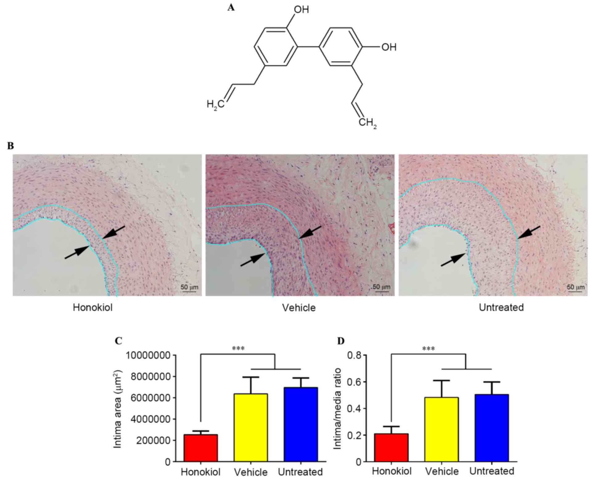
(3′,5-di-(2-propenyl)-1,1′-biphenyl-2,4′-diol) (Fig. 1A) is a natural bioactive product
obtained from plants in the genus Magnolia and has been determined
to have multiple biological activities, including anti-cancer,
anti-angiogenic, anti-inflammatory, anti-hypertensive,
neuroprotective, anti-oxidative and anti-thrombosis properties
(4–7). Honokiol has been shown to have the
ability to protect endothelial cells and stimulate prostacyclin
release in a rat carotid thrombosis model (6), which was revealed to have potential
in preventing intimal hyperplasia (8,9). Lee
et al determined that honokiol treatment inhibited VSMC
growth by activating p38 mitogen activated protein kinase (10). A previous study suggested that
honokiol may suppress tumor necrosis factor-a (TNF-α)-induced rat
aortic smooth muscle cell proliferation via caspase and
mitochondria-dependent apoptosis (11). These studies revealed that honokiol
may have therapeutic potential in the prevention of vascular
remodeling in cardiovascular diseases.
The present study investigated the effect of
honokiol on intimal thickening following vascular balloon injury.
Perivascular drug/gene delivery may effectively prevent intimal
hyperplasia in preclinical experiments (12,13).
Therefore, the present study hypothesized that perivascular
honokiol application may inhibit neointimal hyperplasia in rabbits
following carotid artery balloon injury through anti-proliferative
ability. The present study investigated the effect of perivascular
honokiol application on the expression of collagen, matrix
metalloproteinases (MMPs), the tissue inhibitor of
metalloproteinase-1 (TIMP-1) and the underlying mechanism involved
in this process.
Materials and methods
Materials
F-127 pluronic gel was purchased from Sigma-Aldrich;
Merck Millipore (Darmstadt, Germany). Honokiol was purchased from
Selleck Chemicals (Houston, TX, USA). 3F Fogarty embolectomy
catheters were obtained from Edwards Lifesciences (Irvine, CA,
USA). The primer sequences used in mRNA analysis are presented in
Table I. The primary antibodies
used in western blotting analysis were: Smad2/3 (Santa Cruz
Biotechnology, Inc., Dallas, TX, USA; catalog no. sc-6032),
phosphorylated-Smad2/3 (p-Smad2/3; Santa Cruz Biotechnology, Inc.;
catalog no. sc-11769), transforming growth factor-β1 (TGF-β1; Novus
Biologicals, LLC, Littleton, CO, USA; catalog no. MAB240), TGF-β
receptor type I (TGF-βRI; Santa Cruz Biotechnology, Inc.; catalog
no. sc-398-G), TGF-βRII (Santa Cruz Biotechnology, Inc.; catalog
no. sc-17792) and GAPDH (Abcam, Cambridge, UK; catalog no. ab8245).
Mouse monoclonal α-smooth muscle actin (α-SMA; Abcam; catalog no.
ab7817) and rabbit monoclonal anti-proliferating cell nuclear
antigen (PCNA; Abcam; catalog no. ab19166) were used for
immunohistochemistry analysis. The secondary antibodies were: Mouse
anti-goat IgG horse radish peroxidase (HRP) (Santa Cruz
Biotechnology, Inc.; catalog no. sc-2354) (for Smad2/3, p-Smad2/3,
TGF-βRI), goat anti-mouse IgG HRP (Abcam; catalog no. ab205719)
(for TGF-β1, TGF-βRII, GAPDH and a-SMA), goat anti-rabbit IgG HRP
(Abcam; catalog no. ab205718) (for PCNA). Remaining solvents and
reagents were analytical grade.
 | Table I.Primer pairs used for quantitative
polymerase chain reaction analysis in this study. |
Table I.
Primer pairs used for quantitative
polymerase chain reaction analysis in this study.
| Gene | Forward (5′-3′) | Reverse (5′-3′) |
|---|
| MMP-1 |
AAAGGCCAGTATGCACAGCTTTC |
TTCAACCACTGGGCCACTATTTC |
| MMP-2 |
GAAGAGCGTGAAGGTTGGAA |
TATCAGGTGGGGGTGAGAAG |
| MMP-9 |
TGAGCTTTGACATCCTGCAC |
TTTGTATCCGGCAAACTGGT |
| TIMP-1 |
TTCTCATCGCTGGACAACTG |
AGCGTAGGTCTTGGTGAAGC |
| GAPDH |
GCACCGTCAAGGCTGAGAAC |
TGGTGAAGACGCCAGTGGA |
Preparation of the honokiol-containing
pluronic gel
F-127 pluronic gel (30% w/v) in saline was prepared
and stored at −20°C for future use. Honokiol (10 mg) was dissolved
in 1 ml dimethylsulphoxide (DMSO) and honokiol-containing pluronic
gel was prepared by adding 1 ml of this solution to 9 ml of the
previously prepared 30% pluronic gel at 4°C. Therefore, 1 ml of
this solution contained 1 mg of honokiol. The resulting
honokiol-containing pluronic gel was stored at −20°C for future
use. It is of note that DMSO was used for the dilution because
honokiol is insoluble in water and has a relatively poor solubility
in ethanol.
Balloon endothelial denudation in
rabbit carotid artery and perivascular honokiol application
Male New Zealand White rabbits (3.0–3.5 kg, n=24,
aged 4–5 months) were obtained from the Laboratory Animal Center of
The Ninth People's Hospital, Shanghai Jiao Tong University School
of Medicine (Shanghai, China), animals were housed at a temperature
of 23±1°C and a humidity of 50±20% for 1 week with free access to
food and water. General anesthesia was induced by intravenous
injection of 3% pentobarbital sodium (1 ml/kg). A midline neck
incision was made and the left carotid artery (left common carotid
artery, left external carotid artery, left internal carotid artery)
was exposed. Subsequently, a permanent ligature was made on the
left external carotid artery ~5 mm away from the bifurcation, the
left internal carotid artery was ligated by a bulldog clamp,
another bulldog clamp was also placed on the proximal end of left
common carotid artery. A 3F Fogarty embolectomy catheter was then
introduced into the left common carotid artery through left
external carotid artery to establish the animal model of vascular
balloon injury. The balloon catheter was inflated with ~0.1 ml
saline solution three times to denude the aorta endothelium, the
length was about 3 cm. Immediately following balloon injury, 1 ml
Pluronic gel containing honokiol or DMSO was applied around the
injured carotid artery. Rabbits were randomly divided into three
experimental groups: i) Honokiol treated group (1 mg, n=8); ii)
vehicle (equal quantity of Pluronic gel containing DMSO) treated
group (vehicle alone, n=8); and iii) untreated group (balloon
injury without any treatment, n=8). The left external carotid
artery was ligated immediately after treatment, the bulldog clamps
were removed and the incision was closed. All animals recovered
after 40–60 min surgery and showed no symptoms of stroke. Animals
were euthanized and sacrificed in order to collect the carotid
arteries 14 days post-surgery, three segments were cut from each
artery, one segment was fixed in 10% buffered formalin and the
remaining two segments were stored in liquid nitrogen. The
protocols were approved by the Animal Care and Use Committee of
Shanghai Jiao Tong University School of Medicine.
Histopathological staining and
morphometry
Carotid arteries were fixed in formalin fixative
solution for 24 h, then dehydrated in graded ethanol and
subsequently embedded in paraffin. Transverse 5 µm slices were cut
and then stained with hematoxylin and eosin (H&E) (3 min for
hematoxylin and 30 sec for eosin at room temperature) and Masson
(5–10 min at room temperature) following the manufacturer's
protocol. The cross-sectional intima area, media area and
intima-to-media (I/M) area ratio were calculated from the H&E
stain slices, collagen fibers were calculated on Masson staining
slices (photographed by a Nikon light microscope). All the data was
analyzed and calculated using the Image-Pro Plus version 6.0 (Media
Cybernetics, Inc., Rockville, MD, USA).
Immunohistochemistry staining
Paraffin-embedded tissues were cut into 5-µm thick
slices, the sections were incubated with primary antibodies (α-SMA,
1:2,000 dilution; and PCNA, 1:2,000 dilution) at 4°C overnight and
then stained with a labeled DAB Detection kit (Fuzhou Maixin
Biotech Co., Ltd., Fuzhou, China), followed by counterstaining with
hematoxylin (30 sec at room temperature). Negative control was
treated with PBS. A total of 5 sections of each carotid artery were
selected for quantification, and 4 random fields were imaged at
magnification of ×200 in each section. The number of positively
stained cells was quantified using Image-Pro PLUS version 6.0
(Media Cybernetics, Inc.).
Reverse transcription-quantitative
polymerase chain reaction analysis (RT-qPCR)
TRIzol reagent (Molecular Research Center,
Cincinnati, OH, USA) was used to isolate total RNA from the tissues
according to the manufacturer's protocol. cDNA was synthesized from
1 µg of total RNA using iScript cDNA Synthesis kit (Bio-Rad
Laboratories, Inc., Hercules, CA, USA) at 42°C for 60 min, 70°C for
10 min and 4°C for storage). qPCR was performed using SYBR Premix
Ex Taq kit (Takara, Bio, Inc., Otsu, Japan) on a Stratagene Mx3005
RT-qPCR thermocycler (30 sec at 95°C, followed by 50 cycles of
denaturation at 95°C for 5 sec, annealing step at 60°C for 35 sec
and extension at 72°C for 15 sec). The mRNA expression levels of
samples were measured using the 2−ΔΔCq method (14). The primer sequences used in the
current study are presented in Table
I.
Western blotting analysis
Carotid arteries were homogenized in
radioimmunoprecipitation assay (RIPA) lysis buffer, the protein
concentration was determined using Bicinchoninic acid Protein Assay
reagent (Thermo Fisher Scientific, Inc., Waltham, MA, USA). Equal
quantity protein lysates (30 µg) was separated by 10% sodium
dodecyl sulfate-polyacrylamide gel electrophoresis and then
transferred to polyvinylidene difluoride membrane (EMD Millipore,
Billerica, MA, USA). Membranes were blocked with 5% non-fat milk
for 1 h and subsequently incubated with primary antibodies (Smad2/3
1:1,000 dilution; p-Smad2/3 1:1,000 dilution; TGF-β1 1:1,000
dilution; TGF-βRI 1:200 dilution; TGF-βRII 1:200 dilution; GAPDH
1:1,000 dilution) at 4°C overnight, followed by incubation with a
secondary antibody for 1 h (at room temperature, 1:200 dilution for
all). Protein bands were detected with enhanced chemiluminescence.
Protein bands were determined by Quantity One version 4.62 software
(Bio-Rad Laboratories, Inc., Hercules, CA, USA), and each band was
determined three times.
Statistical analysis
Data are presented as the mean ± standard deviation
and were analyzed by one-way analysis of variance with the SPSS
version 22.0.0.0 (IBM Corporation, Armonk, NY, USA). P<0.05 was
considered to indicate statistically significant difference.
Results
Perivascular honokiol application
inhibited intimal hyperplasia 14 days after vascular injury
H&E staining was used to assess the effect of
honokiol on intimal thickening following vascular injury, as
presented in Fig. 1B. Morphometric
analysis of the three treatment groups revealed that the intima
area was significantly reduced in the honokiol treatment group when
compared with the vehicle and untreated groups (Fig. 1C), the I/M area ratios followed a
similar pattern (Fig. 1C). No
significant difference was identified between intimal area and I/M
area ratios in the vehicle and the untreated groups. These findings
suggested that honokiol was effective in inhibiting intimal
thickening and the vehicle treatment had no effect on intimal
hyperplasia.
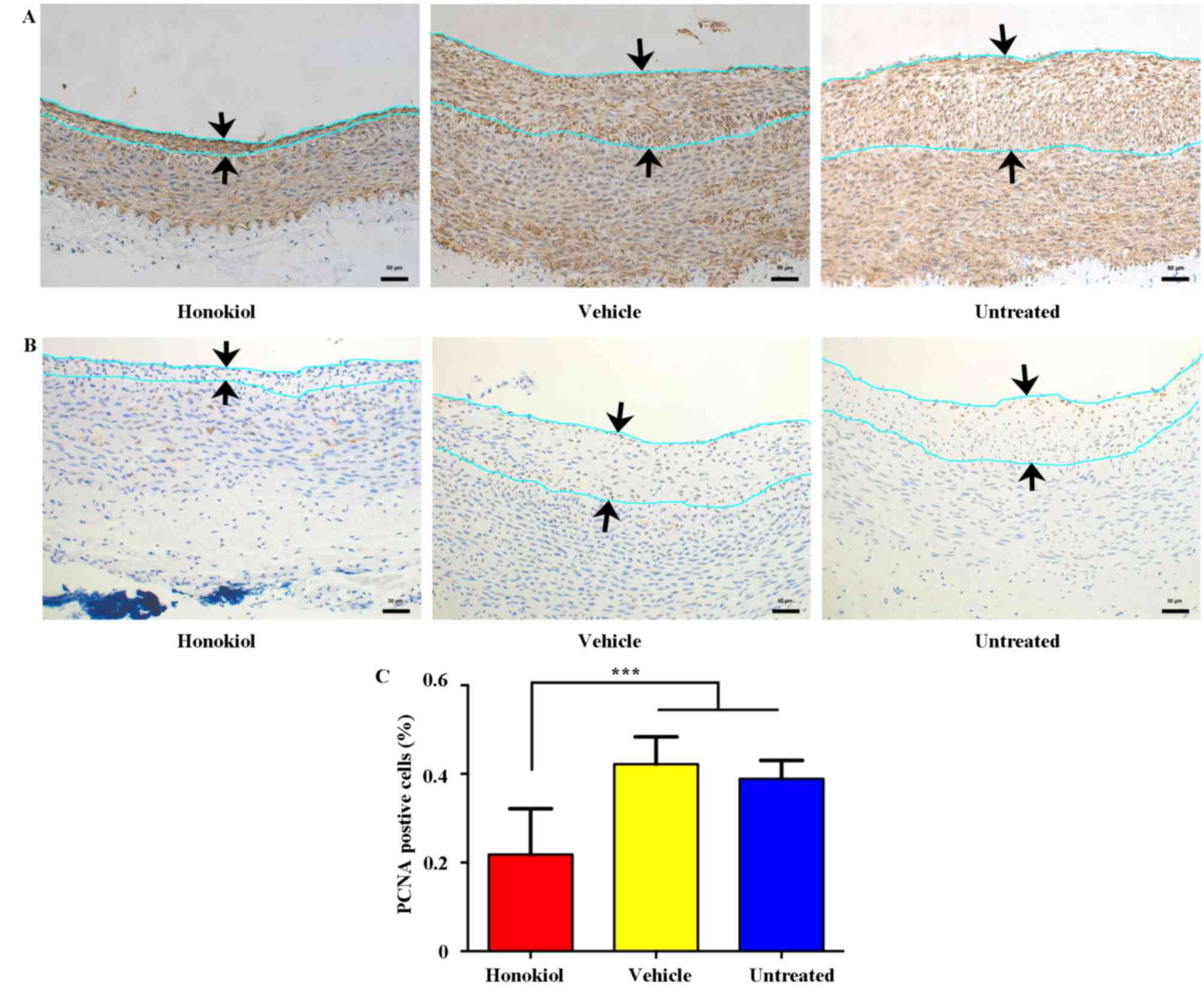
Perivascular honokiol application
reduced intimal VSMC proliferation 14 days after vascular
injury
Immunohistochemistry staining for α-SMA and PCNA was
performed to investigate whether honokiol reduced neointima
formation by inhibiting proliferation of VSMCs. It was determined
that the majority of neointimal cells were positive for α-SMA,
indicating that the cells that proliferated in the intima were
VSMCs (Fig. 2A). Additionally,
intimal proliferation was observed with immunohistochemistry
staining of PCNA (a marker for cell proliferation) and presented in
Fig. 2B, the ratio of
PCNA-positive cells was significantly reduced in the
honokiol-treated group in comparison with vehicle and untreated
groups (Fig. 2C). These findings
indicated that local honokiol treatment significantly inhibited
intimal VSMCs proliferation.
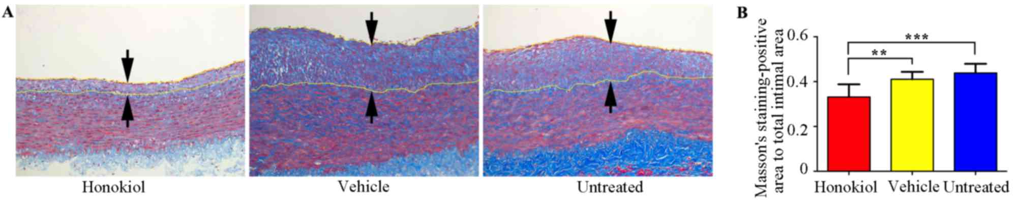
Perivascular honokiol application
reduced collagen deposition following vascular injury
Collagen contents in the arteries 14 days after
vascular injury were assessed with Masson staining (Fig. 3A), as presented in Fig. 3B, honokiol treatment group had a
significantly reduced the collagen content compared with the
vehicle and untreated groups, the vehicle treatment group exhibited
similar collagen deposition compared with the untreated group.
Effects of perivascular honokiol
application on the expression of MMPs and TIMP-1
MMPs and TIMP-1 have essential roles in intimal
thickening following vascular injury; therefore, the expression of
MMPs and TIMP-1 was investigated using RT-qPCR analysis. As
presented in Fig. 4, there was a
significant increase in the MMP-1, MMP-2 and MMP-9 mRNA expression
levels when the honokiol-treated group was compared with the
vehicle and untreated groups. TIMP-1 mRNA expression was
significantly reduced in the honokiol treated group compared with
the vehicle and untreated groups. No significant difference was
identified between the vehicle-treated group and untreated group in
terms of the expression of MMPs and TIMP-1.
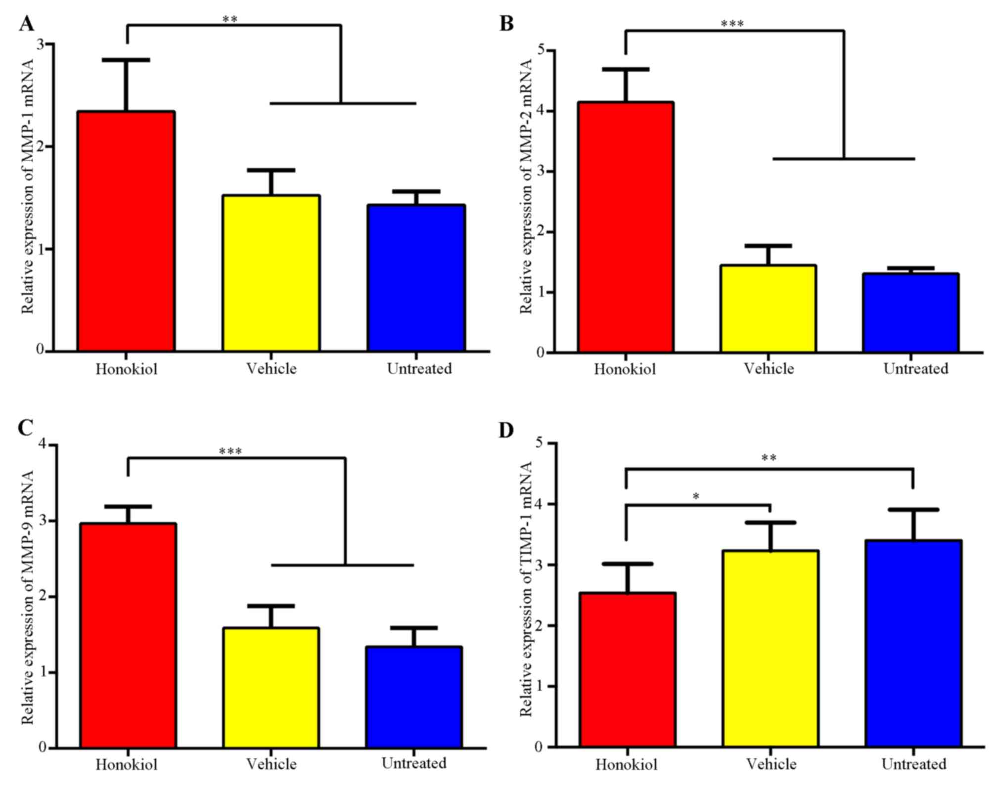 | Figure 4.Effects of perivascular honokiol
application on the expression of MMPs and TIMP-1. (A) Reverse
transcription-quantitative polymerase chain reaction analysis of
(A) MMP-1, (B) MMP-2, (C) MMP-9 and (D) TIMP-1 mRNA expression,
GAPDH was used as an internal standard (n=8). *P<0.05,
**P<0.01, ***P<0.001. MMP-1,-2,-9, matrix
metalloproteinase-1,-2,-9; TIMP-1, tissue inhibitor of
metalloproteinase-1. |
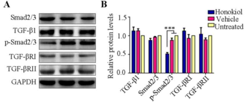
Perivascular honokiol application
downregulated phosphorylation of Smad2/3 expression
TGF-β1/Smad2/3 signaling is a critical regulator in
the pathological process of intimal hyperplasia and inhibition of
the TGF-β1/Smad2/3 signaling pathway may reduce VSMC proliferation
and collagen deposition, thus attenuating intimal hyperplasia
(15–17). Therefore, the present study
investigated whether the reduced VSMC proliferation and collagen
deposition were mediated via TGF-β1/Smad2/3 pathway. It is of note
that western blot analysis revealed that honokiol treatment
significantly reduced the expression of p-Smad2/3, whereas TGF-β1
and Smad2/3 expression showed no obvious change in the honokiol
treatment group compared with vehicle or untreated groups (Fig. 5). TGF-βRII phosphorylates TGF-βRI
and then TGF-βRI in turn phosphorylates Smad2/3 (18); therefore, the expression of TGF-βRI
and TGF-βRII was subsequently analyzed. However, no significant
difference was identified between the honokiol, vehicle or
untreated groups (Fig. 5).
Discussion
The present study demonstrated that perivascular
honokiol treatment inhibited intimal hyperplasia in rabbits 14 days
after carotid artery balloon injury. The findings also revealed
that honokiol treatment reduced VSMC proliferation and collagen
deposition in the arteries following vascular injury. Additionally,
the mRNA expression levels of MMP-1, MMP-2 and MMP-9 were
upregulated, whereas TIMP-1 was downregulated in the honokiol
treatment group. Subsequently the underlying mechanism was
investigated and it was revealed that honokiol treatment reduced
the phosphorylation of Smad2/3.
The adventitia has been considered to be an
important regulator in vascular diseases, including hypertension,
atherosclerosis and restenosis (19). Perivascular drug delivery is an
effective method to prevent intimal hyperplasia as it may obtain
controlled release and reduce the risk of thrombosis (drug released
into the inner wall of vessels can impair reendothelialization,
which would increase the risk of thrombosis) (13,20).
The present study has demonstrated that local application of
honokiol around the injured carotid artery may inhibit intimal
thickening.
VSMCs have an important function in intimal
thickening, and inhibition of VSMC proliferation is a feasible
method to prevent intimal hyperplasia (2). Collagen is the most abundant matrix
protein in ECM, treatments reducing collagen deposition are also
feasible options to inhibit intimal thickening (3). The present study identified that
perivascular honokiol application may reduce VSMCs proliferation 14
days after vascular injury, which was consistent with previous
studies in vitro (10,11).
Collagen content was also reduced in the honokiol-treated group
compared with vehicle and untreated groups (Fig. 3). Additionally, a previous study
determined that honokiol was able to reduce collagen deposition in
a renal fibrosis model (21),
demonstrating a potential antifibrotic effect of honokiol on other
diseases.
MMPs and TIMPs have important functions in intimal
thickening, MMPs may reduce ECM components and promote VSMC
migration/proliferation, whereas TIMP-1 inhibits MMPs activity
(22–24). It has been previously reported that
honokiol may suppress the expression of MMPs in other diseases,
such as cancer (melanoma, non-melanoma, urinary bladder cancer and
gastric cancer) (25,26) and arthritis (27,28).
However, the current study identified increased MMP-1, MMP-2 and
MMP-9 mRNA levels in the honokiol-treated group, whereas TIMP-1
mRNA expression was significantly lower compared with vehicle or
untreated groups (Fig. 4), this
suggested that honokiol may increase the expression of MMPs and
reduce the expression of TIMP-1 in local arteries, one of the
possible reasons for this phenomenon is the difference between the
cell lines and animal models; however, the underlying mechanism
remains to be elucidated.
TGF-β1/Smad2/3 signaling has a key role in intimal
hyperplasia; therefore, inhibition of the TGF-β/Smad2/3 signaling
pathway may reduce VSMCs proliferation and collagen deposition,
thus attenuating intimal hyperplasia (15–17).
It is of note that the present study determined that honokiol
treatment downregulated the protein expression level of p-Smad2/3,
whereas the level of TGF-β1 and total Smad2/3 remained constant.
TGF-β receptors may phosphorylate Smad2/3 (17); therefore, the present study
investigated the expression of TGF-βRI and TGF-βRII. The results
revealed that TGF-βRI and TGF-βRII were not significantly altered
in the honokiol-treatment group. The findings of the present study
revealed that honokiol may attenuate intimal hyperplasia by
suppressing the phosphorylation of Smad2/3. A previous study
determined that a natural compound baicalein may block the
phosphorylation of Smad2 and Smad3 in hypertrophic scar-derived
fibroblasts; however, baicalein did not affect the expression of
TGF-β1, Smad2/3, TGF-βRI and TGF-βRII, the previous study
determined that underlying mechanism involved baicalein binding
directly to the catalytic region of activin receptor-like kinase 5
(ALK5) to inhibit phosphorylation of Smad2 and Smad3 (29). It is possible that in the present
study honokiol blocked the phosphorylation of Smad2/3 via a pathway
that did not involve any change in TGF-β1/Smad2/3 signaling,
similar to ALK5.
In summary, the present study revealed that
perivascular honokiol application reduced intimal thickening
through inhibiting VSMCs proliferation and reducing collagen
deposition in rabbits 14 days after carotid artery injury.
Therefore, honokiol may potentially in reduce intimal hyperplasia
in the pathological process of vascular disorders. To the best of
our knowledge, the current study is the first to determine that
honikiol may increase the expression of MMP-1, MMP-2 and MMP-9 and
reduce TIMP-1 expression in rabbit arteries. Additionally,
perivascular honokiol application inhibited intimal thickening,
most likely through inhibiting the phosphorylation of Smad2/3.
Acknowledgements
The present study was supported by the National
Natural Science Foundation of China for extending financial support
(project no. 81300092).
References
|
1
|
Forte A, Rinaldi B, Berrino L, Rossi F,
Galderisi U and Cipollaro M: Novel potential targets for prevention
of arterial restenosis: Insights from the pre-clinical research.
Clin Sci (Lond). 127:615–634. 2014. View Article : Google Scholar
|
|
2
|
Dzau VJ, Braun-Dullaeus RC and Sedding DG:
Vascular proliferation and atherosclerosis: New perspectives and
therapeutic strategies. Nat Med. 8:1249–1256. 2002. View Article : Google Scholar
|
|
3
|
Osherov AB, Gotha L, Cheema AN, Qiang B
and Strauss BH: Proteins mediating collagen biosynthesis and
accumulation in arterial repair: Novel targets for anti-restenosis
therapy. Cardiovasc Res. 91:16–26. 2011. View Article : Google Scholar
|
|
4
|
Kumar A, Kumar Singh U and Chaudhary A:
Honokiol analogs: A novel class of anticancer agents targeting cell
signaling pathways and other bioactivities. Future Med Chem.
5:809–829. 2013. View Article : Google Scholar
|
|
5
|
Pan J, Lee Y, Wang Y and You M: Honokiol
targets mitochondria to halt cancer progression and metastasis. Mol
Nutr Food Res. 60:1383–1395. 2016. View Article : Google Scholar
|
|
6
|
Hu H, Zhang XX, Wang YY and Chen SZ:
Honokiol inhibits arterial thrombosis through endothelial cell
protection and stimulation of prostacyclin. Acta Pharmacol Sin.
26:1063–1068. 2005. View Article : Google Scholar
|
|
7
|
Zhang GS, Wang RJ, Zhang HN, Zhang GP, Luo
MS and Luo JD: Effects of chronic treatment with honokiol in
spontaneously hypertensive rats. Biol Pharm Bull. 33:427–431. 2010.
View Article : Google Scholar
|
|
8
|
Fetalvero KM, Martin KA and Hwa J:
Cardioprotective prostacyclin signaling in vascular smooth muscle.
Prostaglandins Other Lipid Mediat. 82:109–118. 2007. View Article : Google Scholar
|
|
9
|
Patel SD, Waltham M, Wadoodi A, Burnand KG
and Smith A: The role of endothelial cells and their progenitors in
intimal hyperplasia. Ther Adv Cardiovasc Dis. 4:129–141. 2010.
View Article : Google Scholar
|
|
10
|
Lee B, Kim CH and Moon SK: Honokiol causes
the p21WAF1-mediated G(1)-phase arrest of the cell cycle through
inducing p38 mitogen activated protein kinase in vascular smooth
muscle cells. FEBS Lett. 580:5177–5184. 2006. View Article : Google Scholar
|
|
11
|
Fan S, Li X, Lin J, Chen S, Shan J and Qi
G: Honokiol inhibits tumor necrosis factor-α-stimulated rat aortic
smooth muscle cell proliferation via caspase- and
mitochondrial-dependent apoptosis. Inflammation. 37:17–26. 2014.
View Article : Google Scholar
|
|
12
|
Seedial SM, Ghosh S, Saunders RS,
Suwanabol PA, Shi X, Liu B and Kent KC: Local drug delivery to
prevent restenosis. J Vasc Surg. 57:1403–1414. 2013. View Article : Google Scholar :
|
|
13
|
Chaudhary MA, Guo LW, Shi X, Chen G, Gong
S, Liu B and Kent KC: Periadventitial drug delivery for the
prevention of intimal hyperplasia following open surgery. J Control
Release. 233:174–180. 2016. View Article : Google Scholar :
|
|
14
|
Livak KJ and Schmittgen TD: Analysis of
relative gene expression data using real-time quantitative PCR and
the 2(-Delta Delta C(T)) method. Methods. 25:402–408. 2001.
View Article : Google Scholar
|
|
15
|
Chen W, Chu Y, Zhu D, Yan C, Liu J, Ji K
and Gao P: Perivascular gene transfer of dominant-negative N19RhoA
attenuates neointimal formation via inhibition of TGF-beta1-Smad2
signaling in rats after carotid artery balloon injury. Biochem
Biophys Res Commun. 389:217–223. 2009. View Article : Google Scholar
|
|
16
|
Suwanabol PA, Kent KC and Liu B: TGF-β and
restenosis revisited: A Smad link. J Surg Res. 167:287–297. 2011.
View Article : Google Scholar :
|
|
17
|
Lu P, Wang S, Cai W and Sheng J: TGF-beta
1/Smad3 expression and its effects on carotid intimal hyperplasia.
Front Biosci (Elite Ed). 4:2022–2028. 2012. View Article : Google Scholar
|
|
18
|
Kamato D, Burch ML, Piva TJ, Rezaei HB,
Rostam MA, Xu S, Zheng W, Little PJ and Osman N: Transforming
growth factor-β signalling: Role and consequences of Smad linker
region phosphorylation. Cell Signal. 25:2017–2024. 2013. View Article : Google Scholar
|
|
19
|
Sartore S, Chiavegato A, Faggin E, Franch
R, Puato M, Ausoni S and Pauletto P: Contribution of adventitial
fibroblasts to neointima formation and vascular remodeling: From
innocent bystander to active participant. Circ Res. 89:1111–1121.
2001. View Article : Google Scholar
|
|
20
|
Luscher TF, Steffel J, Eberli FR, Joner M,
Nakazawa G, Tanner FC and Virmani R: Drug-eluting stent and
coronary thrombosis: Biological mechanisms and clinical
implications. Circulation. 115:1051–1058. 2007. View Article : Google Scholar
|
|
21
|
Chiang CK, Sheu ML, Lin YW, Wu CT, Yang
CC, Chen MW, Hung KY, Wu KD and Liu SH: Honokiol ameliorates renal
fibrosis by inhibiting extracellular matrix and pro-inflammatory
factors in vivo and in vitro. Br J Pharmacol. 163:586–597. 2011.
View Article : Google Scholar :
|
|
22
|
Newby AC: Dual role of matrix
metalloproteinases (matrixins) in intimal thickening and
atherosclerotic plaque rupture. Physiol Rev. 85:1–31. 2005.
View Article : Google Scholar
|
|
23
|
Azevedo A, Prado AF, Antonio RC, Issa JP
and Gerlach RF: Matrix metalloproteinases are involved in
cardiovascular diseases. Basic Clin Pharmacol Toxicol. 115:301–314.
2014. View Article : Google Scholar
|
|
24
|
Jacob MP: Extracellular matrix remodeling
and matrix metalloproteinases in the vascular wall during aging and
in pathological conditions. Biomed Pharmacother. 57:195–202. 2003.
View Article : Google Scholar
|
|
25
|
Ahn KS, Sethi G, Shishodia S, Sung B,
Arbiser JL and Aggarwal BB: Honokiol potentiates apoptosis,
suppresses osteoclastogenesis, and inhibits invasion through
modulation of nuclear factor-kappaB activation pathway. Mol Cancer
Res. 4:621–633. 2006. View Article : Google Scholar
|
|
26
|
Zhang Q, Zhao W, Ye C, Zhuang J, Chang C,
Li Y, Huang X, Shen L, Li Y, Cui Y, et al: Honokiol inhibits
bladder tumor growth by suppressing EZH2/miR-143 axis. Oncotarget.
6:37335–37348. 2015. View Article : Google Scholar :
|
|
27
|
Kim KR, Park KK, Chun KS and Chung WY:
Honokiol inhibits the progression of collagen-induced arthritis by
reducing levels of pro-inflammatory cytokines and matrix
metalloproteinases and blocking oxidative tissue damage. J
Pharmacol Sci. 114:69–78. 2010. View Article : Google Scholar
|
|
28
|
Chen YJ, Tsai KS, Chan DC, Lan KC, Chen
CF, Yang RS and Liu SH: Honokiol, a low molecular weight natural
product, prevents inflammatory response and cartilage matrix
degradation in human osteoarthritis chondrocytes. J Orthop Res.
32:573–580. 2014. View Article : Google Scholar
|
|
29
|
Zhang YF, Zhou SZ, Cheng XY, Yi B, Shan
SZ, Wang J and Li QF: Baicalein attenuates hypertrophic scar
formation via inhibition of the transforming growth
factor-β/Smad2/3 signalling pathway. Br J Dermatol. 174:120–130.
2016. View Article : Google Scholar
|