Introduction
Colorectal cancer (CRC) has become a fatal threat
with the 4th highest morbidity and 5th highest mortality in all
cancers (1), and incidence of CRC
also increased dramatically in China (2,3). Due
to difficulty of detecting this disease at early stage, the
majority of patients were diagnosed as being at the progressive
stage at the first visit to hospital. Although the chemotherapy,
one of the main treatments for CRC, is being updated, the outcome
remains unsatisfactory and results in poor prognosis (4–10).
Cancer cells can develop resistance to chemotherapeutic drugs, such
as oxaliplatin (L-OHP), and this has been demonstrated as the
crucial factor of chemotherapy failure (11–13).
Given this fact, reversing the drug resistance of cancer cells has
been regarded as a practical method for enhancing chemotherapy
efficacy and improving the prognosis of patients.
Salidroside is the active ingredient of Rhodiolae, a
plant has been used in the Far East and Russia traditional medicine
for a variety of diseases (14–16).
Evidence has demonstrated that salidroside can inhibit the
proliferation and invasion of abnormal cells in renal clear cell
cancer (17), lung cancer
(18), breast cancer (19), and colon cancer (20). However, the effect of salidroside
on CRC cells against L-OHP resistance remains unclear.
In present study, we assessed the effect of
salidroside on the activity, cycle and apoptosis of L-OHP
resistance CRC cells in vitro. We also detected the
expression variation of drug-resistant genes before and after
salidroside intervention, in terms of MRP-1, P-gp, LOXL2, Survivin,
Livin, Bcl-2 and Bax, and also investigated the molecular mechanism
and potential clinical value of salidroside.
Materials and methods
Cell lines and cell culture
CRC cell line HT-29 was obtained from the Institute
of Biochemistry and Cell Biology, Shanghai Institutes for
Biological Sciences, Chinese Academy of Sciences (Shanghai, China).
L-OHP-resistant CRC line HT-29/L-OHP was cultivated and identified
by our research team using increased concentration, which reached a
resistant index (RI) of 4.26 for L-OHP. HT-29 and HT-29/L-OHP were
cultivated under 5% CO2 and 37°C with Dulbecco's
modified Eagle's medium (DMEM) (Gibco; Thermo Fisher Scientific,
Inc., Waltham, MA, USA), which contains 10% fetal calf serum (FCS).
Trypsin (0.25%) (Gibco; Thermo Fisher Scientific, Inc.) with 0.02%
EDTA was used for the digestive transfer. L-OHP (1.5 µg/ml) was
added into the substrate of HT-29/L-OHP to maintain the
drug-resistant phenotype until 2 weeks before the experiment.
Detection of cell activity with SRB
assay
Single-cell suspension was prepared and adjusted to
106/ml whereas salidroside (Sigma-Aldrich; Merck KGaA,
Darmstadt, Germany) and L-OHP (Laboratoires Thissen, Belgium) were
added after the cell attachment. Cell fixation was conducted using
50% TCA (50 µl) under 4°C for 1 h which was followed by elution (5
times) using ultrapure water and air drying. 100 µl sulphorhodamine
B (SRB; Pierce, Rockford, IL, USA) were added to each well for 10
min in dark place (25°C) and then discarded. The optical density
(OD) value was detected using microplate reader under 545 nm. The
cell activity was presented as the OD ratio which is defined as OD
ratio in experimental group/OD ration in control groups. This
experiment was repeated for 3 times.
Detection of the expressions of
targeted proteins
After quantification (ABC assay), 60 µg proteins
from specimens in each group was purified by polyacrylamide gel
electrophoresis (PAGE) and then transferred to PVDF membrane for
overnight (4°C) after adding first antibody or β-actin
(Sigma-Aldrich; Merck KGaA). Elution was performed using TBST for 3
times and then the second antibody marked by horseradish peroxidase
(HRP) was added. After incubation under 25°C for 1 h,
chemiluminescence was applied for coloration following by stripe
scanning.
Detection of cell cycle
After centrifugation, cells were fixed using 70%
alcohol under 4°C. The cell cycle was determined by flow cytometry
(FCM; Becton-Dickinson, San Jose, CA, USA) with 100 µl cell
suspension (1×10⁶/ml) which was pretreated by PBS
(containing 50 µg/ml propidium iodide, 100 µg/ml RNase A and 0.2%
Triton X-100) under 4°C for 30 min in dark.
Detection of cell apoptosis
The cell apoptosis rate was detected by Annexin
V-FITC/propidium iodide (PI) detection kit (Jiamei North Biological
Technology Co., Ltd., Beijing, China) with 100 µl cell suspension
(1×10⁶/ml) which was pretreated using 5 µl Annexin
V-FITC and 10 µl PI under 25°C for 15 min in dark.
Cell scratch assay
Single-cell suspension was adjusted to
5×10⁴/ml and then transferred to the 24-well plate for
cultivating during a period of 48 h. After discarding DMEM,
scratching was conducted using the 200 µl tips which was followed
by 24-h culture using serum-free medium. Wound healing was
inspected under microscope after washing out the scratched cells.
The distance and ratio of cell migration were calculated.
Transwell chamber assay
The pore size of the polycarbonate membrane of the
Transwell chamber is 8 µm where the upper chamber was membraned
using 100 µl Matrigel under ultraviolet radiation for 2 h. Cells
were inoculated to the 6-well plate for 1×10⁶/ml per
well and intervened for 24 h when growing up to 60–70%. Single-cell
suspension (200 µl) was inoculated to the upper chamber and then
placed to the 24-well plate, while the lower chamber was cultivated
using DMEM (containing 10% FCS). After 24 h culture, the Matrigel
and cells in the upper chamber were removed using aseptic swab. The
polycarbonate membrane was fixed using methanol for 10 min and then
colored by crystal violet. Cell counts were calculated by
inspecting 5 random fields under high magnification. The inhibition
rate of invasion was defined as (1 – counts of invasive cells in
experimental group/counts of invasive cells in control group) ×
100%. The procedure was repeated for 3 times.
Real-time PCR assay
Total RNA of cells were extracted and retransformed
into cDNA, which was then amplified by PCR for detecting the mRNA
expressions of targeted molecules. The primers are shown in
Table I. The amplified results
were identified using agarose gel electrophoresis. The results of
fluorogenic quantitative PCR was calculated using 2−ΔΔCt
method with β-actin working as internal reference.
 | Table I.Primer sequences for quantitative
real-time PCR. |
Table I.
Primer sequences for quantitative
real-time PCR.
| Genes | Forward primer
(5′-3′) | Reverse primer
(5′-3′) |
|---|
| MRP-1 |
CATCAGCAGGCACCACAAC |
TTCCAGGTCTCCTCCTTCTTG |
| MDR1 |
GAATGTTCAGTGGCTCCGAG |
ACAATCTCTTCCTGTGACACC |
| LOXL2 |
CACCCACTATGACCTGCTGA |
TCTTCTGGATGTCTCCTTCACA |
| Survivin |
GCCAGATTTGAATCGCGGGA |
GCAGTGGATGAAGCCAGCCT |
| Livin |
TCCACAGTGTGCAGGAGACT |
ACGGCACAAAGACGATGGAC |
| Bcl-2 |
TGTGTGGAGAGCGTCAACC |
TGGATCCAGGTGTGCAGGT |
| Bax |
TTTCTGACGGCAACTTCAAC |
AGTCCAATGTCCAGCCCAT |
| GAPDH |
GACCCCTTCATTGACCTCAAC |
CGCTCCTGGAAGATGGTGAT |
Detection of caspase-3 activity
N-acetyl-Asp-Glu-Val-Asp-7-amino-4-trifluorom
ethylcoumarin (Ac-DEVD-AFC; Calbiochem, San Diego, CA, USA) was
utilized to detect the activity of caspase-3, caspase-3 analysis
buffer (50 mM HEPES, pH 7.4, 100 mM NaCl, 1 mM EDTA and 10 mM DTT)
was added into 15 µl protein extracts. After incubation under 37°C
for 2 h, specimens were treated using 15 µl Ac-DEVD-AFC solution (2
mM) and then detected by ELIASA reader under 505 nm.
Statistical analysis
Mean ± SD was recorded to demonstrate the results of
cell activity, western blot analysis, cell cycle, apoptosis,
invasion/metastasis, values of fluorogenic quantitative PCR test
and caspase-3 activity. The heterogeneities between different
groups were detected using one-way ANOVA and Dunnett's test by SPSS
19.0 (SPSS, Inc., Chicago, IL, USA). P<0.05 was considered to
indicate a statistically significant difference.
Results
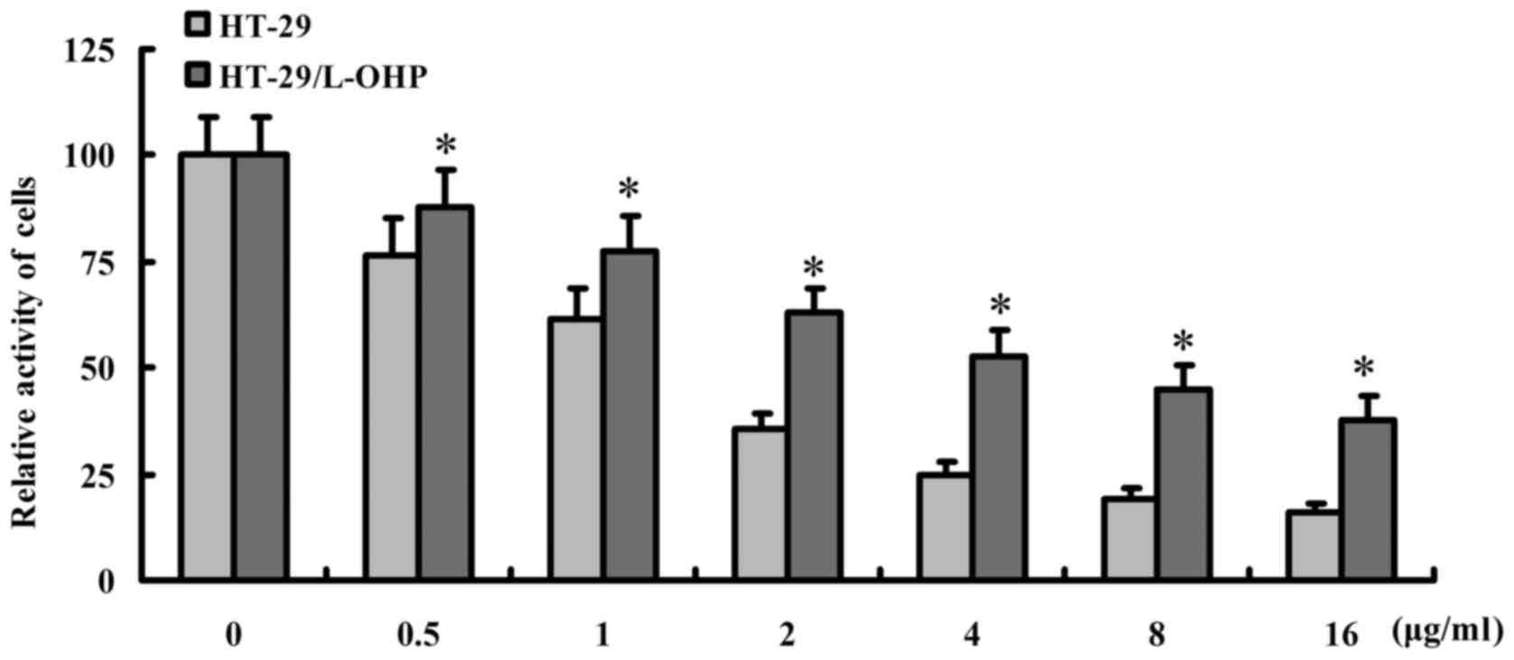
The effect of L-OHP on HT-29 and
HT-29/L-OHP cells
To evaluate the effect of L-OHP on HT-29 and
HT-29/L-OHP cells, different dosages of L-OHP were applied for
intervention and the cell activity was detected by SRB assay. As
shown in Fig. 1, the activity of
HT-29 was less obvious than HT-29/L-OHP after the intervention
(P<0.05), suggesting a stronger resistance of HT-29/L-OHP
against L-OHP, and 0.5 µg/ml L-OHP was selected for further
studies.
The expressions of drug-resistant
proteins in HT-29 and HT-29/L-OHP cells
Western blot analysis was applied to examine the
expressions of drug-resistant proteins in HT-29 and HT-29/L-OHP
cells, including MRP-1, P-gp, LOXL2, Survivin, Livin, Bcl-2 and Bax
proteins. The result showed higher expressions of MRP-1, P-gp,
LOXL2, Survivin, Livin and Bcl-2 but lower expression of Bax in
HT-29 than in HT-29/L-OHP (P<0.01) (Fig. 2), which suggested the expression
difference of drug-resistant genes between the two cell lines may
contribute to the stronger tolerance of HT-29/L-OHP than HT-29
towards L-OHP. Therefore, HT-29/L-OHP was selected to assess the
impacts of salidroside in the following experiments.
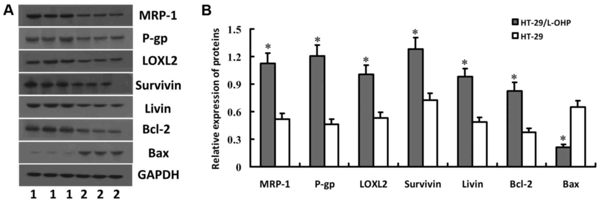 | Figure 2.Expression of MRP-1, P-gp, LOXL2,
Survivin, Livin, Bcl-2 and Bax proteins in HT-29 and HT-29/L-OHP
cells. HT-29 and HT-29/L-OHP cells were subjected to western blot
analysis to detect the expression of MRP-1, P-gp, LOXL2, Survivin,
Livin, Bcl-2 and Bax proteins. Gel electrophoresis map was shown in
(A) and relative protein levels were shown in (B). *P<0.05 vs.
HT-29 cells. |
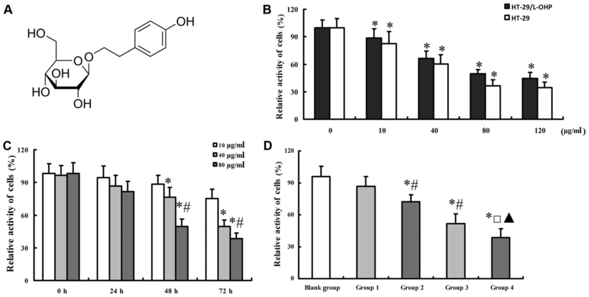
The effect of salidroside on the cell
activity of HT-29/L-OHP and HT-29 cells
The molecular formula of salidroside was shown as
Fig. 3A. To explore the effect of
salidroside on the cell activity of HT-29/L-OHP cells, different
dosages of salidroside were imposed on HT-29 and HT-29/L-OHP cells.
Results showed that either of increasing dosage and longer
application of salidroside decreased cell activity in HT-29/L-OHP
and HT-29 cells (Fig. 3B and C),
based on which the accurate dosage and treatment period (48 h) of
salidroside were determined. The cell activity decreased further
with the combined use of salidroside and L-OHP (Fig. 3D).
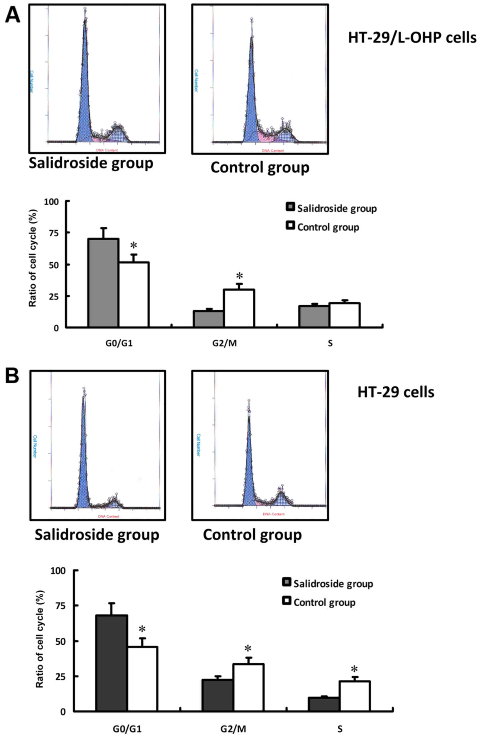
The effect of salidroside on the cell
cycle of HT-29/L-OHP and HT-29 cells
In this study, concentration of 80 µg/ml salidroside
with 48 h treatment was applied to evaluate the effect of
salidroside on the cell cycle of HT-29/L-OHP and HT-29 cells. The
result of FCM was shown in Fig.
4A. Compared with control group, the intervention resulted in
more HT-29/L-OHP and HT-29 cells arrested in
G0/G1 stage, less in G2/M stage
(P<0.01), whereas almost the same proportion in S stage
(Fig. 4B), suggesting salidroside
could inhibit the proliferation of HT-29/L-OHP and HT-29 cells.
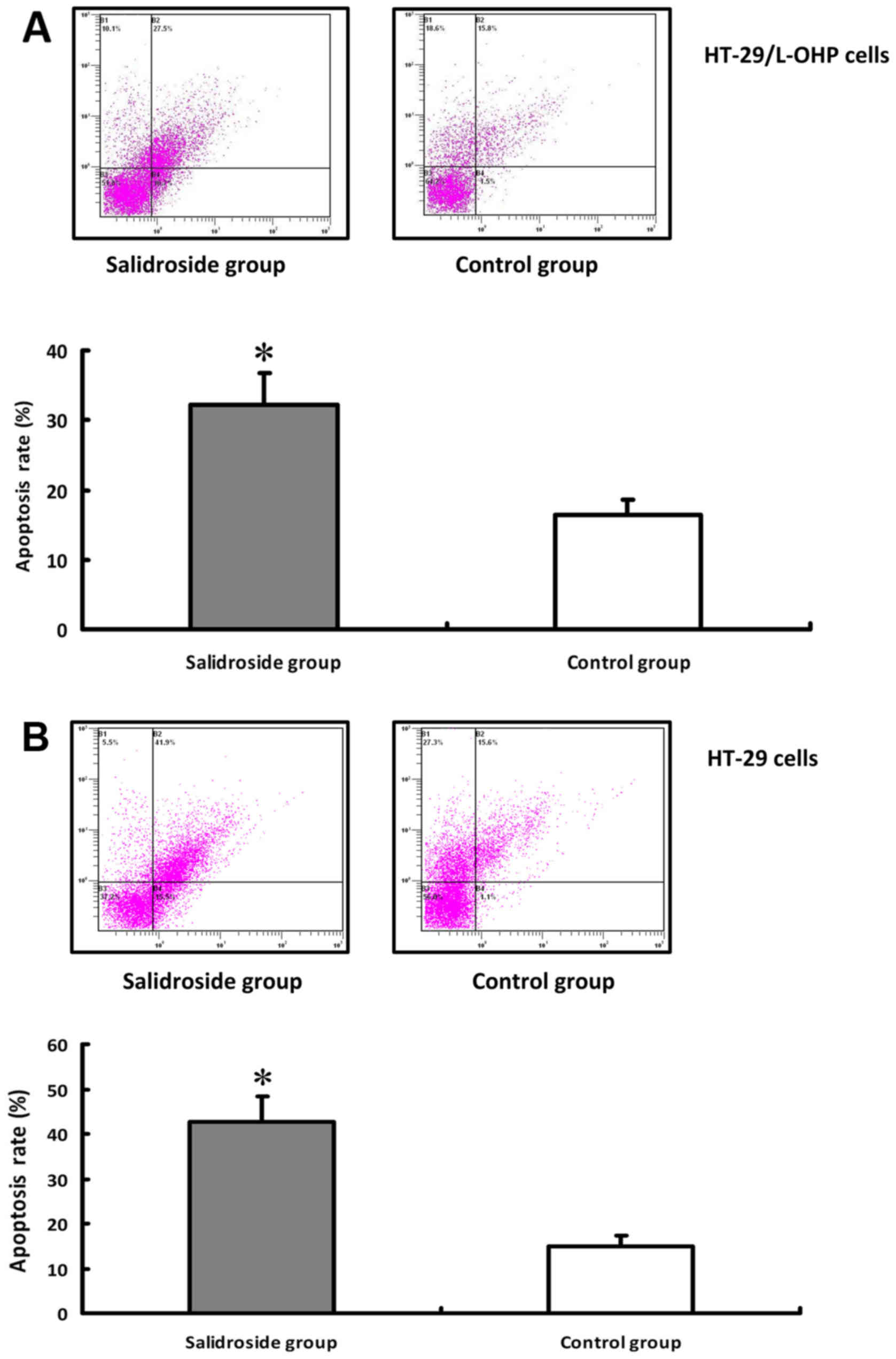
The effect of salidroside on apoptosis
of HT-29/L-OHP and HT-29 cells
The effects of salidroside on the apoptosis of
HT-29/L-OHP and HT-29 cells were detected under the same
experimental dosage and time. The result of FCM was shown in
Fig. 5A. In comparison with the
control group, the apoptosis rate was higher in HT-29/L-OHP and
HT-29 cells after salidroside intervention (P<0.01) (Fig. 5B), suggesting that salidroside
could promote the apoptosis of HT-29/L-OHP and HT-29 cells.
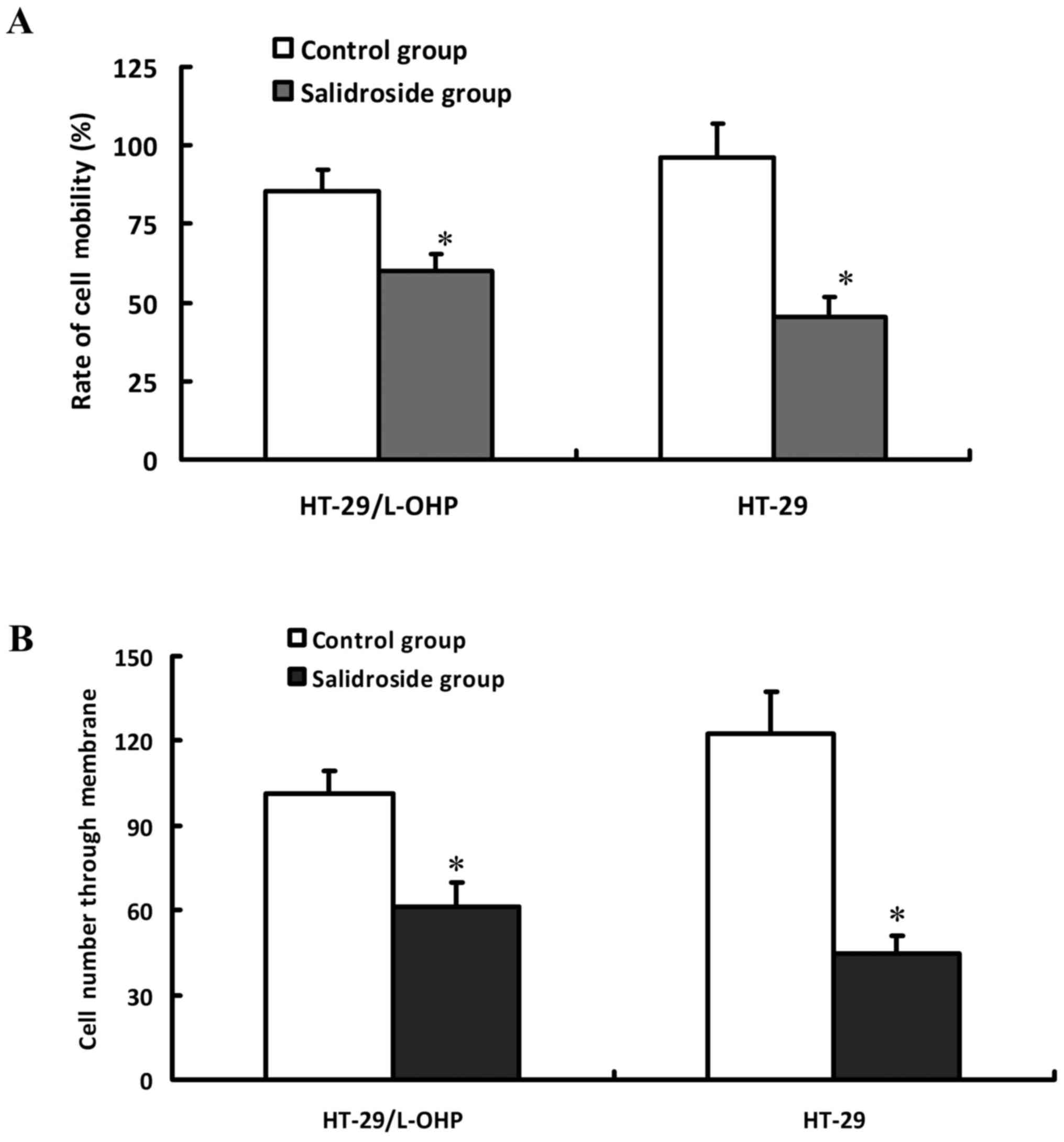
The effect of salidroside on the
invasion and migration of HT-29/L-OHP and HT-29 cells
HT-29/L-OHP and HT-29 cells were pretreated by 80
µg/ml salidroside for 48 h respectively, and then detected with
scratch test to evaluate the cell migration activity and Transwell
chamber assay to assess the cell invasion capacity. As shown in
Fig. 6, the inhibition ratio and
invasion descended in a dose-dependent manner after the
intervention, indicating that salidroside could inhibit the
invasion and migration of HT-29/L-OHP and HT-29 cells.
The effect of salidroside on the
expressions of drug-resistant genes and caspase-3 activity in
HT-29/L-OHP and HT-29 cells
To explore the molecular mechanism of salidroside on
L-OHP resistance cells, the expressions of drug-resistant genes in
HT-29/L-OHP were detected. The results indicated decreased
expressions of MRP-1, MDR1/P-gp, LOXL2, Survivin, Livin and Bcl-2
but increased Bax in HT-29/L-OHP cells, compared with Control group
(P<0.05) (Fig. 7A-C), which
suggested that salidroside could strengthen the sensitivity of
HT-29/L-OHP to chemotherapeutic drugs by regulating the expressions
of drug-resistant genes. Our results suggested salidroside could
inhibit the expressions of Survivin and Livin, two anti-apoptosis
proteins that could affect the function of caspase-3. Results
indicated that the activity of caspase-3 in HT-29/L-OHP also
increased after the intervention, implying salidroside may exert by
suppressing the expressions of Survivin and Livin and enhancing
caspase-3 activity (Fig. 7D).
HT-29 cells were also treated with salidroside, and then
drug-resistant genes and caspase-3 activity were also tested.
Results of salidroside on HT-29 cells were similar to those of
salidroside on HT-29/L-OHP cells (Fig.
8).
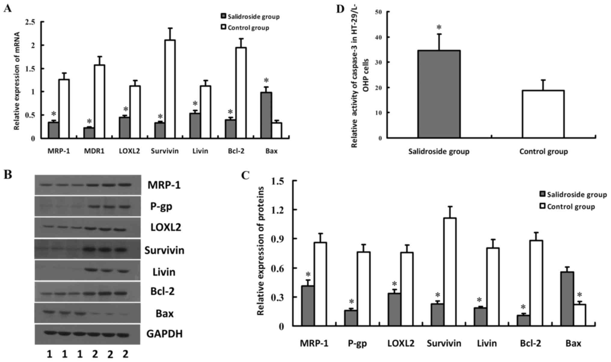 | Figure 7.Effect of salidroside on the
expressions of MRP-1, MDR1/P-gp, LOXL2, Survivin, Livin, Bcl-2, Bax
genes and caspase-3 activity in HT-29/L-OHP cells. HT-29/L-OHP
cells were treated with salidroside, and then were subjected to
real-time quantitative PCR and western blot analyses, to detect the
mRNA or protein expression levels of MRP-1, MDR1/P-gp, LOXL2,
Survivin, Livin, Bcl-2, Bax genes. Results showed that expressions
of MRP-1, MDR1/P-gp, LOXL2, Survivin, Livin and Bcl-2 decreased but
Bax increased in HT-29/L-OHP cells, which was illustrated in (A-C).
Activity of caspase-3 in HT-29/L-OHP also increased after
salidroside intervention, as shown in (D). *P<0.05 vs. control
group. |
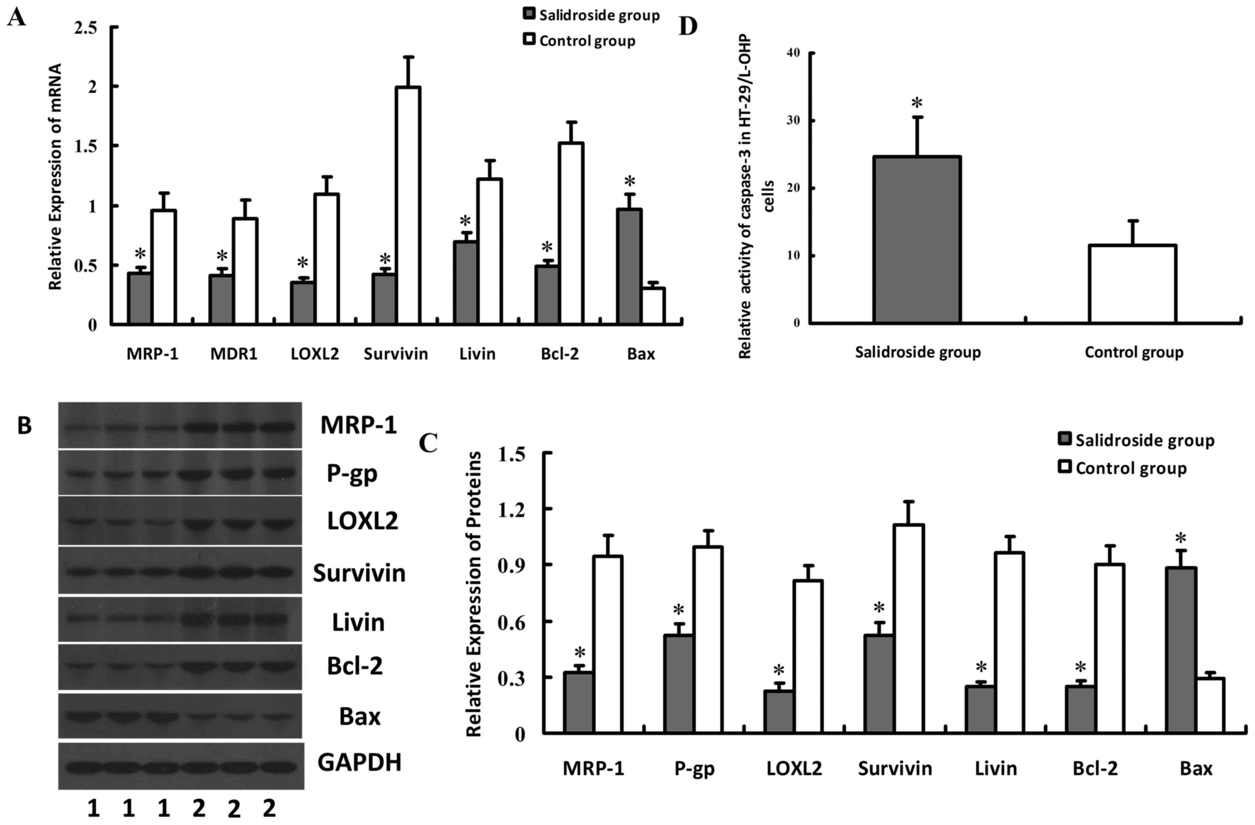 | Figure 8.Effect of salidroside on the
expressions of MRP-1, MDR1/P-gp, LOXL2, Survivin, Livin, Bcl-2, Bax
genes and caspase-3 activity in HT-29 cells. HT-29 cells were
treated with salidroside, and then were subjected to real-time
quantitative PCR and western blot analyses, to detect the mRNA or
protein expression levels of MRP-1, MDR1/P-gp, LOXL2, Survivin,
Livin, Bcl-2, Bax genes. Results showed that expressions of MRP-1,
MDR1/P-gp, LOXL2, Survivin, Livin and Bcl-2 decreased but Bax
increased in HT-29 cells, which was illustrated in (A-C). Activity
of caspase-3 in HT-29 also increased after salidroside
intervention, as shown in (D). *P<0.05 vs. control group. |
Discussion
CRC is a common malignant digestive tract tumor in
China, which is characterized by the dissatisfactory therapeutic
efficacy and poor prognosis (21,22).
Chemotherapy plays an important role in the treatment of CRC,
which, however, frequently fails and results in recurrence and
metastasis due to the drug resistance of CRC cells (23,24).
L-OHP, a 3rd generation platinum-based anticancer drug, is the
primary component in the first-line chemotherapy for CRC. The
cytotoxic and anti-cancer effect of L-OHP is to inhibiting DNA
synthesis in the cancer cells by creating crosslink within and
between DNA chains (25,26). Although the effect of L-OHP is
better than DDP, the resistance of CRC cells against L-OHP
decreases the clinical outcomes (27,28).
In this study, the cell activity of CRC cells was lower than L-OHP
resistance CRC cells after L-OHP intervention, demonstrating the
resistance of CRC cells to L-OHP (29). Therefore, a combination of
innovative drugs and L-OHP may improve the effectiveness of
chemotherapy.
Rhodiolae, a traditional medicinal plant, is widely
used in Far East and Russia (15,16)
for immunity improvement (30),
anti-oxidation/cancer (31,32),
cardiovascular protection (33),
glycol-/lipid-metabolism enhancement and neuron protection
(34,35). Salidroside is the active ingredient
of Rhodiolae, which has obvious suppression on renal clear cell
cancer, lung cancer and breast cancer (17–19).
Recent evidence has demonstrated that salidroside could inhibit the
proliferation of CRC cells by regulating the JAK2/STAT3 signaling.
However, whether Rhodiolae can relieve the resistance of CRCs
against L-OHP remains unclear. In this study, we observed higher
sensitivity of CRC cells to L-OHP after salidroside intervention,
suggesting the positive effect of salidroside on the resistance of
CRC cells against L-OHP. The results indicated that salidroside can
impede the transition of HT-29/L-OHP and HT-29 cells from
G0/G1 stage to G2/M stage, whereas
it also promotes the apoptosis and inhibits the invasion and
migration of HT-29/L-OHP and HT-29 cells. Our results revealed
multiple pathways of salidroside's suppression on HT-29/L-OHP cells
and suggested the combined use of salidroside in the L-OHP oriented
chemotherapy can improve the clinical effect.
Previous studies revealed the participations of
agent pump protein, detoxification pathway and DNA repair in the
L-OHP resistance (36–39). To explore the mechanism of
salidroside on HT-29/L-OHP and HT-29 cells, we compared the
expressions of MRP-1, P-gp, LOXL2, Survivin, Livin and Bcl-2 before
and after salidroside intervention. MRP-1 and P-gp, located at the
membrane of cancer cells, could pump chemotherapeutic drugs out via
endoergic reaction (40,41). Recent study has demonstrated that
LOXL2 can inhibit the diffusion of gemcitabine in the pancreatic
ductal adenocarcinoma (PDAC) (42). Survivin (43), Livin (44) and Bcl-2 could enhance the
anti-apoptosis of cancer cells, while in contrast Bax can promote
apoptosis. Therefore, the ratio of Bcl-2/Bax in the dimer composed
by the two proteins determines the apoptosis capacity of cancer
cells (45,46). In this study, we observed declines
of MRP-1, MDR1/P-gp, LOXL2, Survivin, Livin and Bcl-2, whereas
increasing expression of Bax after salidroside intervention,
suggesting salidroside can relieve the resistance of CRC cells to
L-OHP by regulating these drug-resistant genes. Results of
salidroside on HT-29 cells were similar to those of salidroside on
HT-29/L-OHP cells. However, more studies are needed to explore the
interaction between specific molecules, and in further study we'll
investigate effects of salidroside on other CRC cell lines.
In conclusion, our study revealed that salidroside
could decrease the activity and invasion capacity of HT-29/ L-OHP,
and treatment of salidroside related to apoptosis of cancer cells
by regulating the expressions of related genes, possibly due to
inhibiting the expressions of MRP-1, MDR1/P-gp, LOXL2, Survivin,
Livin and Bcl-2, whereas promoting the expression of Bax genes.
Therefore, we suggest the combined application of salidroside in
the L-OHP oriented chemotherapy in order to improve the clinical
efficacy for CRC patients. However, further researches are needed
to confirm this conclusion via in vivo experiments and
large-scale clinical studies.
References
|
1
|
Favoriti P, Carbone G, Greco M, Pirozzi F,
Pirozzi RE and Corcione F: Worldwide burden of colorectal cancer: A
review. Updates Surg. 68:7–11. 2016. View Article : Google Scholar : PubMed/NCBI
|
|
2
|
Chen W, Zheng R, Zeng H and Zhang S: The
incidence and mortality of major cancers in China, 2012. Chin J
Cancer. 35:732016. View Article : Google Scholar : PubMed/NCBI
|
|
3
|
Bode AM, Dong Z and Wang H: Cancer
prevention and control: Alarming challenges in China. Natl Sci Rev.
3:117–127. 2016. View Article : Google Scholar : PubMed/NCBI
|
|
4
|
Rogowski W and Sulżyc-Bielicka V: Optimal
duration of a first-line palliative chemotherapy in disseminated
colorectal cancer-a review of the literature from a developing
country perspective. Contemp Oncol (Pozn). 20:210–214.
2016.PubMed/NCBI
|
|
5
|
Zhao L, Liu R, Zhang Z, Li T, Li F, Liu H
and Li G: Oxaliplatin/fluorouracil-based adjuvant chemotherapy for
locally advanced rectal cancer after neoadjuvant chemoradiotherapy
and surgery: A systematic review and meta-analysis of randomized
controlled trials. Colorectal Dis. 18:763–772. 2016. View Article : Google Scholar : PubMed/NCBI
|
|
6
|
Betge J, Barat A, Murphy V, Hielscher T,
Van Grieken NC, Belle S, Zhan T, Härtel N, Kripp M, Bacon O, et al:
Outcome of colorectal cancer patients treated with combination
bevacizumab therapy: A pooled retrospective analysis of three
european cohorts from the angiopredict initiative. Digestion.
94:129–137. 2016. View Article : Google Scholar : PubMed/NCBI
|
|
7
|
Suzuki S, Shimazaki J, Morishita K, Koike
N, Harada N, Hayashi T and Suzuki M: Efficacy and safety of
oxaliplatin, bevacizumab and oral S-1 for advanced recurrent
colorectal cancer. Mol Clin Oncol. 5:391–394. 2016. View Article : Google Scholar : PubMed/NCBI
|
|
8
|
Zhang X, Chen Y, Hao L, Hou A, Chen X, Li
Y, Wang R, Luo P, Ruan Z, Ou J, et al: Macrophages induce
resistance to 5-fluorouracil chemotherapy in colorectal cancer
through the release of putrescine. Cancer Lett. 381:305–313. 2016.
View Article : Google Scholar : PubMed/NCBI
|
|
9
|
Garborg K: Colorectal cancer screening.
Surg Clin North Am. 95:979–989. 2015. View Article : Google Scholar : PubMed/NCBI
|
|
10
|
De Divitiis C, Nasti G, Montano M,
Fisichella R, Iaffaioli RV and Berretta M: Prognostic and
predictive response factors in colorectal cancer patients: Between
hope and reality. World J Gastroenterol. 20:15049–15059. 2014.
View Article : Google Scholar : PubMed/NCBI
|
|
11
|
Wang W, Liu J, Qi J, Zhang J, Zhu Q, Ma J
and Qin C: Downregulation of RLIP76 is associated with vincristine
resistance in human colorectal cancer HCT-8/VCR cells. Int J Oncol.
49:1505–1512. 2016.PubMed/NCBI
|
|
12
|
Oddo D, Sennott EM, Barault L, Valtorta E,
Arena S, Cassingena A, Filiciotto G, Marzolla G, Elez E, van Geel
RM, et al: Molecular landscape of acquired resistance to targeted
therapy combinations in BRAF-mutant colorectal cancer. Cancer Res.
76:4504–4515. 2016. View Article : Google Scholar : PubMed/NCBI
|
|
13
|
Wang Z, Zhang L, Ni Z, Sun J, Gao H, Cheng
Z, Xu J and Yin P: Resveratrol induces AMPK-dependent MDR1
inhibition in colorectal cancer HCT116/L-OHP cells by preventing
activation of NF-κB signaling and suppressing cAMP-responsive
element transcriptional activity. Tumour Biol. 36:9499–9510. 2015.
View Article : Google Scholar : PubMed/NCBI
|
|
14
|
Li F, Tang H, Xiao F, Gong J, Peng Y and
Meng X: Protective effect of salidroside from Rhodiolae radix on
diabetes-induced oxidative stress in mice. Molecules. 16:9912–9924.
2011. View Article : Google Scholar : PubMed/NCBI
|
|
15
|
Zheng KY, Guo AJ, Bi CW, Zhu KY, Chan GK,
Fu Q, Xu SL, Zhan JY, Lau DT, Dong TT, et al: The extract of
Rhodiolae Crenulatae Radix et Rhizoma induces the accumulation of
HIF-1α via blocking the degradation pathway in cultured kidney
fibroblasts. Planta Med. 77:894–899. 2011. View Article : Google Scholar : PubMed/NCBI
|
|
16
|
Li F, Xiao F, Gong J and Yu T: Applied
orthogonal experiment design for the optimum microwave-assisted
extraction conditions of polysaccharides from Rhodiolae radix. Afr
J Tradit Complement Altern Med. 10:179–185. 2013.PubMed/NCBI
|
|
17
|
Lv C, Huang Y, Liu ZX, Yu D and Bai ZM:
Salidroside reduces renal cell carcinoma proliferation by
inhibiting JAK2/STAT3 signaling. Cancer Biomark. 17:41–47. 2016.
View Article : Google Scholar : PubMed/NCBI
|
|
18
|
Wang J, Li JZ, Lu AX, Zhang KF and Li BJ:
Anticancer effect of salidroside on A549 lung cancer cells through
inhibition of oxidative stress and phospho-p38 expression. Oncol
Lett. 7:1159–1164. 2014.PubMed/NCBI
|
|
19
|
Zhao G, Shi A, Fan Z and Du Y: Salidroside
inhibits the growth of human breast cancer in vitro and in vivo.
Oncol Rep. 33:2553–2560. 2015. View Article : Google Scholar : PubMed/NCBI
|
|
20
|
Sun KX, Xia HW and Xia RL: Anticancer
effect of salidroside on colon cancer through inhibiting JAK2/STAT3
signaling pathway. Int J Clin Exp Pathol. 8:615–621.
2015.PubMed/NCBI
|
|
21
|
Kraus S, Nabiochtchikov I, Shapira S and
Arber N: Recent advances in personalized colorectal cancer
research. Cancer Lett. 347:15–21. 2014. View Article : Google Scholar : PubMed/NCBI
|
|
22
|
Berge E, Thompson C and Messersmith W:
Development of novel targeted agents in the treatment of metastatic
colorectal cancer. Clin Colorectal Cancer. 10:266–278. 2011.
View Article : Google Scholar : PubMed/NCBI
|
|
23
|
Temraz S, Mukherji D, Alameddine R and
Shamseddine A: Methods of overcoming treatment resistance in
colorectal cancer. Crit Rev Oncol Hematol. 89:217–230. 2014.
View Article : Google Scholar : PubMed/NCBI
|
|
24
|
Bian Z, Feng Y, Xue Y, Hu Y, Wang Q, Zhou
L, Liu Z, Zhang J, Yin Y, Gu B and Huang Z: Down-regulation of SNX1
predicts poor prognosis and contributes to drug resistance in
colorectal cancer. Tumour Biol. 37:6619–6625. 2016. View Article : Google Scholar : PubMed/NCBI
|
|
25
|
Suzuki S and Tanigawara Y: Forced
expression of S100A10 reduces sensitivity to oxaliplatin in
colorectal cancer cells. Proteome Sci. 12:262014. View Article : Google Scholar : PubMed/NCBI
|
|
26
|
Toki MI, Saif MW and Syrigos KN:
Hypersensitivity reactions associated with oxaliplatin and their
clinical management. Expert Opin Drug Saf. 13:1545–1554. 2014.
View Article : Google Scholar : PubMed/NCBI
|
|
27
|
Bi J, Bai Z, Ma X, Song J, Guo Y, Zhao J,
Yi X, Han S and Zhang Z: Txr1: An important factor in oxaliplatin
resistance in gastric cancer. Med Oncol. 31:8072014. View Article : Google Scholar : PubMed/NCBI
|
|
28
|
Chen CC, Chen LT, Tsou TC, Pan WY, Kuo CC,
Liu JF, Yeh SC, Tsai FY, Hsieh HP and Chang JY: Combined modalities
of resistance in an oxaliplatin-resistant human gastric cancer cell
line with enhanced sensitivity to 5-fluorouracil. Br J Cancer.
97:334–344. 2007. View Article : Google Scholar : PubMed/NCBI
|
|
29
|
Hu CJ, Wang B, Tang B, Chen BJ, Xiao YF,
Qin Y, Yong X, Luo G, Zhang JW, Zhang D, et al: The FOXM1-induced
resistance to oxaliplatin is partially mediated by its novel target
gene Mcl-1 in gastric cancer cells. Biochim Biophys Acta.
1849:290–299. 2015. View Article : Google Scholar : PubMed/NCBI
|
|
30
|
Bocharova OA, Serebriakova RV and Bodrova
NB: Preventive effect of Rhodiolae rosea in spontaneous
liver carcinogenesis in a mice model of high-tumor strain. Vestn
Ross Akad Med Nauk. 5:41–43. 1994.(In Russian).
|
|
31
|
Lee OH, Kwon YI, Apostolidis E, Shetty K
and Kim YC: Rhodiola-induced inhibition of adipogenesis involves
antioxidant enzyme response associated with pentose phosphate
pathway. Phytother Res. 25:106–115. 2011. View Article : Google Scholar : PubMed/NCBI
|
|
32
|
Bassa LM, Jacobs C, Gregory K, Henchey E,
Ser-Dolansky J and Schneider SS: Rhodiola crenulata induces
an early estrogenic response and reduces proliferation and
tumorsphere formation over time in MCF7 breast cancer cells.
Phytomedicine. 23:87–94. 2016. View Article : Google Scholar : PubMed/NCBI
|
|
33
|
Yu L, Qin Y, Wang Q, Zhang L, Liu Y, Wang
T, Huang L, Wu L and Xiong H: The efficacy and safety of Chinese
herbal medicine, Rhodiola formulation in treating ischemic heart
disease: A systematic review and meta-analysis of randomized
controlled trials. Complement Ther Med. 22:814–825. 2014.
View Article : Google Scholar : PubMed/NCBI
|
|
34
|
Wang J, Rong X, Li W, Yang Y, Yamahara J
and Li Y: Rhodiola crenulata root ameliorates derangements
of glucose and lipid metabolism in a rat model of the metabolic
syndrome and type 2 diabetes. J Ethnopharmacol. 142:782–788. 2012.
View Article : Google Scholar : PubMed/NCBI
|
|
35
|
Xiao L, Li H, Zhang J, Yang F, Huang A,
Deng J, Liang M, Ma F, Hu M and Huang Z: Salidroside protects
Caenorhabditis elegans neurons from polyglutamine-mediated
toxicity by reducing oxidative stress. Molecules. 19:7757–7769.
2014. View Article : Google Scholar : PubMed/NCBI
|
|
36
|
Helmbach H, Kern MA, Rossmann E, Renz K,
Kissel C, Gschwendt B and Schadendorf D: Drug resistance towards
etoposide and cisplatin in human melanoma cells is associated with
drug-dependent apoptosis deficiency. J Invest Dermatol.
118:923–932. 2002. View Article : Google Scholar : PubMed/NCBI
|
|
37
|
Hunakova L, Gronesova P, Horvathova E,
Chalupa I, Cholujova D, Duraj J and Sedlak J: Modulation of
cisplatin sensitivity in human ovarian carcinoma A2780 and SKOV3
cell lines by sulforaphane. Toxicol Lett. 230:479–486. 2014.
View Article : Google Scholar : PubMed/NCBI
|
|
38
|
Murphy RF, Komlodi-Pasztor E, Robey R,
Balis FM, Farrell NP and Fojo T: Retained platinum uptake and
indifference to p53 status make novel transplatinum agents active
in platinum-resistant cells compared to cisplatin and oxaliplatin.
Cell Cycle. 11:963–973. 2012. View Article : Google Scholar : PubMed/NCBI
|
|
39
|
Gourdier I, Del Rio M, Crabbé L, Candeil
L, Copois V, Ychou M, Auffray C, Martineau P, Mechti N, Pommier Y
and Pau B: Drug specific resistance to oxaliplatin is associated
with apoptosis defect in a cellular model of colon carcinoma. FEBS
Lett. 529:232–236. 2002. View Article : Google Scholar : PubMed/NCBI
|
|
40
|
Sui H, Fan ZZ and Li Q: Signal
transduction pathways and transcriptional mechanisms of
ABCB1/Pgp-mediated multiple drug resistance in human cancer cells.
J Int Med Res. 40:426–435. 2012. View Article : Google Scholar : PubMed/NCBI
|
|
41
|
Wang Y, Liu L, Liu X, Zhang H, Liu J, Feng
B, Shang Y, Zhou L, Wu K, Nie Y, et al: Shugoshin1 enhances
multidrug resistance of gastric cancer cells by regulating MRP1,
Bcl-2 and Bax genes. Tumour Biol. 34:2205–2214. 2013. View Article : Google Scholar : PubMed/NCBI
|
|
42
|
Le Calvé B, Griveau A, Vindrieux D,
Maréchal R, Wiel C, Svrcek M, Gout J, Azzi L, Payen L, Cros J, et
al: Lysyl oxidase family activity promotes resistance of pancreatic
ductal adenocarcinoma to chemotherapy by limiting the intratumoral
anticancer drug distribution. Oncotarget. 7:32100–32112. 2016.
View Article : Google Scholar : PubMed/NCBI
|
|
43
|
Véquaud E, Desplanques G, Jézéquel P, Juin
P and Barillé-Nion S: Survivin contributes to DNA repair by
homologous recombination in breast cancer cells. Breast Cancer Res
Treat. 155:53–63. 2016. View Article : Google Scholar : PubMed/NCBI
|
|
44
|
Hsieh CH, Lin YJ, Wu CP, Lee HT, Shyu WC
and Wang CC: Livin contributes to tumor hypoxia-induced resistance
to cytotoxic therapies in glioblastoma multiforme. Clin Cancer Res.
21:460–470. 2015. View Article : Google Scholar : PubMed/NCBI
|
|
45
|
Eimani Golestani B, Sanati MH, Houshmand
M, Ataei M, Akbarian F and Shakhssalim N: Expression and prognostic
significance of bcl-2 and bax in the progression and clinical
outcome of transitional bladder cell carcinoma. Cell J. 15:356–363.
2014.PubMed/NCBI
|
|
46
|
Wu S, Liu B, Zhang Q, Liu J, Zhou W, Wang
C, Li M, Bao S and Zhu R: Dihydromyricetin reduced Bcl-2 expression
via p53 in Human Hepatoma HepG2 Cells. PLoS One. 8:e768862013.
View Article : Google Scholar : PubMed/NCBI
|