Introduction
Chronic prostatitis (CP) is a common disease in
urology, and voiding dysfunction is the primary clinical
manifestation of CP (1), which
affects the quality of life of patients with CP. Urinary tract
obstruction was previously reported in patients with CP upon
urodynamic examination (2).
Prostate fibrosis is a contributing factor to lower urinary tract
symptom (LUTS) etiology (3).
Tissue fibrosis is reported to be associated with
tissue damage caused by various factors, including aging (4), infection (5), tumors (6) and additional secondary diseases in
various organs, which leads to dysfunction. The mechanism of
fibrosis involves the accumulation of myofibroblasts, collagen
deposition, remodeling of the extracellular matrix and elevated
tissue rigidity. High levels of collagen in tissue leads to organ
dysfunction as tissue elasticity and compliance are reduced
(4,5). However, the role of fibrosis in the
development of CP is poorly understood. Therefore, the present
study investigated whether CP with urinary dysfunction was
associated with collagen accumulation in the prostate.
Mast cells (MCs) are important effector cells in
tissue fibrosis (7,8). MC activation and degranulation leads
to the release of inflammatory and profibrotic mediators, which
promotes tissue fibrosis. MCs express serine proteases, tryptase
and chymase, which are associated with fibrosis in certain diseases
(9,10). However, the involvement of these
proteases in CP is poorly understood. Therefore, the role of
tryptase and chymase in the pathogenesis of CP was investigated in
the present study. The pleiotropic cytokine, transforming growth
factor-β (TGF-β), serves numerous physiological roles in
vivo, including the initiation and control of fibrosis
(11). The profibrotic effects of
MCs are closely associated with TGF-β (12). Experimental studies have associated
increased MC numbers and staining intensity with increased TGF-β
production and interstitial fibrosis (12,13).
The Wnt/β-catenin signaling pathway has a vital role in
TGF-β-induced fibrosis (14).
Therefore, the potential role of the Wnt/β-catenin/TGF-β signaling
pathway in the pathogenesis of CP was also determined in the
present study.
Resveratrol (15)
(trans-3,4,5-trihydroxy stilbene) is present in various plants and
foods, including Cassia, pine trees, grapes, wine, mulberry
and peanuts. Resveratrol was originally used as a phytoalexin
(16), and the reliable and
extensive anti-inflammatory effects of resveratrol gained the
attention of researchers (17).
Resveratrol treatment represses and reverses prostate fibroblast to
myofibroblast phenoconversion in vitro (18). Our previous studies identified that
resveratrol can improve the outcome of prostatitis (19,20).
However, to the best of our knowledge, no reports have investigated
the effect of resveratrol on mast cell induced fibrosis in
prostatitis. Antifibrotic therapeutics may be efficacious for the
treatment of LUTS (18).
Over the past decades, a variety of animal models of
prostatitis have been established with the aim of deciphering the
pathogenesis of CP. The present study employed a rat model of
experimental autoimmune prostatitis, which has been widely used
(21,22). This model exhibits the majority of
the characteristics of human chronic autoimmune prostatitis, as
specific lymphocytes against prostate antigens and histological
lesions are observed in the prostate (23).
The present study investigated whether prostate
fibrosis was associated with urinary dysfunction in CP and whether
resveratrol improved urinary dysfunction. The activity of the
c-kit/stem cell factor (SCF) and TGF-β/Wnt/β-catenin signaling
pathways, the number of MC infiltrates and the expression levels of
tryptase, chymase and α-smooth muscle actin (α-SMA) in CP rats was
investigated.
Materials and methods
Chemicals
Resveratrol of >99% purity was purchased from
Dalian Meilun Biotech Co., Ltd. (Dalian, China). The
pertussis-diphtheria-tetanus (DPT) vaccine was obtained from the
Wuhan Institute of Biological Products Co., Ltd. (Wuhan, China).
All other chemicals used in the present study were of analytical
grade and commercially available.
Animals
A total of 24 male Sprague-Dawley (SD) rats (180±20
g) were purchased from the Experimental Animal Center of Dalian
Medical University (Dalian, China). The rats were housed in a
controlled temperature of 22±2°C with 60±5% relative humidity, a 12
h light/dark cycle and ad libitum access to water and food;
rats were fasted overnight and provided with water prior to
surgery. All animal experiments were approved by the ethics
committee of Dalian Medical University and performed in accordance
with the institutional guidelines.
Rat model of CP
For the purification of rat prostatic protein, male
SD rats (240–300 g) were sacrificed and prostate tissue was removed
under sterile conditions and washed with saline solution. Prostate
tissue was placed into a physiological saline solution containing
0.5% Triton X-100 and homogenized in an ice-water bath with a glass
homogenizer. The homogenized samples were centrifuged (10,000 × g
at 4°C) for 10 min and the protein was measured by using a
bicinchoninic acid assay (Beijing Solarbio Science and Technology
Co., Ltd., Beijing, China) and diluted to 15 mg/ml in PBS
buffer.
Rats were subcutaneously injected with the DPT
vaccine (0.5 ml/kg) at day 0, and then injected at multipoint sites
(right foot pad, left foot pad and base of the tail and shoulders)
with a mixture (volume, 1 ml) of purified rat prostatic protein and
Freund's Complete Adjuvant (1:1 ratio) at 0, 15 and 30 days.
Control rats were injected with same volume of PBS. The rat model
of CP was established after 45 days. Rats were randomly divided
into the following three groups (n=8/group): Control group, rats
orally administered with saline for 10 days without CP induction;
CP group, CP rats orally administered with saline for 10 days
starting on day 45; resveratrol group, CP rats orally administered
with resveratrol (10 mg/kg, dissolved in saline) for 10 days
starting on day 45.
Bladder pressure and volume tests in
rats
Rats were anesthetized using an intraperitoneal
injection of pentobarbital (60 mg/kg) prior to surgery. Rats were
fixed in the operating frame, and the upper edge of the pubic
symphysis skin was incised. The bladder was exposed and placed on
the incision to avoid the effect of abdominal pressure on the
detrusor pressure. Two 24G tubes were inserted into the bladder and
fixed, and the bladder was irrigated with saline (0.4 ml/min) via
one of the 24G tubes. Another tube was connected to a MedLab
biological signal acquisition system via a pressure transducer. The
maximum capacity of the bladder, residual urine volume and maximum
voiding pressure were measured.
Morphological changes
The rats were sacrificed at the end of the bladder
pressure and volume test. The prostate was removed, fixed in 10%
(v/v) neutral formalin for 48 h, then embedded in paraffin and
processed using standard histological techniques. sections (4 µm)
were stained at room temperature with hematoxylin (15 min) and
eosin (3 min; H&E), and examined for morphological changes
using light microscope (8 fields of view analyzed per sample). The
protein expression of tryptase, chymase, TGF-β, Wnt and α-SMA were
determined in the samples by western blot analysis or
immunohistochemical staining, as described below.
Western blot analysis
Proteins were extracted from rat prostate using a
protein extraction kit (Nanjing KeyGen Biotech, Co., Ltd., Nanjing,
China) according to the manufacturer's instructions. As a positive
control (+) for chymase, a protein sample from the hearts of rats
was used. Proteins were measured by using a bicinchoninic acid
assay (Beijing Solarbio Science and Technology Co., Ltd., Beijing,
China). Proteins (20 µg) were resuspended in electrophoresis sample
buffer (Beijing Solarbio Science and Technology Co., Ltd.)
containing β-mercaptoethanol and separated by electrophoresis on a
pre-cast 10% SDS-polyacrylamide gel (Bio-Rad Laboratories, Inc.,
Hercules, CA, USA), followed by electrotransfer to a polyvinylidene
difluoride membrane (EMD Millipore, Billerica, MA, USA). Membranes
were blocked using 5% non-fat milk in Tris-buffered saline with
0.1% Tween-20 (TBST) for 2 h at 37°C. β-actin served as the loading
control. Membranes were incubated overnight at 4°C with a 1:1,000
dilution of polyclonal antibodies for tryptase (sc-32889), TGF-β
(sc-146) and β-catenin (sc-7199; Santa Cruz Biotechnology, Inc.,
Dallas, TX, USA) and a 1:1,500 dilution of a monoclonal antibody
for chymase (bs-2353R), α-SMA (bs-0189R) and β-actin (bsm-33036M;
BIOSS, Beijing, China). Membranes were subsequently washed with
TBST and incubated with 1:1,000 secondary antibodies (sc-2004;
Santa Cruz Biotechnology, Inc.) for 1 h at 37°C. Blots were
extensively washed with TBST and exposed to Enhanced
Chemiluminescence Plus reagent (Beyotime Institute of
Biotechnology), according to the manufacturer's instructions.
Emitted light was documented using a BioSpectrum-410 multispectral
imaging system with a Chemi HR camera 410 (Bio-Rad Laboratories,
Inc.). Protein bands were visualized and images were captured under
transmitted ultraviolet light. The images (three images per animal)
were used for semi-quantitative measurements based on band
densitometry. Blots were semi-quantified by densitometric analysis
using the Image Lab software version 4.0 (Bio-Rad Laboratories,
Inc.).
Immunohistochemical staining
The prostates were fixed in 10% (v/v) neutral
formalin for 48 h, then embedded in paraffin, Histological sections
of rat prostate (4 µm) were mounted onto poly-L-lysine-coated
slides. Slides were deparaffinized in xylene and rehydrated in
graded alcohols. Sections were pretreated with citrate buffer (0.01
mol/l citric acid, pH 6.0) for 20 min at 95°C and immersed in PBS
containing 3% H2O2 for 10 min at room
temperature. Sections were exposed to 10% normal goat serum
(streptavidin/peroxidase; SP-9001; ZSGB-BIO, Beijing, China) in PBS
for 30 min at room temperature and incubated at 4°C overnight with
rabbit polyclonal anti-tryptase or anti-chymase antibodies
mentioned above (1:100 dilution; Santa Cruz Biotechnology, Inc.).
Sections were rinsed with PBS, incubated with biotinylated goat
anti-rabbit IgG (SP-9001; ZSGB-BIO) for 20 min at room temperature
and treated with 3,3-diaminobenzidine chromogen for 5 min at room
temperature. Sections were counterstained under light microscope
with hematoxylin (C0107, Beyotime Institute of Biotechnology) for 2
min at room temperature.
MC assessment
Two independent investigators, who were blinded to
the study, assessed MC infiltration. Each examiner analyzed five
randomized high-power fields in the tissues following
immunohistochemical staining in each group, and analysis was
performed at different magnifications. The number and morphology of
the MCs were evaluated. The 5 areas with the highest number of MCs
were identified at low power (×100), and the number of MCs in the
selected fields were counted at a higher power (×400). The number
of MCs was recorded as the total number in 5 different areas at
high power (×400). The location and morphology of tryptase (+)
cells was considered while counting. Tryptase (+) cells with a
round to oval shape and granular nuclei were considered to be
MCs.
Collagen determination
The prostates were fixed in 10% (v/v) neutral
formalin for 48 h, then embedded in paraffin and 4 µm thick
sections mounted onto poly-L-lysine-coated slides. Picrosirius red
staining was performed on serially sectioned tissues.
Paraffin-embedded tissues were deparaffinized in xylene, rehydrated
in graded alcohols and incubated in a 0.1% picrosirius red solution
for 1 h at room temperature. Sections were counterstained with
hematoxylin for 2 min at room temperature. The sections were
examined under a light microscope.
Statistical analysis
Statistical analysis was performed using SPSS
version 13.0 software (SPSS, Inc., Chicago, IL, USA). Data are
presented as the mean + standard deviation. Statistically
significant differences between two datasets were determined using
one-way analysis of variance followed by Tukeys post hoc test.
P<0.05 was considered to indicate a statistically significant
difference.
Results
Histological examinations
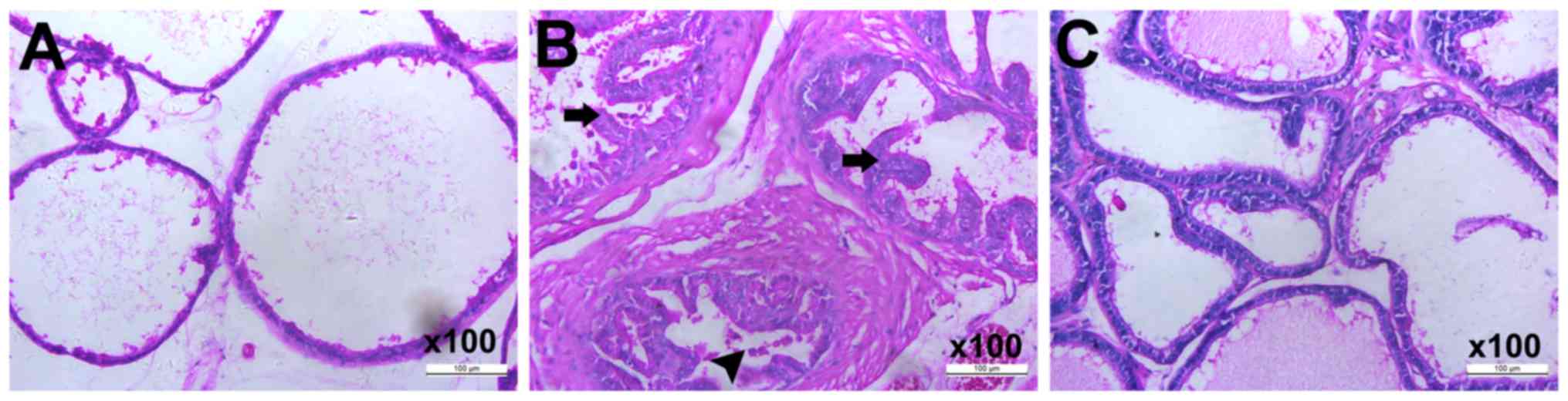
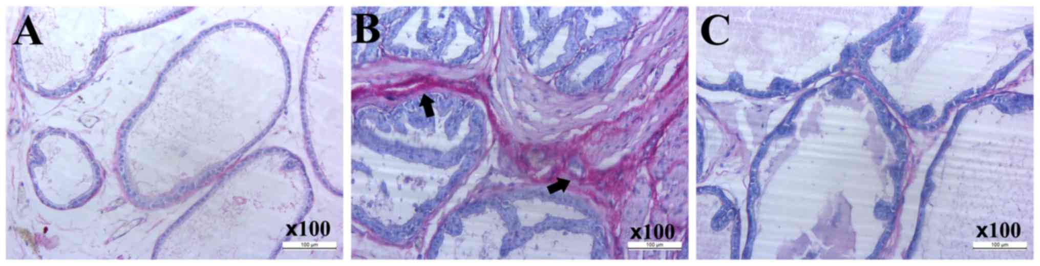
The extent of inflammatory cell infiltration, acinar
alterations and interstitial fibrosis were evaluated following
treatment. Compared with the control group (Fig. 1A), the CP model group (Fig. 1B) exhibited severe diffuse chronic
inflammation, characterized by leukocyte infiltration and papillary
frond protrusion into the gland cavities and a notable increase in
prostatic epithelial height. Gland lumen diameter was also markedly
smaller in the lateral lobe of the prostate. However, these changes
were suppressed in the rats treated with resveratrol (Fig. 1C).
Resveratrol improves the overactive
bladder in CP rats
The results of the bladder pressure and volume test
revealed that the maximum capacity of the bladder, maximum voiding
pressure and residual urine volume in the control group were 0.51
ml, 27.24 cm H2O and 0.17 ml, respectively (Table I). The maximum capacity of the
bladder, maximum voiding pressure and residual urine volume in the
CP group increased significantly compared with the control group
(Table I), which indicated that
the CP model was successfully established. Resveratrol treatment of
CP rats markedly reversed the maximum capacity, maximum voiding
pressure and residual urine volume of the bladder compared with the
CP group (Table I). These results
indicate that resveratrol effectively improved an overactive
bladder in CP rats.
 | Table I.Resveratrol improves overactive
bladder in CP rats. |
Table I.
Resveratrol improves overactive
bladder in CP rats.
| Group | Maximum capacity,
ml | Maximum voiding
pressure, cm H2O | Residual urine
volume, ml |
|---|
| Control |
0.51±0.05 |
27.24±1.52 |
0.17±0.03 |
| CP |
0.94±0.17a |
35.64±2.61a |
0.52±0.03a |
| Resveratrol |
0.72±0.40a,b |
30.51±1.15a,b |
0.28±0.05a,b |
Resveratrol reduces tryptase (+) MC
infiltration in CP rats, but chymase (+) MCs are not detected
MCs have an essential role in fibrosis, and their
phenotype and function during resveratrol treatment for CP was
investigated in the present study. Rat prostates were immediately
acquired following sacrifice for western blot analysis and
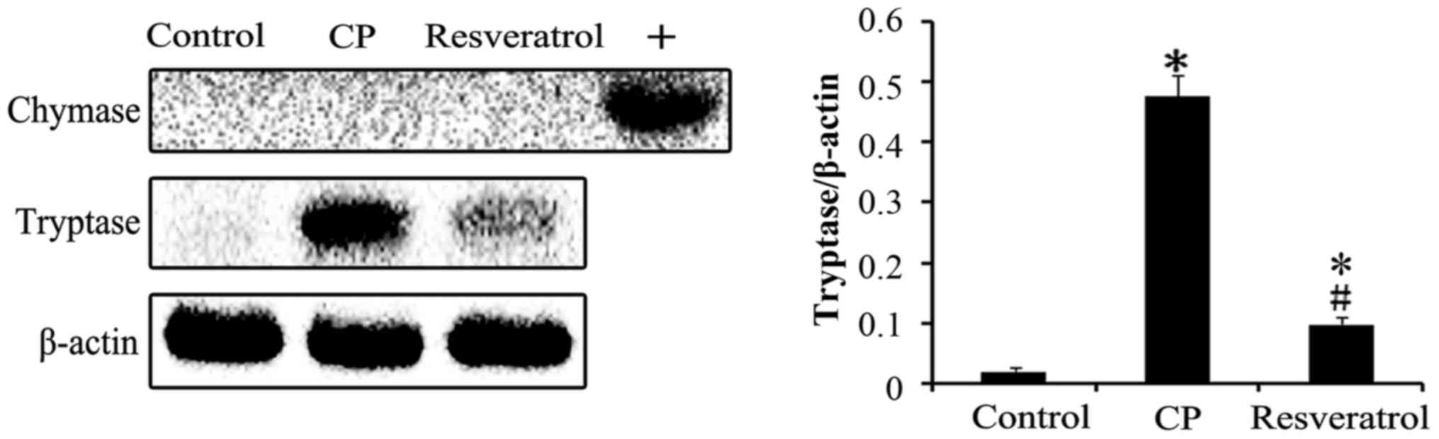
immunohistochemical assays. Western blotting revealed that the
expression levels of tryptase protein in the prostate of the CP
group was increased significantly compared with the control group
(Fig. 2). However, resveratrol
treatment of CP rats significantly decreased expression of this
protein compared with the CP group (Fig. 2). The expression of chymase was not
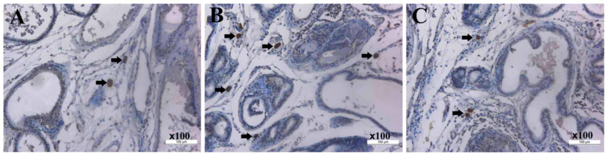
detected using western blotting in any of the groups (Fig. 2). Immunohistochemical staining
indicated that, compared with the control group (Fig. 3A and Table II), the number of tryptase (+) MCs
was significantly higher in the CP group (Fig. 3B and Table II). Resveratrol treatment of CP
rats resulted in a significant reduction in MC number in prostate
tissue (Fig. 3C and Table II). Chymase (+) MCs were not
detected using immunohistochemistry in the prostates from all
groups.
| Table II.Mast cell counts using
immunohistochemical staining in each group. |
Table II.
Mast cell counts using
immunohistochemical staining in each group.
|
| Counts using
immunohistochemical staining |
|---|
|
|
|
|---|
| Groups | Examiner 1 | Examiner 2 | Mean |
|---|
| Control |
3.17±1.17 |
3.50±1.05 |
3.33±1.07 |
| CP |
13.50±1.38 |
13.67±1.03 |
13.58±1.16a |
| Resveratrol |
8.33±0.82 |
8.83±1.17 |
8.58±1.00a,b |
Resveratrol inhibits the activity of
TGF-β and the Wnt/β-catenin signaling pathway
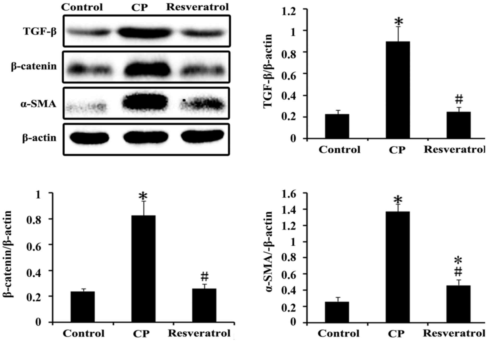
TGF-β is a profibrotic cytokine that induces
macrophage and fibroblast proliferation via the induction of other
growth factors. Western blot analysis revealed an increased
expression level of TGF-β in the CP group compared with the control
group, which was significantly decreased following resveratrol
treatment (Fig. 4). The
Wnt/β-catenin signaling pathway is vital in TGF-β-induced fibrosis.
An increase in β-catenin and α-SMA expression was observed in CP
tissues compared with the control group, and resveratrol treatment
of CP rats significantly reduced the expression of β-catenin and
α-SMA (Fig. 4).
Resveratrol reduces collagen
accumulation in CP tissues
Picrosirius red staining indicated that, compared
with the control group (Fig. 5A),
there was a higher collagen content in the CP group (Fig. 5B), which was decreased in CP rats
following resveratrol treatment (Fig.
5C).
Discussion
Ma et al (3)
reported that prostatic fibrosis was associated with LUTS; however,
the underlying mechanism of LUTS promotion was not investigated.
Chronic prostatitis is a common disease in urology and voiding
dysfunction is the primary clinical manifestation. The presence of
urinary tract obstruction was revealed in patients with CP
following urodynamic examination (2). The maximum capacity of the bladder is
assessed as a parameter of storage function, and the residual urine
volume and maximum voiding pressure are evaluated as parameters of
voiding function. In the present study, the maximum capacity of the
bladder, residual urine volume and maximum voiding pressure in rats
in the CP group were increased significantly compared with the
control group. However, following treatment with resveratrol, these
changes were suppressed.
We hypothesized that increased collagen accumulation
in the prostate during CP progression is a consequence of fibrosis,
which increases tissue stiffness. Periurethral tissue fibrosis may
reduce the flexibility and capacity of the prostatic urethra to
expand to accommodate urinary flow, which may lead to symptoms
associated with obstruction. Fibrosis is a connective tissue
remodeling process that is characterized by the activation and
accumulation of myofibroblasts (24), of which α-SMA is a marker. The
present study revealed that α-SMA expression was significantly
higher in the CP group compared with the control group, and
resveratrol treatment of CP rats decreased α-SMA expression.
Picrosirius red staining demonstrated markedly increased collagen
content in the CP group compared with the control group, and the
maximum capacity of the bladder, residual urine volume and maximum
voiding pressure in the CP group increased significantly compared
with the control group. Furthermore, H&E staining in CP rats
revealed high leukocyte infiltration and papillary fronds that
protruded into the gland cavities, and there was a notable increase
in prostatic epithelial height and a smaller gland lumen diameter.
However, resveratrol treatment suppressed these alterations.
MCs are important effector cells in tissue fibrosis
that express the serine proteases, tryptase and chymase, which are
the most abundant proteins in MCs. Tryptase and chymase have been
reported to stimulate the proliferation of fibroblasts, induce
collagen synthesis in fibroblast cultures and induce collagen
fibril formation (25,26). However, limited information is
known regarding the involvement of these proteases in CP. The
results of the present study demonstrated that the number of
tryptase (+) MCs was significantly higher in the CP group compared
with the control group. Resveratrol administration to CP rats
significantly reduced the number of tryptase (+) MC in prostate
tissues. However, chymase (+) MCs were not detected in the
prostates from all groups which indicated that mast cell chymase
was not involved in prostatitis progression. As previously reported
(27), the primary fibrotic role
of tryptase in the heart is the induction of MC degranulation,
which induces the release of chymase and promotes fibrosis.
However, Beghdadi et al (28) reported that MC protease 4, the
functional counterpart of human chymase, exhibited antifibrotic
potential in acutely induced obstructive nephropathy. The present
study indicated that tryptase may individually induce prostate
fibrosis in CP in the absence of chymase. The molecular phenotypic
heterogeneity of MC makes them attractive therapeutic targets in
various diseases (29,30). The
tryptase+/chymase− MC may serve a critical
role in CP. Therefore, the fibrosis-inducing mechanism, which MC is
dependent on, is complex and further studies are required.
The profibrotic effects of MCs are closely
associated with TGF-β. Studies have demonstrated that MC numbers
and staining intensity were associated with increased TGF-β
production and interstitial fibrosis (12,13).
TGF-β-induced fibrosis process primarily occurs via the Smad
pathway, which serves a critical role in transmitting TGF-β
signals. The Wnt/β-catenin signaling pathway is also important in
the genesis of fibrosis in various organ systems. The Wnt/β-catenin
signaling pathway was reported to be a downstream regulatory
pathway of the TGF-β pathway (31). TGF-β activates canonical Wnt
signaling, which is implicated in the profibrotic effects of TGF-β
(32). The Wnt/β-catenin signaling
pathway is vital in TGF-β-induced fibrosis (14). Wnt signaling has been reported to
skew TGF-β signaling towards dominant signaling via the Smad
pathway (33). Therefore,
interactions of Wnt with TGF-β/Smad signaling requires further
investigation in different situations. CP tissues in the present
study exhibited increased TGF-β and β-catenin expression, which was
reduced in the resveratrol treatment group. Control tissue sections
exhibited limited positive expression.
Resveratrol (15),
a botanical compound that is derived primarily from the skins of
red grapes, has been extensively employed in traditional medicine
and also as a dietary supplement. A previous study demonstrated
that resveratrol may inhibit tissue fibrosis in various organs
(34). In addition, resveratrol
repressed and reversed prostate fibroblast to myofibroblast
phenoconversion in vitro (18). Consistent with these findings, the
results of the present study demonstrated that resveratrol
treatment markedly inhibited the overactivity of MCs, the
TGF-β/Wnt/β-catenin pathway and tissue fibrosis in the CP tissues,
therefore improving voiding dysfunction in CP rats.
In conclusion, the progression of urinary
dysfunction in CP may be induced by fibrosis in the prostate.
Notably, in the present study, tissues from CP rats revealed
increased MC infiltration and an upregulated activity of the
TGF-β/Wnt/β-catenin pathway, which were reduced following
resveratrol treatment. The results of the present study
demonstrated the antifibrotic effect of resveratrol. Therefore,
resveratrol may be considered as a candidate for the treatment of
CP. MCs are not only important for the periurethral tissue fibrosis
in CP, but are also a potential target for therapy.
Acknowledgements
The study of the present study was technically
supported by the College of Pharmacy of Dalian Medical University
and the Second Affiliated Hospital of Dalian Medical University
(Dalian, China). The study was funded by a project supported by the
Natural Science Foundation of Liaoning province (grant no.
201601229).
References
|
1
|
Tyagi P, Kashyap M, Pore S, Wang Z and
Yoshimura N: MP25-07 prostatic inflammation evokes upergulation of
neurotrophins in sensory ganglia: Possible contribution to
dysfunctional voiding. J Urol. 193:e2872015. View Article : Google Scholar
|
|
2
|
Liao LM, Shi BY and Liang CQ: Ambulatory
urodynamic monitoring of external urethral sphincter behavior in
chronic prostatitis patients. Asian J Androl. 1:215–217.
1999.PubMed/NCBI
|
|
3
|
Ma J, Gharaee-Kermani M, Kunju L,
Hollingsworth JM, Adler J, Arruda EM and Macoska JA: Prostatic
fibrosis is associated with lower urinary tract symptoms. J Urolo.
188:1375–1381. 2012. View Article : Google Scholar
|
|
4
|
Karsdal MA, Genovese F, Madsen EA,
Manon-Jensen T and Schuppan D: Collagen and tissue turnover as a
function of age: Implications for fibrosis. J Hepatol. 64:103–109.
2016. View Article : Google Scholar : PubMed/NCBI
|
|
5
|
Fabre V, Wu H, PondTor S, Coutinho H,
Acosta L, Jiz M, Olveda R, Cheng L, White ES, Jarilla B, et al:
Tissue inhibitor of matrix-metalloprotease1 predicts risk of
hepatic fibrosis in human schistosoma japonicum infection. J Infect
Dis. 203:707–714. 2011. View Article : Google Scholar : PubMed/NCBI
|
|
6
|
Trujillo KA, Heaphy CM, Mai M, Vargas KM,
Jones AC, Vo P, Butler KS, Joste NE, Bisoffi M and Griffith JK:
Markers of fibrosis and epithelial to mesenchymal transition
demonstrate field cancerization in histologically normal tissue
adjacent to breast tumors. Int J Cancer. 129:1310–1321. 2011.
View Article : Google Scholar : PubMed/NCBI
|
|
7
|
Silver RB: Role of mast cells in renal
fibrosis. Kidney Int. 84:2142013. View Article : Google Scholar : PubMed/NCBI
|
|
8
|
Cruse G and Bradding P: Mast cells in
airway diseases and interstitial lung disease. Eur J Pharma.
778:125–138. 2016. View Article : Google Scholar
|
|
9
|
Gaca MDA, Arthur MJP and Benyon RC: Mast
cell protease and stem cell factor expression in rat liver
fibrosis. J Hepatol. 28:811998. View Article : Google Scholar
|
|
10
|
Yadav A, Desai RS, Bhuta BA, Singh JS,
Mehta R and Nehete AP: Altered immunohistochemical expression of
mast cell tryptase and chymase in the pathogenesis of oral
submucous fibrosis and malignant transformation of the overlying
epithelium. PLoS One. 9:e987192014. View Article : Google Scholar : PubMed/NCBI
|
|
11
|
Zerr P, Palumbo-Zerr K, Huang J, Tomcik M,
Sumova B, Distler O, Schett G and Distler JH: Sirt1 regulates
canonical TGF-(β) signalling to control fibroblast activation and
tissue fibrosis. Ann Rheum Dis. 75:226–233. 2016. View Article : Google Scholar : PubMed/NCBI
|
|
12
|
Summers SA, Gan PY, Dewage L, Ma FT, Ooi
JD, O'Sullivan KM, Nikolic-Paterson DJ, Kitching AR and Holdsworth
SR: Mast cell activation and degranulation promotes renal fibrosis
in experimental unilateral ureteric obstruction. Kidney Int.
82:676–685. 2012. View Article : Google Scholar : PubMed/NCBI
|
|
13
|
Jeong DH, Lee GP, Jeong WI, Do SH, Yang
HJ, Yuan DW, Park HY, Kim KJ and Jeong KS: Alterations of mast
cells and TGF-beta1 on the silymarin treatment for CCI(4)-induced
hepatic fibrosis. World J Gastroenterol. 11:1141–1148. 2005.
View Article : Google Scholar : PubMed/NCBI
|
|
14
|
Akhmetshina A, Palumbo K, Dees C, Bergmann
C, Venalis P, Zerr P, Horn A, Kireva T, Beyer C, Zwerina J, et al:
Activation of canonical Wnt signalling is required for
TGF-β-mediated fibrosis. Nat Commun. 3:7352012. View Article : Google Scholar : PubMed/NCBI
|
|
15
|
Torres P, Poveda A, Jimenez-Barbero J,
Ballesteros A and Plou FJ: Regioselective lipase-catalyzed
synthesis of 3-o-acyl derivatives of resveratrol and study of their
antioxidant properties. J Agric Food Chem. 58:807–813. 2010.
View Article : Google Scholar : PubMed/NCBI
|
|
16
|
Sharma S, Anjaneyulu M, Kulkarni SK and
Chopra K: Resveratrol, a polyphenolic phytoalexin, attenuates
diabetic nephropathy in rats. Pharmacology. 76:69–75. 2006.
View Article : Google Scholar : PubMed/NCBI
|
|
17
|
Csiszar A: Anti-inflammatory effects of
resveratrol: Possible role in prevention of age-related
cardiovascular disease. Ann N Y Acad Sci. 1215:117–122. 2011.
View Article : Google Scholar : PubMed/NCBI
|
|
18
|
Gharaee-Kermani M, Moore BB and Macoska
JA: Resveratrol-mediated repression and reversion of prostatic
myofibroblast phenoconversion. PLoS One. 11:e01583572016.
View Article : Google Scholar : PubMed/NCBI
|
|
19
|
He Y, Zeng H, Yu Y, Zhang J, Zeng X, Gong
F, Duan X, Liu Q and Yang B: Resveratrol improved the progression
of chronic prostatitis via the downregulation of c-kit/SCF by
activating Sirt1. J Agric Food Chem. 65:5668–5673. 2017. View Article : Google Scholar : PubMed/NCBI
|
|
20
|
He Y, Zeng HZ, Yu Y, Zhang JS, Duan X,
Zeng XN, Gong FT, Liu Q and Yang B: Resveratrol improves prostate
fibrosis during progression of urinary dysfunction in chronic
prostatitis. Environ Toxicol Pharmacol. 54:120–124. 2017.
View Article : Google Scholar : PubMed/NCBI
|
|
21
|
Wong L, Done JD, Schaeffer AJ and
Thumbikat P: Experimental autoimmune prostatitis induces microglial
activation in the spinal cord. Prostate. 75:50–59. 2015. View Article : Google Scholar : PubMed/NCBI
|
|
22
|
Zheng T, Wang R, Zhang TB, Jia DH, Wang
CL, Sun Y and Zhang WX: AB036. Effects and its potential mechanisms
of Cox-2 inhibitors on ejaculation latency of rat with experimental
autoimmune prostatitis. Transl Androl Urol. 5 Suppl 1:AB0362016.
View Article : Google Scholar :
|
|
23
|
Rivero VE, Motrich RD, Maccioni M and
Riera CM: Autoimmune etiology in chronic prostatitis syndrome: An
advance in the understanding of this pathology. Crit Rev Immunol.
27:33–46. 2007. View Article : Google Scholar : PubMed/NCBI
|
|
24
|
Fraga F, Sturny M, Ruoccolo R, Santos R,
Silva R and Stergiopulos N: 139 Angiotensin-(1–7) inhibits
cavernosal fibrosis via attenuation of fibroblast differentiation
to myofibroblast. J Sex Med. 14 Suppl 1:S392017. View Article : Google Scholar
|
|
25
|
Abe M, Kurosawa M, Ishikawa O, Miyachi Y
and Kido H: Mast cell tryptase stimulates both human dermal
fibroblast proliferation and type I collagen production. Clin Exp
Allergy. 28:1509–1517. 1998. View Article : Google Scholar : PubMed/NCBI
|
|
26
|
Kofford MW, Schwartz LB, Schechter NM,
Yager DR, Diegelmann RF and Graham MF: Cleavage of type I
procollagen by human mast cell chymase initiates collagen fibril
formation and generates a unique carboxyl-terminal propeptide. J
Biol Chem. 272:7127–7131. 1997. View Article : Google Scholar : PubMed/NCBI
|
|
27
|
Li J, Jubair S, Levick SP and Janicki JS:
The autocrine role of tryptase in pressure overload-induced mast
cell activation, chymase release and cardiac fibrosis. IJC
Metabolic and Endocr. 10:16–23. 2016. View Article : Google Scholar
|
|
28
|
Beghdadi W, Madjene LC, Claver J, Pejler
G, Beaudoin L, Lehuen A, Daugas E and Blank U: Mast cell chymase
protects against renal fibrosis in murine unilateral ureteral
obstruction. Kidney Int. 84:317–326. 2013. View Article : Google Scholar : PubMed/NCBI
|
|
29
|
Globa T, Saptefrţi L, Ceauşu RA, Gaje P,
Cimpean AM and Raica M: Mast cell phenotype in benign and malignant
tumors of the prostate. Pol J Pathol. 65:147–153. 2014. View Article : Google Scholar : PubMed/NCBI
|
|
30
|
Qi L, Song W, Liu Z, Zhao X, Cao W and Sun
B: Wnt3a promotes the vasculogenic mimicry formation of colon
cancer via wnt/β-catenin signaling. Int J Mol Sci. 16:18564–18579.
2015. View Article : Google Scholar : PubMed/NCBI
|
|
31
|
Conidi A, van den Berghe V and Huylebroeck
D: Aptamers and their potential to selectively target aspects of
EGF, Wnt/β-catenin and TGFβ-smad family signaling. Int J Mol Sci.
14:6690–6719. 2013. View Article : Google Scholar : PubMed/NCBI
|
|
32
|
Pons M, Ali L, Beghdadi W, Danelli L,
Alison M, Madjène LC, Calvo J, Claver J, Vibhushan S, Åbrink M, et
al: Mast cells and MCPT4 chymase promote renal impairment after
partial ureteral obstruction. Front Immunol. 8:4502017. View Article : Google Scholar : PubMed/NCBI
|
|
33
|
van den Bosch MH, Blom AB, van Lent PL,
van Beuningen HM, Blaney Davidson EN, van der Kraan PM and van den
Berg WB: Canonical Wnt signaling skews TGF-beta signaling in
chondrocytes towards signaling via ALK1 and Smad 1/5/8. Cell
Signal. 26:951–958. 2014. View Article : Google Scholar : PubMed/NCBI
|
|
34
|
Xiao Z, Chen C, Meng T, Zhang W and Zhou
Q: Resveratrol attenuates renal injury and fibrosis by inhibiting
transforming growth factor-β pathway on matrix metalloproteinase 7.
Exp Biol Med (Maywood). 241:140–146. 2016. View Article : Google Scholar : PubMed/NCBI
|