Introduction
Isoflurane and sevoflurane are commonly used
anesthetics in clinical surgery (1). However, increasing evidence from
epidemiological studies indicates that children <4 years that
are exposed to anesthetics may be at an increased risk of
developing cognitive disabilities and learning difficulties
(2–4). It has been reported that sevoflurane
treatment induces neuroapoptosis and degeneration in neonatal rats,
which consequently leads to pathological alternations in the
hippocampus (5,6). The safety of anesthetics in surgical
procedures has received a large amount of attention, however, the
underlying molecular mechanism by which sevoflurane induces
neurodegeneration remains largely unknown.
Accumulating evidence has demonstrated that
cognitive impairments induced by surgical anesthesia are associated
with alterations in the expression of a variety of genes involved
in brain development (6,7). In recent years, dysregulation of
microRNAs (miRNAs/miRs) has been implicated in
anesthesia-associated cognitive dysfunction (8–10).
MiRNAs are small, noncoding RNAs that negatively regulate gene
expression through inhibiting the translation of transcripts or
inducing the degradation of target mRNA (11–15).
It has been well-documented that aberrant expression of miRNAs is
associated with the development of human cancers, and miRNAs
therefore serve as potential biomarkers for cancer diagnosis and
treatment (13,16–23).
Additionally, an increasing number of miRNAs, including miR-665,
miR-572 and miR-34a, have been demonstrated to be associated with
anesthetic-induced hippocampal apoptosis (24–26).
As miRNAs have important roles in the regulation of cell death and
differentiation, the identification of novel miRNAs that
participate in the neurodegeneration caused by anesthetics
exposure, and understanding the functional molecular mechanisms of
these miRNAs, is critical.
A recent study by Zhou et al (6) screened the differentially expressed
miRNAs following sevoflurane exposure. Among these, the expression
of miRNA-34c was significantly downregulated in the hippocampus of
developing rat brains following sevoflurane treatment. miR-34c is
reported to be a direct downstream target of p53 and to regulate
the expression of Bcl-2 and Bcl-2-associated X in response to the
sevoflurane administration (25).
In addition to the downregulated miRNAs, miR-188-3p has been
identified as one of the most significantly increased miRNAs
following sevoflurane treatment; however, the functional molecular
mechanism of miR-188-3p in sevoflurane anesthesia-induced remains
largely unknown.
In the present study, the expression level of
miR-188-3p in the hippocampus of rats following exposure to
sevoflurane was detected. To determine the functional mechanism of
miR-188-3p, the downstream targets of miR-188-3p were predicted by
databases. The results of the present study demonstrated that the
expression of miR-188-3p was significantly increased following
sevoflurane exposure, and that miR-188-3p may be implicated in
sevoflurane-induced apoptosis in the rat brain through negative
regulation of the MDM2 proto-oncogene (MDM2)-p53 pathway.
Materials and methods
Cell lines and animals
SH-SY5Y neuroblastoma cells, which are subcloned
from the SK-N-SH neuroblastoma cell line, were obtained from
Leibniz Institut DSMV-Deutsche Sammlung von Mikroorganismen und
Zellkulturen GmbH (DSMZ, Braunschweig, Germany). The cells were
cultured in a mixture of Dulbecco's modified Eagle's medium (DMEM)
and F12 (1:1) supplemented with 10% fetal bovine serum (all from
Thermo Fisher Scientific, Inc., Waltham, MA, USA). Cells were
cultured at 37°C in a humidified atmosphere of 5% CO2.
Animal experiments were performed with Sprague-Dawley rats (n=120;
male; 7-weeks-old; weight, 240±10 g) that were obtained from
Beijing Vital River Laboratory Animal Technology Co., Ltd.
(Beijing, China). The animals were housed under a 12 h light/dark
cycle with a room temperature (21±1°C) and humidity (60±5%). Rats
were allowed ad libitum access to water and food. The
experiments were approved by the Animal Care and Ethical Committee
of Cangzhou Central Hospital (Cangzhou, China) and was performed in
accordance with the Guide for the Care and Use of Laboratory
Animals (27).
Sevoflurane exposure and animal
transduction
The rats were randomly divided into two groups: A
control group (n=8) receiving regular air inhalation for 6 h and a
sevoflurane group (n=8) receiving 2.5% sevoflurane (Shanghai
Hengrui Pharmaceutical Co., Ltd., Shanghai, China) at 600 µg/kg/min
in 100% O2 at the identical conditions for 6 h in a
plastic container. The levels of sevoflurane and oxygen were
detected by the gas monitor. The lentivirus expressing miR-188-3p
or miR-188-3p inhibitor was purchased from the Guangzhou RiboBio
Co., Ltd. (Guangzhou, China). For transduction of animals with
miR-188-3p mimics, miR-188-3p inhibitor or respective controls, 10
µl lentivirus-miR-188-3p mimics/inhibitor or control miRNA (20 µM)
was given intracerebroventricularly using a Hamilton
microsurgically gauged syringe into the left lateral cerebral
ventricles with a pre-drilled skull micro-hole (10×10 µm).
Following transfection for 120 h, the following experiments were
performed.
Morris water maze task
The learning and memory ability of rats exposed to
sevoflurane and regular air was assessed by the Morris water maze
task, as previously described (28). Briefly, rats in each group were
trained to swim for 5 days in a circular water pool (200 cm in
diameter, 60 cm in depth). A hidden circular platform of ~10 cm in
diameter and 2 cm below the water surface was submerged in the pool
filled with warm water. Rats were forced to swim and find the
platform within 60 sec. When rats failed to locate the hidden
platform within the time limit, animals were gently guided. The
time taken to locate the platform, the number of platform crossings
and the swimming speed of each rat on their first attempt on day 5,
which was recorded with the ANY-maze Video Tracking System version
5.1 (Stoelting Co., Wood Dale, IL, USA), was used to evaluate the
learning ability of the rats.
Cell transfection
miR-188-3p mimics (5′-CUCCCACAUGCAGGGUUUGCA) and
negative control (NC) miRNA (5′-UUUGUACUACACAAAAGUACUG) (Guangzhou
RiboBio Co., Ltd.) was dissolved with RNase-free water. SH-SY5Y
cells were seeded in 6-well plates at a density of
1×10⁵/well overnight at 37°C. miRNAs were transfected
using the NanoFectin Transfection Reagent (Shanghai ExCell Biology,
Inc., Shanghai, China), with a final concentration of 100 nM. The
mixture of transfection reagent and miRNAs was incubated at room
temperature for 25 min prior to addition to the cell culture.
Following transfection for 96 h, the cells were harvested and the
expression level of miR-188-3p was detected by RT-qPCR.
miR-188-3p target prediction
TargetScan (www.targetscan.org, release 7.1) and miRNA.org (http://www.microrna.org, August 2010 release) were
used for scanning the candidate targets of miR-188-3p. Basic
information of miR-188-3p was submitted online and the potential
targets of miR-188-3p were presented.
Luciferase assay
The wild-type (WT) and mutant 3′-untranslated region
(3′-UTR) of MDM2, containing the predicted binding site of
miR-188-3p, were constructed into the pMIR-REPORT firefly
luciferase vector (Ambion; Thermo Fisher Scientific, Inc.). For the
luciferase experiments, cells were seeded into the 96-well plate at
the density of 1,000 cells/well. Cells were then co-transfected
with 100 nM miR-188-3p or NC miRNA in the presence of 50 µg
luciferase vector containing either WT or mutant forms of the
3′-UTR of MDM2 using Lipofectamine® 2000 (Thermo Fisher
Scientific, Inc.). Following transfection for 36 h, the luciferase
activity was determined and normalized with the activity of
Renilla luciferase using a Luciferase Reporter assay kit
(BioVision, Inc., Milpitas, CA, USA). The experiment was performed
in triplicate.
Reverse transcription-quantitative
polymerase chain reaction (RT-qPCR) assay
The rats were sacrificed 6 h after the last
sevoflurane exposure by decapitation. The hippocampal tissues were
dissected and stored in the liquid nitrogen. For the RT-qPCR assay,
the tissues were ground into powder. Total RNA and miRNA extraction
from the hippocampal tissues was performed with TRIzol reagent
(Invitrogen; Thermo Fisher Scientific, Inc.) and an miRNeasy Mini
kit (Qiagen GmbH, Hilden, Germany), respectively. The RNA quality
and quantity were detected using the NanoDrop ND-1000
spectrophotometer (NanoDrop Technologies; Thermo Fisher Scientific,
Inc., Wilmington, DE, USA). RT was performed at 42°C for 15 min
using a stem-loop RT primer (miRcute Plus miRNA First-Strand cDNA
Synthesis kit, KR211-01; Tiangen Biotech Co., Ltd., Beijing, China)
that hybridized to miR-188-3p. qPCR was performed using the
SsoFast™ EvaGreen® Supermix kit (Bio-Rad Laboratories,
Inc., Hercules, CA, USA) and the iQ5 Real-Time PCR System (Bio-Rad
Laboratories, Inc.). GAPDH and U6 were used as the internal control
for mRNA and miRNA respectively and quantification used the
2−∆∆Cq method (29).
The reaction conditions were as follows: 95°C for 10 min, followed
by 40 cycles of 95°C for 10 sec and 60°C for 1 min. The following
primer sequences were used: miR-188-3p,
5′-ATTATTGGCTCCCACATGCAGGG-3′ (forward) and
5′-ATCCAGTGCAGGGTCCGAGG-3′ (reverse); MDM2,
5′-TACAGGGACGCCATCGAATC-3′ (forward) and
5′-TGAAGTGCATTTCCAATAGTCAGC-3′ (reverse); p53,
5′-TACTCCCCTGCCTCAACAAG-3′ (forward) and 5′-CGCTATCTGAGCAGCGCTCAT3′
(reverse); U6, 5′-GCTTCGGCAGCACATATACTAA-3′ (forward) and
5′-AACGCTTCACGAATTTGCGT-3′ (reverse); and GAPDH,
5′-TGTGTCCGTCGTGGATCTGA-3′ (forward) and
5′-CCTGCTTCACCACCTTCTTGA-3′ (reverse). The experiment was repeated
three times.
Terminal
deoxynucleotidyl-transferase-mediated dUTP nick end labeling
(TUNEL) assay
Hippocampal tissues from rats exposed to the
sevoflurane treatment for 6 h were fixed with 4% paraformaldehyde
at room temperature for 10 min. Tissues were dehydrated in a grade
series of ethanol and embedded in paraffin. Sections (3 µm) were
obtained by cutting the tissues with the microtome (JungRM 2035;
Leica Microsystems GmbH, Wetzlar, Germany). The paraffin-embedded
tissues were dewaxed by incubating the slides at 55°C for 30 min,
and then washed in a series of ethanol (100, 95, 80, 75 and 50%)
for 2 min, respectively. The slides were incubated with 20 µg/ml
proteinase K for 30 min at room temperature and then rinsed with
PBS for 10 min at room temperature. TUNEL reaction mixture (50 µl,
TUNEL kit; Roche Diagnostics GmbH, Mannheim, Germany) was added to
the tissues and incubated at room temperature for 2 h.
Subsequently, tissues were washed twice with pre-cooled PBS
followed by staining with 25 µg/ml propidium iodide (PI)
(Sigma-Aldrich; Merck KGaA, Darmstadt, Germany) for 30 min at room
temperature. Then 10 fields of view were observed from each sample
with an Olympus FLUOVIEW FV10i confocal microscope (Olympus
Corporation, Tokyo, Japan).
Western blot analysis
The hippocampal tissues of rats exposed to
sevoflurane treatment for 6 h were homogenized in NP-40 lysis
buffer (Beyotime Institute of Biotechnology, Shanghai, China)
containing 1% phenylmethylsulfonyl fluoride. The samples were
centrifuged at 13,000 × g for 15 min at 4°C and the supernatant was
collected. The protein concentration was determined by a BCA
protein assay (Bio-Rad Laboratories, Inc.). Protein samples (50
mg/lane) were separated by 15% SDS-PAGE. Subsequently, proteins
were transferred onto nitrocellulose membranes. The membranes were
blocked with 5% non-fat milk at room temperature for 1 h and
subsequently incubated with the indicated primary antibodies at
room temperature for 2 h, followed by incubation with secondary
antibodies at room temperature for 1 h. Protein bands were detected
using the Pierce ECL Plus Western Blotting Substrate system
(Pierce; Thermo Fisher Scientific, Inc.) and visualized with
ImageMaster 2D Platinum 7.0 software (GE Healthcare Bio-Sciences,
Pittsburgh, PA, USA). The antibodies used in the current study were
as follows: Rabbit anti-GAPDH (1:2,000, cat. no. ab70699), rabbit
anti-MDM2 (1:2,000, cat. no. ab137413), mouse anti-p53 (DO-1,
1:500, cat. no. ab1101) (all from Abcam, Cambridge, UK), goat
anti-rabbit IgG (H+L)-HRP conjugated secondary antibody (cat. no.
170–6515, 1:5,000), and goat anti-mouse IgG (H+L)-HRP conjugated
secondary antibody (cat. no. 170–6516, 1:5,000) (both from Bio-Rad
Laboratories, Inc.).
Exposure of SH-SY5Y cells to
sevoflurane and cell apoptosis
SH-SY5Y cells (2×10⁵) were incubated with
or without 10 µM pifithrin-α (04–0040; Yeasen Biotechnology, Co.,
Ltd., Shanghai, China) for 12 h at 37°C. Then the cells were
treated with 5 µM sevoflurane for 30 min at 37°C and the cell
apoptosis was determined using an Alexa Fluor 488 Annexin V/Dead
Cell Apoptosis kit (Thermo Fisher Scientific, Inc.), according to
the manufacturer's instructions. Briefly, cells were harvested and
washed with pre-cooled PBS. Cells were centrifuged at 1,000 × g for
5 min at 4°C and resuspended in annexin-binding buffer.
Subsequently, Annexin V-fluorescein isothiocyanate (5 µl) was added
to ~1×10⁵ cells and incubated at room temperature for 15
min, followed by the addition of 1 µl PI (100 µg/ml). Cell
apoptosis wad detected with a BD FACSCalibur™ flow cytometer and
analyzed with the CFlow Plus Software, version l.0.172.9 (BD
Biosciences, Franklin Lakes, NJ, USA).
Statistical analysis
Data are presented as the mean ± standard deviation
of three independent experiments. Statistical analysis was
performed by Student's t-test or one-way analysis of variance
followed by Tukey's post hoc comparison test using SPSS 19.0
software (IBM Corp., Armonk, NY, USA). The plots were generated
using GraphPad Prism 6.0 (GraphPad Software, Inc., La Jolla, CA,
USA). P<0.05 was considered to indicate a statistically
significant difference.
Results
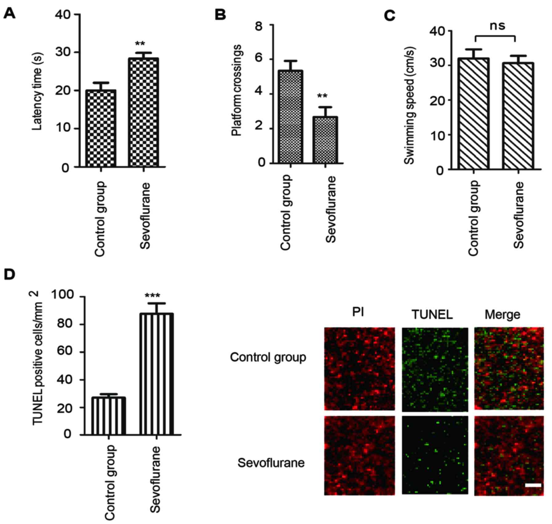
Sevoflurane anesthesia induces spatial
memory impairments in rats
The Morris water maze test is considered to be a
highly sensitive method of determining cognitive function to
examine hippocampus-dependent learning and memory (30). To determine the effect of
sevoflurane exposure on the memory ability of rats, the animals in
each group were trained to locate the hidden platform. As
demonstrated in Fig. 1A, the
latency time taken to locate the hidden platform was significantly
higher for rats treated with sevoflurane compared with the control
group. Consistently, a decreased number of platform crossings in
the sevoflurane group was observed in comparison with the control
group (Fig. 1B). However, no
significant differences in swimming speed were observed between the
two groups of rats (Fig. 1C).
These results demonstrated that sevoflurane exposure led to
cognitive impairment in rats.
To determine whether sevoflurane-induced cognitive
dysfunction in rats was associated with neuroapoptosis, apoptosis
was investigated by TUNEL staining of the hippocampal tissues
following sevoflurane exposure. The results demonstrated that,
compared with the control group, sevoflurane administration for 6 h
significantly increased the proportion of TUNEL-positive cells in
the hippocampus of rats (Fig. 1D).
These results indicated that sevoflurane exposure induced
neuroapoptosis and subsequent cognitive impairment in rats.
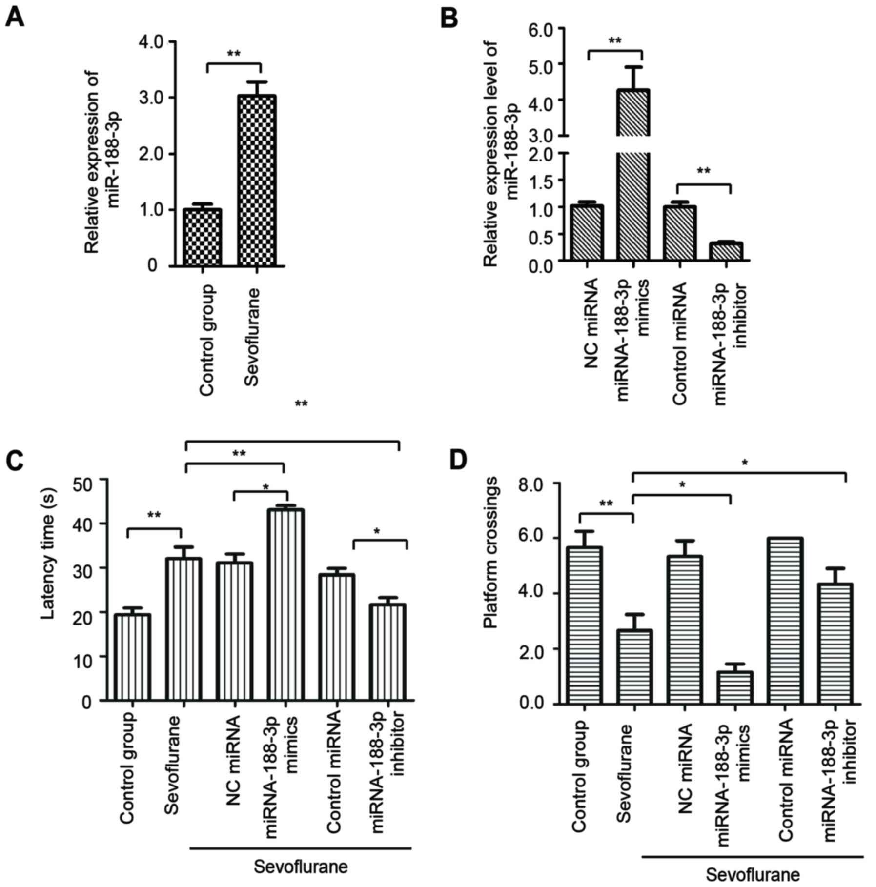
Sevoflurane anesthesia upregulates the
expression of miR-188-3p
As the present study reported sevoflurane-induced
cognitive dysfunction in rats, the potential underlying mechanisms
of this effect were subsequently investigated. It has been
well-documented that miRNAs have important roles in the regulation
of gene expression in response to sevoflurane treatment (6,31–35),
and bioinformatics analyses identified miR-188-3p as one of the
most significantly upregulated miRNAs following sevoflurane
administration (6). To confirm
this observation, the expression level of miR-188-3p in the
hippocampal tissue of rats with or without sevoflurane treatment
was determined by RT-qPCR. As demonstrated in Fig. 2A, the expression of miR-188-3p was
significantly increased in the hippocampus of rats following
sevoflurane exposure, compared with the control group.
To validate the association between upregulated
miR-188-3p expression in the hippocampus and sevoflurane-induced
cognitive impairments, rats were transfected with lentiviruses
containing miR-188-3p mimics or inhibitor. The expression level of
miR-188-3p following transduction with miR-188-3p mimics,
miR-188-3p inhibitor and the respective controls was confirmed by
RT-qPCR (Fig. 2B). The Morris
water maze test was performed to determine the learning and memory
ability of rats harboring overexpression or depletion of miR-188-3p
following sevoflurane treatment. The data indicated that, following
exposure to sevoflurane, rats with overexpression of miR-188-3p
exhibited an increased latency time compared with the rats
transduced with NC miRNA and the sevoflurane-only group (Fig. 2C). By contrast, the rats treated
with miR-188-3p inhibitor exhibited a lower latency time compared
with the control miRNA group and the sevoflurane-only group
(Fig. 2C). Similarly,
overexpression of miR-188-3p in rats treated with sevoflurane
decreased the number of platform crossings, while depletion of
miR-188-3p significantly increased the number of platform
crossings, compared with the sevoflurane-only group (Fig. 2D). These results demonstrated the
critical involvement of miR-188-3p in sevoflurane-induced cognitive
dysfunction in rats.
MDM2 is a direct target of
miR-188-3p
To further investigate the underlying molecular
mechanism by which miR-188-3p may regulate sevoflurane-induced
cognitive dysfunction, the downstream targets of miR-188-3p were
predicted using TargetScan and miRNA databases. Among the putative
candidates, MDM2 was predicted as one of the downstream targets of
miR-188-3p (Fig. 3A). To confirm
this result, a luciferase assay was performed to investigate the
binding between miR-188-3p and the 3′-UTR of MDM2. As indicated in
Fig. 3B, decreased luciferase
activity was observed in SH-SY5Y neuroblastoma cells that were
co-transfected with miR-188-3p mimics and the WT 3′-UTR of MDM2,
compared with those co-transfected with NC miRNA and the WT 3′UTR
(Fig. 3B). No significant
difference was observed between cells co-transfected with the
mutant 3′-UTR of MDM2 and miR-188-3p mimics or NC miRNA (Fig. 3B). These results confirmed that
miR-188-3p specifically bound to the 3′-UTR of MDM2 mRNA.
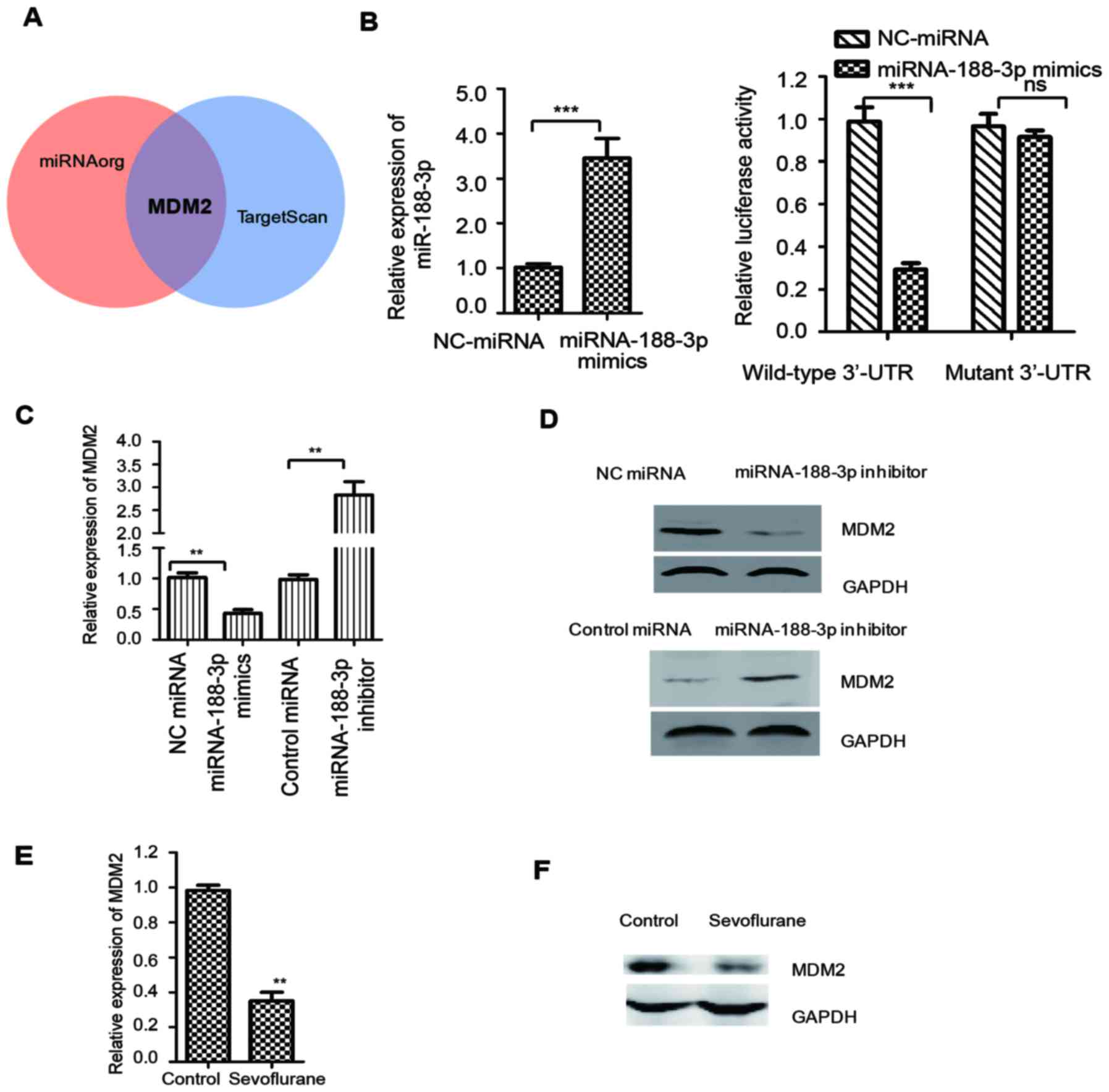 | Figure 3.MDM2 is a downstream target of
miR-188-3p. (A) Candidate targets of miR-188-3p were predicted
using miRNA and TargetScan databases. MDM2 was selected as a
putative target of miR-188-3p. (B) SH-SY5Y neuroblastoma cells were
co-transfected with a luciferase vector containing a wild-type or
mutant version of the MDM2 3′UTR and NC miRNA or miR-188-3p mimics,
and the luciferase activity was measured. The expression level of
miR-188-3p was detected by RT-qPCR as shown in left panel. (C) mRNA
expression of MDM2 was detected in the hippocampal tissues of rats
that were treated with miR-188-3p mimics or miR-188-3p inhibitor
after sevoflurane administration, n=8/group. (D) Protein expression
of MDM2 in the hippocampal tissues of rats transduced with
miR-188-3p mimics, miR-188-3p inhibitor and the respective controls
was determined by western blot analysis. GAPDH was used as the
loading control, n=8/group. (E) mRNA and (F) protein levels of MDM2
in the hippocampal tissues of rats with or without sevoflurane
treatment were detected by reverse transcription-quantitative
polymerase chain reaction and western blot analysis, respectively,
n=8/group. **P<0.01 and ***P<0.001 as indicated. MDM2, MDM2
proto-oncogene; miR/miRNA, microRNA; UTR, untranslated region; NC,
negative control; ns, not significant. |
To confirm this conclusion, the expression of MDM2
in the hippocampal tissues of rats treated with miR-188-3p mimics
or inhibitor, and the respective controls, was determined by
RT-qPCR and western blotting. As demonstrated in Fig. 3C, compared with the NC miRNA group,
the mRNA abundance of MDM2 was significantly decreased in rats
harboring miR-188-3p mimics. Consistent with this result, the mRNA
level of MDM2 was increased in rats that were treated with
miR-188-3p inhibitor, compared with the control miRNA group
(Fig. 3C). Western blot analysis
was performed to determine the protein levels of MDM2 in
hippocampal tissues from rats treated with miR-188-3p mimics or
inhibitor. The results demonstrated that overexpression of
miR-188-3p suppressed the expression of MDM2, while downregulation
of miR-188-3p increased the expression of MDM2, compared with their
relative control groups (Fig. 3D).
These results indicated that miR-188-3p may target MDM2 and
negatively regulate its expression. To further confirm the
involvement of MDM2 in sevoflurane-induced neuroapoptosis, the
expression level of MDM2 was detected following treatment of rats
with sevoflurane. As demonstrated in Fig. 3E and F, the mRNA and protein
expression of MDM2 was markedly decreased following sevoflurane
treatment, compared with the control group. These results indicate
that sevoflurane treatment may negatively regulate the expression
of MDM2.
miR-188-3p regulates the MDM2-p53
signaling pathway
It has been well-documented that MDM2 is an E3
ubiquitin ligase that mediates the polyubiquitylation and
degradation of p53 (36). To
determine whether the negative regulation of MDM2 by miR-188-3p
affects the expression of p53, the protein and mRNA expression of
p53 was determined in hippocampal tissues from rats transduced with
miR-188-3p mimics or NC miRNA. Western blotting results
demonstrated that overexpression of miR-188-3p enhanced the protein
expression of p53, compared with the NC miRNA group (Fig. 4A). However, the mRNA expression
level of p53 was not significantly altered following overexpression
of miR-188-3p (Fig. 4B). To
investigate the effect of sevoflurane on the expression of p53, the
protein and mRNA expression of p53 in the hippocampal tissues of
rats with or without sevoflurane treatment was also determined. The
results demonstrated that the protein expression, but not the mRNA
expression, of p53 was increased following sevoflurane treatment,
compared with the control group (Fig.
4C and D). These results indicated that the activation of p53
may be increased following sevoflurane exposure.
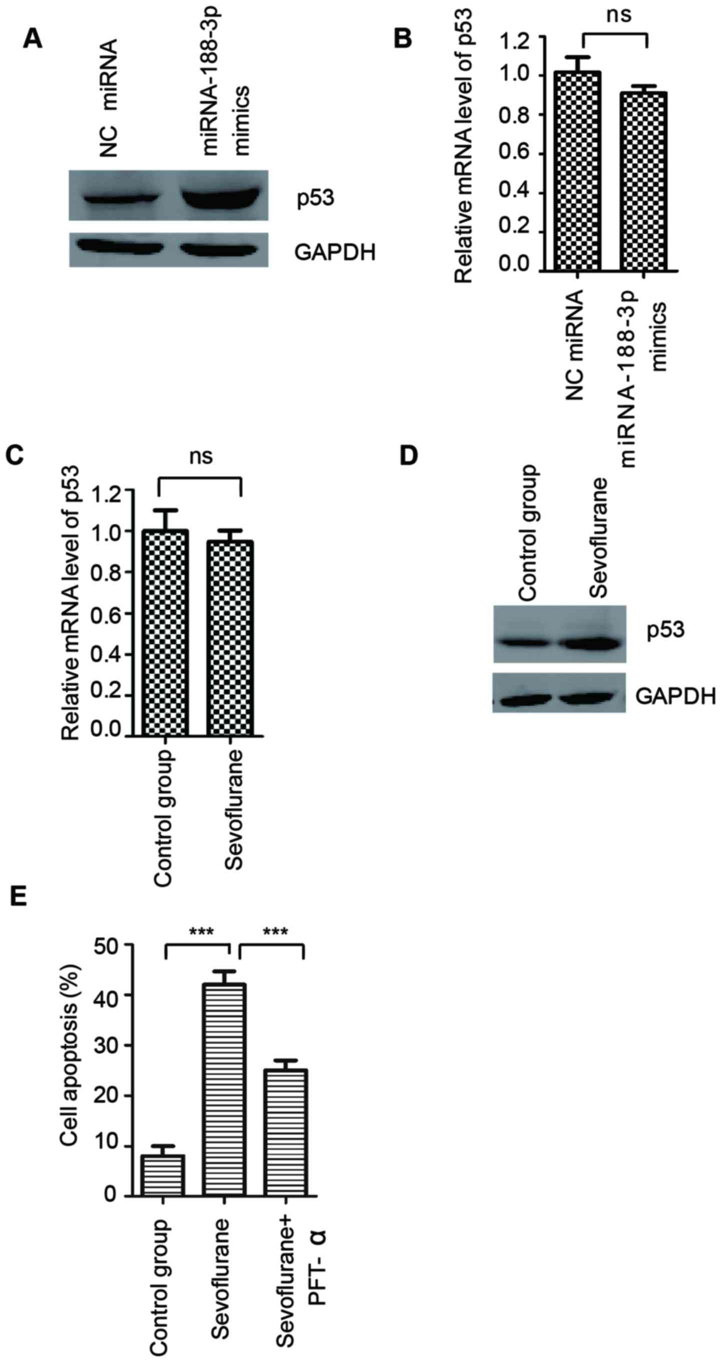 | Figure 4.miR-188-3p overexpression stabilized
p53 and contributed to the neuroapoptosis induced by sevoflurane.
(A) Protein and (B) mRNA levels of p53 were detected by western
blot analysis and RT-qPCR, respectively, in the hippocampal tissues
of rats that were treated with miR-188-3p mimics or NC miRNA,
n=8/group. (C) mRNA and (D) protein levels of p53 in the
hippocampal tissues of rats with or without sevoflurane treatment
were detected by RT-qPCR and western blot analysis, respectively,
n=8/group. (E) SH-SY5Y neuroblastoma cells were exposed to
sevoflurane with or without the specific inhibitor of p53, PFT-α,
and the cell apoptosis rate was determined by flow cytometry.
***P<0.001 as indicated. miR/miRNA, microRNA; RT-qPCR, reverse
transcription-quantitative polymerase chain reaction; NC, negative
control; PFT-α, pifithrin-α; ns, not significant. |
Enhanced expression of p53 is associated with cell
apoptosis. To determine whether the increased stability of p53
induced by miR-188-3p contributed to sevoflurane-induced
neuroapoptosis, SH-SY5Y neuroblastoma cells exposed to sevoflurane
were treated with pifithrin-α, the specific inhibitor of p53, and
the rate of cell apoptosis rate was measured by flow cytometry. The
results demonstrated that sevoflurane treatment induced cell
apoptosis compared with the control group, while inhibition of p53
suppressed sevoflurane-induced cell apoptosis (Fig. 4E). These results indicated that
sevoflurane enhanced the expression of miR-188-3p, which negatively
regulated the MDM2-p53 pathway and the stabilization of p53 and may
partially contribute to the cell apoptosis induced by
sevoflurane.
Discussion
Anesthesia-induced cognitive impairment is
considered to be the most common type of postoperative cognitive
impairment. Previous studies have demonstrated that extended
exposure to anesthetics resulted in excessive apoptosis of neurons
and decreased the self-renewal capacity of neural stem cells, which
consequently led to learning and memory deficits in animals
(37–41). miRNA dysregulation has been
previously associated with neurodegeneration induced by anesthesia
(6,15,24).
The present study demonstrated that miR-188-3p was upregulated in
the hippocampus of rats following sevoflurane administration.
Furthermore, the results indicated that overexpression of
miR-188-3p may promote cognitive dysfunction by targeting MDM2 and
increasing the stability of p53, as inhibition of the activity of
p53 suppressed sevoflurane-induced neuroapoptosis.
Numerous miRNAs have been reported to be associated
with anesthesia-induced cognitive dysfunction. Among these, miR-665
antagonized sevoflurane anesthesia-induced neurodegeneration in
rats via the phosphatidylinositol 3-kinase/Akt signaling pathway by
targeting insulin-like growth factor 2 (24). In addition, miR-34c was reported to
be involved in sevoflurane-induced neural apoptosis through the
mitochondrial pathway in the hippocampus of developing rat brains
(6) and miR-572 was demonstrated
to improve early postoperative cognitive dysfunction by
downregulating the expression of neural cell adhesion molecule 1
(25). The present study
investigated the function of miR-188-3p in sevoflurane-induced
cognitive impairment, as this miRNA was reported to be one of the
most significantly upregulated miRNAs following sevoflurane
exposure in a previous study (6).
The results of the present study demonstrated that increased
expression of miR-188-3p was observed in the hippocampal tissues of
rats that received sevoflurane treatment compared with control
rats. It has been reported that the expression of miR-34c was
decreased in response to sevoflurane treatment, and bioinformatics
analyses indicated that miR-34c was a direct downstream target of
p53 (6). The upstream regulators
that mediate the overexpression of miR-188-3p in sevoflurane
exposure require further investigation.
Using TargetScan and miRNA databases, MDM2 was
predicted and confirmed as a downstream target of miR-188-3p. MDM2
is a well-established oncogene and an E3 ubiquitin-protein ligase
that is responsible for the ubiquitylation of p53 (42,43).
Increased expression of MDM2 was reported to promote the
degradation of p53 and attenuate p53-dependent cell cycle arrest
and cell apoptosis (43).
Additionally, a p53-independent role of MDM2, via the Notch1
signaling pathway, was also reported to be associated with
apoptosis inhibition and cell proliferation (44). In the present study, miR-188-3p
overexpression reduced the expression of MDM2, which consequently
upregulated the protein expression of p53. Furthermore, a specific
inhibitor of p53 significantly reversed the cell apoptosis induced
by sevoflurane. These results indicated that regulation of the
MDM2-p53 axis by miR-188-3p may have a critical role in
sevoflurane-induced neurodegeneration.
In conclusion, the results of the present study
demonstrated that sevoflurane treatment enhances the expression of
miR-188-3p, which may subsequently regulate the MDM2-p53 pathway
and contribute to cell apoptosis following sevoflurane anesthesia.
Therefore, small interfering RNA targeting miR-188-3p may have
therapeutic potential and alleviate anesthetic-induced neuronal
toxicity.
References
|
1
|
Sinner B, Becke K and Engelhard K: General
anaesthetics and the developing brain: An overview. Anaesthesia.
69:1009–1022. 2014. View Article : Google Scholar : PubMed/NCBI
|
|
2
|
DiMaggio C, Sun LS and Li G: Early
childhood exposure to anesthesia and risk of developmental and
behavioral disorders in a sibling birth cohort. Anesth Analg.
113:1143–1151. 2011. View Article : Google Scholar : PubMed/NCBI
|
|
3
|
Ing C, DiMaggio C, Whitehouse A, Hegarty
MK, Brady J, von Ungern-Sternberg BS, Davidson A, Wood AJ, Li G and
Sun LS: Long-term differences in language and cognitive function
after childhood exposure to anesthesia. Pediatrics. 130:e476–e485.
2012. View Article : Google Scholar : PubMed/NCBI
|
|
4
|
Li W, Li DY, Zhao SM, Zheng ZJ, Hu J, Li
ZZ and Xiong SB: Rutin attenuates isoflurane-induced neuroapoptosis
via modulating JNK and p38 MAPK pathways in the hippocampi of
neonatal rats. Exp Ther Med. 13:2056–2064. 2017. View Article : Google Scholar : PubMed/NCBI
|
|
5
|
Feng X, Liu JJ, Zhou X, Song FH, Yang XY,
Chen XS, Huang WQ, Zhou LH and Ye JH: Single sevoflurane exposure
decreases neuronal nitric oxide synthase levels in the hippocampus
of developing rats. Br J Anaesth. 109:225–233. 2012. View Article : Google Scholar : PubMed/NCBI
|
|
6
|
Zhou X, Xian D, Xia J, Tang Y, Li W, Chen
X, Zhou Z, Lu D and Feng X: MicroRNA-34c is regulated by p53 and is
involved in sevoflurane-induced apoptosis in the developing rat
brain potentially via the mitochondrial pathway. Mol Med Rep.
15:2204–2212. 2017. View Article : Google Scholar : PubMed/NCBI
|
|
7
|
Pan Z, Lu XF, Shao C, Zhang C, Yang J, Ma
T, Zhang LC and Cao JL: The effects of sevoflurane anesthesia on
rat hippocampus: A genomic expression analysis. Brain Res.
1381:124–133. 2011. View Article : Google Scholar : PubMed/NCBI
|
|
8
|
Goto G, Hori Y, Ishikawa M, Tanaka S and
Sakamoto A: Changes in the gene expression levels of microRNAs in
the rat hippocampus by sevoflurane and propofol anesthesia. Mol Med
Rep. 9:1715–1722. 2014. View Article : Google Scholar : PubMed/NCBI
|
|
9
|
Luo T, Yin S, Shi R, Xu C, Wang Y, Cai J,
Yue Y and Wu A: miRNA expression profile and involvement of
Let-7d-APP in aged rats with isoflurane-induced learning and memory
impairment. PLoS One. 10:e01193362015. View Article : Google Scholar : PubMed/NCBI
|
|
10
|
Barry G: Integrating the roles of long and
small non-coding RNA in brain function and disease. Mol Psychiatry.
19:410–416. 2014. View Article : Google Scholar : PubMed/NCBI
|
|
11
|
Fu L, Jin L, Yan L, Shi J, Wang H, Zhou B
and Wu X: Comprehensive review of genetic association studies and
meta-analysis on miRNA polymorphisms and rheumatoid arthritis and
systemic lupus erythematosus susceptibility. Hum Immunol. 77:1–6.
2016. View Article : Google Scholar : PubMed/NCBI
|
|
12
|
Ma H, Wu Y, Yang H, Liu J, Dan H, Zeng X,
Zhou Y, Jiang L and Chen Q: MicroRNAs in oral lichen planus and
potential miRNA-mRNA pathogenesis with essential cytokines: A
review. Oral Surg Oral Med Oral Pathol Oral Radiol. 122:164–173.
2016. View Article : Google Scholar : PubMed/NCBI
|
|
13
|
Mizuguchi Y, Takizawa T, Yoshida H and
Uchida E: Dysregulated miRNA in progression of hepatocellular
carcinoma: A systematic review. Hepatol Res. 46:391–406. 2016.
View Article : Google Scholar : PubMed/NCBI
|
|
14
|
Organista-Nava J, Gomez-Gomez Y,
Illades-Aguiar B and Leyva-Vazquez MA: Regulation of the miRNA
expression by TEL/AML1, BCR/ABL, MLL/AF4 and TCF3/PBX1 oncoproteins
in acute lymphoblastic leukemia (Review). Oncol Rep. 36:1226–1232.
2016. View Article : Google Scholar : PubMed/NCBI
|
|
15
|
Wen MM: Getting miRNA therapeutics into
the target cells for neurodegenerative diseases: A mini-review.
Front Mol Neurosci. 9:1292016. View Article : Google Scholar : PubMed/NCBI
|
|
16
|
Wang QX, Zhu YQ, Zhang H and Xiao J:
Altered MiRNA expression in gastric cancer: A systematic review and
meta-analysis. Cell Physiol Biochem. 35:933–944. 2015. View Article : Google Scholar : PubMed/NCBI
|
|
17
|
Ganju A, Khan S, Hafeez BB, Behrman SW,
Yallapu MM, Chauhan SC and Jaggi M: miRNA nanotherapeutics for
cancer. Drug Discov Today. 22:424–432. 2017. View Article : Google Scholar : PubMed/NCBI
|
|
18
|
Liu C, Zhang YH, Deng Q, Li Y, Huang T,
Zhou S and Cai YD: Cancer-Related triplets of mRNA-lncRNA-miRNA
revealed by integrative network in uterine corpus endometrial
carcinoma. Biomed Res Int. 2017:38595822017.PubMed/NCBI
|
|
19
|
Nalluri JJ, Barh D, Azevedo V and Ghosh P:
miRsig: A consensus-based network inference methodology to identify
pan-cancer miRNA-miRNA interaction signatures. Sci Rep.
7:396842017. View Article : Google Scholar : PubMed/NCBI
|
|
20
|
Lodewijk L, Prins AM, Kist JW, Valk GD,
Kranenburg O, Rinkes IH and Vriens MR: The value of miRNA in
diagnosing thyroid cancer: A systematic review. Cancer Biomark.
11:229–238. 2012. View Article : Google Scholar : PubMed/NCBI
|
|
21
|
Srivastava K and Srivastava A:
Comprehensive review of genetic association studies and
meta-analyses on miRNA polymorphisms and cancer risk. PLoS One.
7:e509662012. View Article : Google Scholar : PubMed/NCBI
|
|
22
|
Harquail J, Benzina S and Robichaud GA:
MicroRNAs and breast cancer malignancy: An overview of
miRNA-regulated cancer processes leading to metastasis. Cancer
Biomark. 11:269–280. 2012. View Article : Google Scholar : PubMed/NCBI
|
|
23
|
Lu X, Lv S, Mi Y, Wang L and Wang G:
Neuroprotective effect of miR-665 against sevoflurane
anesthesia-induced cognitive dysfunction in rats through PI3K/Akt
signaling pathway by targeting insulin-like growth factor 2. Am J
Transl Res. 9:1344–1356. 2017.PubMed/NCBI
|
|
24
|
Ratert N, Meyer HA, Jung M, Lioudmer P,
Mollenkopf HJ, Wagner I, Miller K, Kilic E, Erbersdobler A, Weikert
S and Jung K: miRNA profiling identifies candidate mirnas for
bladder cancer diagnosis and clinical outcome. J Mol Diagn.
15:695–705. 2013. View Article : Google Scholar : PubMed/NCBI
|
|
25
|
Lu X, Lv S, Mi Y, Wang L and Wang G:
Neuroprotective effect of miR-665 against sevoflurane
anesthesia-induced cognitive dysfunction in rats through PI3K/Akt
signaling pathway by targeting insulin-like growth factor 2. Am J
Transl Res. 9:1344–1356. 2017.PubMed/NCBI
|
|
26
|
Yu X, Liu S, Li J, Fan X, Chen Y, Bi X,
Liu S and Deng X: MicroRNA-572 improves early post-operative
cognitive dysfunction by down-regulating neural cell adhesion
molecule 1. PLoS One. 10:e01185112015. View Article : Google Scholar : PubMed/NCBI
|
|
27
|
Jiang XL, Du BX, Chen J, Liu L, Shao WB
and Song J: MicroRNA-34a negatively regulates anesthesia-induced
hippocampal apoptosis and memory impairment through FGFR1. Int J
Clin Exp Pathol. 7:6760–6767. 2014.PubMed/NCBI
|
|
28
|
Committee for the Update of the Guide for
the Care and Use of Laboratory Animals, Institute for Laboratory
Animal Research, Division on Earth and Life Studies, National
Research Council of the National Academies, . Guide for the Care
and Use of Laboratory Animals. 8th. The National Academies Press;
Washington, DC: 2011, PubMed/NCBI
|
|
29
|
Vorhees CV and Williams MT: Morris water
maze: Procedures for assessing spatial and related forms of
learning and memory. Nat Protoc. 1:848–858. 2006. View Article : Google Scholar : PubMed/NCBI
|
|
30
|
Livak KJ and Schmittgen TD: Analysis of
relative gene expression data using real-time quantitative PCR and
the 2(-Delta Delta C(T)) method. Methods. 25:402–408. 2001.
View Article : Google Scholar : PubMed/NCBI
|
|
31
|
Zhang S, Hu X, Guan W, Luan L, Li B, Tang
Q and Fan H: Isoflurane anesthesia promotes cognitive impairment by
inducing expression of beta-amyloid protein-related factors in the
hippocampus of aged rats. PLoS One. 12:e01756542017. View Article : Google Scholar : PubMed/NCBI
|
|
32
|
Liu X, Liu X, Wang R, Luo H, Qin G, Wang
LU, Ye Z, Guo Q and Wang E: Circulating microRNAs indicate
cardioprotection by sevoflurane inhalation in patients undergoing
off-pump coronary artery bypass surgery. Exp Ther Med.
11:2270–2276. 2016. View Article : Google Scholar : PubMed/NCBI
|
|
33
|
Wang Q, Li G, Li B, Chen Q, Lv D, Liu J,
Ma J, Sun N, Yang L, Fei X and Song Q: Sevoflurane represses the
self-renewal ability by regulating miR-7a, 7b/Klf4 signalling
pathway in mouse embryonic stem cells. Cell Prolif. 49:609–617.
2016. View Article : Google Scholar : PubMed/NCBI
|
|
34
|
Ye J, Zhang Z, Wang Y, Chen C, Xu X, Yu H
and Peng M: Altered hippocampal microRNA expression profiles in
neonatal rats caused by sevoflurane anesthesia: MicroRNA profiling
and bioinformatics target analysis. Exp Ther Med. 12:1299–1310.
2016. View Article : Google Scholar : PubMed/NCBI
|
|
35
|
Yi W, Li D, Guo Y, Zhang Y, Huang B and Li
X: Sevoflurane inhibits the migration and invasion of glioma cells
by upregulating microRNA-637. Int J Mol Med. 38:1857–1863. 2016.
View Article : Google Scholar : PubMed/NCBI
|
|
36
|
Jiang J, Chen Z, Yang Y, Yan J and Jiang
H: Sevoflurane downregulates IGF1 via microRNA98. Mol Med Rep.
15:1863–1868. 2017. View Article : Google Scholar : PubMed/NCBI
|
|
37
|
Jin F and Wang Y, Wang X, Wu Y, Wang X,
Liu Q, Zhu Y, Liu E, Fan J and Wang Y: Bre Enhances osteoblastic
differentiation by promoting the Mdm2-mediated degradation of p53.
Stem Cells. 35:1760–1772. 2017. View Article : Google Scholar : PubMed/NCBI
|
|
38
|
Yang Z, Lv J, Li X, Meng Q, Yang Q, Ma W,
Li Y and Ke ZJ: Sevoflurane decreases self-renewal capacity and
causes c-Jun N-terminal kinase-mediated damage of rat fetal neural
stem cells. Sci Rep. 7:463042017. View Article : Google Scholar : PubMed/NCBI
|
|
39
|
Kodama M, Satoh Y, Otsubo Y, Araki Y,
Yonamine R, Masui K and Kazama T: Neonatal desflurane exposure
induces more robust neuroapoptosis than do isoflurane and
sevoflurane and impairs working memory. Anesthesiology.
115:979–991. 2011. View Article : Google Scholar : PubMed/NCBI
|
|
40
|
Tagawa T, Sakuraba S, Kimura K and
Mizoguchi A: Sevoflurane in combination with propofol, not
thiopental, induces a more robust neuroapoptosis than sevoflurane
alone in the neonatal mouse brain. J Anesth. 28:815–820. 2014.
View Article : Google Scholar : PubMed/NCBI
|
|
41
|
Yang ZJ, Wang YW, Li CL, Ma LQ and Zhao X:
Pre-treatment with a Xingnaojing preparation ameliorates
sevoflurane-induced neuroapoptosis in the infant rat striatum. Mol
Med Rep. 11:1615–1622. 2015. View Article : Google Scholar : PubMed/NCBI
|
|
42
|
Liu B, Xia J, Chen Y and Zhang J:
Sevoflurane-Induced endoplasmic reticulum stress contributes to
neuroapoptosis and BACE-1 expression in the developing brain: The
role of eIF2α. Neurotox Res. 31:218–229. 2017. View Article : Google Scholar : PubMed/NCBI
|
|
43
|
Bond GL, Hu W and Levine AJ: MDM2 is a
central node in the p53 pathway: 12 years and counting. Curr Cancer
Drug Targets. 5:3–8. 2005. View Article : Google Scholar : PubMed/NCBI
|
|
44
|
Haupt Y, Maya R, Kazaz A and Oren M: Mdm2
promotes the rapid degradation of p53. Nature. 387:296–299. 1997.
View Article : Google Scholar : PubMed/NCBI
|
|
45
|
Pettersson S, Sczaniecka M, McLaren L,
Russell F, Gladstone K, Hupp T and Wallace M: Non-degradative
ubiquitination of the Notch1 receptor by the E3 ligase MDM2
activates the Notch signalling pathway. Biochem J. 450:523–536.
2013. View Article : Google Scholar : PubMed/NCBI
|