Introduction
In developing countries, the morbidity and mortality
of lung cancer has grown rapidly with the increasing prevalence of
smoking and worsening air pollution. The most recent
epidemiological data indicate that in the majority of
unindustrialized regions, lung cancer is the main cause of
mortality among the most common malignant tumors (1). According to data in the ‘China Cancer
Registration Annual Report 2015’ released by the Chinese National
Cancer Center, lung cancer is the most common malignant disease,
which accounts for ~1/4 of all cancer cases in adult men in China
(2). Non-small cell lung cancer
(NSCLC) is the most common type of lung cancer, representing ~85%
of all lung cancer cases (1).
Unfortunately, the majority of patients with lung cancer (~70%) are
diagnosed at a late stage, and NSCLC accounts for 80% of these
cases (2). Malignant pleural
effusion (MPE) is one of the most common complications of advanced
NSCLC, which is diagnosed by the presence of malignant cells in
pleural effusion (3,4). The occurrence of MPE in patients with
NSCLC often indicates an average life expectancy of ~3.3 months,
depending on the subtypes of the tumor and its clinical stage
(3,4). There is strong evidence to suggest
that patients with lung cancer and MPE have a shorter survival
time, whereas patients with MPE caused by other malignant diseases,
including ovarian cancer and carcinoma of unknown primary, usually
survive longer (3,4). Current therapeutic approaches for MPE
in patients with NSCLC include chest puncture drainage, thoracic
catheter drainage and intracavity chemotherapy (3,4);
however, the clinical effects of these approaches are often poor
and unsatisfactory.
Vascular endothelial growth factor (VEGF) is a
family of endothelial growth factors, which includes VEGFA, -B, -C,
-D and -E, and placental growth factor (5). As early as 1939, Ide et al
(6) speculated that tumor cells
may produce and release proangiogenic factors. Recognizing the role
of the tumor microenvironment in tumorigenesis, in 1971, Folkman
(7) proposed that ‘tumor growth is
angiogenesis dependent’. In addition, it was suggested that: i)
Virtually all tumors would be restricted to a microscopic size in
the absence of angiogenesis; ii) tumors would be revealed to
secrete diffusible angiogenic molecules; and, iii) tumor dormancy
would result from suppressed angiogenesis. In 1983, Senger et
al (8) confirmed that the
speculated ‘vascular permeability factor’ is a VEGF. Ferrara and
Henzel (9) successfully isolated
and purified the first VEGF in 1989 and demonstrated a critical
role for VEGF as an important proangiogenic factor. Later in 1992,
De Vries et al (10)
identified the first receptor for VEGF. VEGF has now been
recognized as the most important regulatory factor in tumor
angiogenesis, participating in the entire process of tumor growth
through its ability to stimulate tumor angiogenesis, activate host
vascular endothelial cells and promote malignant proliferation with
the increase of local essential oxygen and nutrients for tumor
metastasis (11). It has been
reported that VEGF is not only the most important angiogenic
factor, but also a potent stimulator that increases vascular
permeability and triggers endothelial cell migration (11,12).
High expression levels of VEGF have been confirmed in various
normal human tissues and an increased level of VEGF has been
reported in the serum of patients with numerous types of cancer and
in pleural effusions due to malignant diseases (12). Specifically, VEGF levels in MPE are
closely associated with the clinical prognosis of patients with
NSCLC; therefore, VEGF may be a critical pathological factor in the
occurrence and development of MPE in patients with NSCLC (12,13).
Notably, NSCLC cells can produce and secrete VEGF, promoting
pleural effusion formation, angiogenesis and tumor metastatic
progression (14). Improved
understanding of the pathogenic mechanisms, coupled with novel
local and/or systemic administration of drugs targeting VEGF, has
the potential to improve the efficacy of current management
strategies for MPE. The present review examined the role of VEGF in
the pathophysiology, diagnosis and management of MPE in patients
with NSCLC.
Diagnostic value of VEGF for MPE in patients
with NSCLC
The gold standard for the diagnosis of MPE remains
the detection of malignant cells in pleural effusion or in the
tissues of a pleural biopsy (4,15).
The MPE diagnostic methods include chest imaging, pleural fluid
cytology and detection of tumor markers, pathological evaluation of
pleural biopsy and molecular biotechnology (Table I). The sensitivity and specificity
of these methods vary and possess certain limitations (4,15–26);
more specific diagnostic methods with a higher sensitivity are
therefore required.
 | Table I.Diagnostic methods for MPE. |
Table I.
Diagnostic methods for MPE.
| Author (year) | Method | Key
measurement | (Refs.) |
|---|
| Cell counts |
|
|
|
| Nam
(2014) and Light (2011) | Lymphocytes | >50% of cases of
MPE have higher lymphocyte count. Lymphocyte count >85% suggests
tuberculous pleurisy, lymphoma, sarcoidosis, chronic rheumatoid
pleurisy, yellow syndrome, or chylous pleurisy | (21,25) |
| Nam
(2014) and Light (2011) | Red blood
cells | Severe bloody
effusions suggest MPE, in addition to benign asbestos pleurisy,
heart injury syndrome, trauma and pulmonary infarction | (21,25) |
| Nam
(2014) and Light (2011) | Eosinophils | 12–24% of
eosinophilic effusions (count >10%) are due to malignant
tumors | (21,25) |
| Chemical
analysis |
|
|
|
| Nam
(2014) and Light (2011) | Proteins and
LDH | Light's criteria:
Most MPE has secretions, 3–10% for exudative; however, LDH
>1,000 IU/l indicates empyema, rheumatic pleurisy and
paragonimiasis | (21,25) |
| Nam
(2014) and Light (2011) | Glucose | 15–20% of cases of
MPE have a glucose level <60 mg/dl, which is also common for
rheumatoid pleurisy, complicated pneumonia parietal effusions,
tuberculous pleurisy, lupus pleurisy, or esophageal rupture. Lower
levels of blood glucose in patients with MPE suggest a poor
prognosis | (21,25) |
| Nam
(2014) and Light (2011) | pH | 30% of cases of MPE
are associated with pH <7.30, low glucose levels are usually
associated with a lower pH | (21,25) |
| Nam
(2014) and Light (2011) | Amylase | 10% of cases of MPE
have MPE exhibit high amylase levels (>100 IU/l), which are
associated with short-term survival. Not recommended as a routine
test unless for differential diagnosis of pancreatic diseases or
rupture of the esophagus | (21,25) |
| Tumor markers |
|
|
|
| Sriram
et al (2011), Nam (2014) and Liu (2013) | CEA | High levels can
rule out mesothelioma, with a sensitivity of 54% and specificity of
94% for MPE | (18,21,26) |
| Sriram
et al (2011), Nam (2014) and Liu (2013) | CA153, CA199 and
CYFRA21-1 | CA153, CA199 and
CYFRA21-1 exhibit a high specificity; however, the sensitivity is
relatively low for MPE | (18,21,26) |
| Sriram
et al (2011), Nam (2014) and Liu (2013) | VEGF | Sensitivity ~75%
and specificity ~70% | (18,21,26) |
| Cytology and
biopsy |
|
|
|
| Nam
(2014) | Cytology | An ideal
combination of immunohistochemical markers for MPE is not feasible;
sensitivity varies for these immunohistochemical immunological
markers | (21) |
| Nam
(2014) | Biopsy | Recommended if
cytology is negative. Image-guided biopsy puncture can improve the
diagnostic rate, as it is possible to obtain biopsy tissues from
pleural <5 mm thick | (21) |
| Molecular
biology |
|
|
|
| Nam
(2014) and Palaoro et al (2007) | DNA copy number and
sequence | The sensitivity of
silver-stained nucleolar organizer protein is ~95%. The cytogenetic
specificity is high but takes time and requires specialized cell
culture and technical expertise. The fluorescence in situ
hybridization technique includes chromosome karyotype analysis, CGH
and CGH arrays | (21,24) |
| Jiang
et al (2008) | TTF-1 | The detection rate
of TTF-1 mRNA in MPE is ~70% and absent in benign disease. In
tumors associated with MPE, TTF-1 mRNA sensitivity was 93%,
specificity was 100%, and accuracy was 96.6% | (23) |
| Jiang
et al (2008) | VEGF | VEGF and endostatin
RNA levels are significantly higher in MPE than in benign
effusions; a higher VEGF RNA was 82.6% sensitive and 84.3%
accurate, and higher endostatin RNA was 100% specific | (23) |
| Nam
(2014) | miRNA | Diagnostic value of
miRNAs in MPE has not been investigated | (21) |
The diagnostic methods for MPE measure total cell
counts, individual cell counts, protein levels, lactate
dehydrogenase, glucose and pH, in addition to microbiological and
cytological measurements. A recent meta-analysis report summarized
these routine tumor markers in pleural effusion for the diagnosis
of MPE, including carcinoembryonic antigen (CEA), carbohydrate
antigen (CA) 153, CA 19-9, CA 125 and cytokeratin 19 fragment
(CYFRA 21-1) (17). In terms of
specificity, a higher level of CEA in pleural effusion may rule out
the possibility of malignant mesothelioma; CA 153, CA 19-9 and
CYFRA 21-1 may have a high specificity but their sensitivity is
relatively low (17). However, the
combination of two or more tumor markers in pleural effusion can
usually increase the sensitivity of the diagnosis (17). Recent developments in modern
molecular biological technology have provided novel markers for the
diagnosis of MPE. For example, an elevated level of thyroid
transfection factor-1 (TTF-1) mRNA was detected in 73.2% of
patients with MPE, with a sensitivity of 93% and a specificity up
to 100%, whereas a high level of TTF-1 mRNA has not been reported
in the pleural effusions of non-malignant patients (21–24).
Due to its high level in the pleural effusions of
patients with MPE, VEGF has been implicated as an important marker
with significant diagnostic value (17). Elevated mRNA expression levels of
VEGF and endostatin in pleural effusion are more frequently
detected in patients with MPE than in pleural effusions caused by
non-malignant diseases (17); the
sensitivity and specificity for elevated VEGF mRNA expression in
MPE are 82.6 and 84.3%, respectively. The specificity for elevated
endostatin mRNA expression in MPE is almost 100%. A fluorescence
in situ hybridization-based approach has recently been
established to reliably detect the copy number of VEGF mRNA in
pleural cells from patients with MPE (20). In addition, the mRNA expression
levels of VEGF in MPE samples from patients with NSCLC are usually
significantly increased compared with in pleural effusion samples
from patients with non-malignant diseases (17). In addition, increased VEGF mRNA
expression, coupled with VEGF receptor (VEGFR) expression in
pleural effusion, can significantly increase the diagnostic
sensitivity of MPE in patients with NSCLC (18). It has been reported that a
combination of elevated levels of VEGF and endostatin in pleural
fluid can increase the diagnostic sensitivity of MPE in patients
with NSCLC, particularly for the differential diagnosis of
tuberculous pleural effusion (19). A combination of elevated levels of
VEGF and CEA in pleural effusion can also increase the diagnostic
sensitivity of MPE in patients with NSCLC (26). In addition, the serum levels of
VEGF are associated with its level in pleural effusions in patients
with NSCLC and MPE (17).
The role of VEGF in the pathogenesis of
MPE
MPE is the result of integrated interactions between
host and tumor cells, as summarized by Stathopoulos and Kalomenidis
(27). Many effector molecules,
from either the host or tumor cells, are involved in its
pathogenesis. These effectors can generally be classified into two
categories: Immunoregulatory effectors and modulators that increase
vascular permeability (Fig. 1).
The immunoregulatory factors include interleukin (IL)-2, tumor
necrosis factor (TNF) and interferons. Important modulators that
induce vascular permeability are VEGF, matrix metalloproteinases
(MMPs) and numerous others (28–30).
Of these effector modulators, VEGF serves a central role in the
accumulation of pleural effusion.
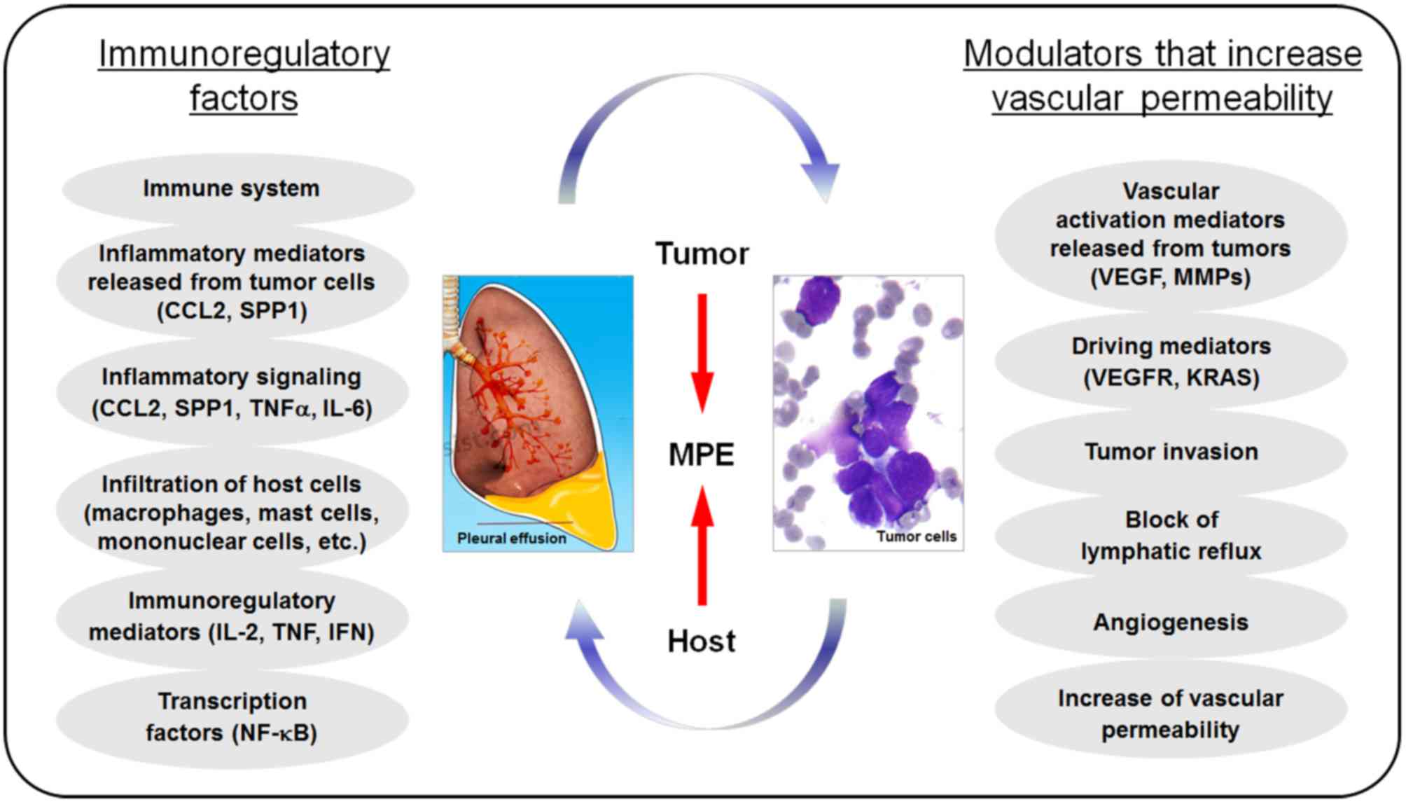 | Figure 1.Pathogenesis of MPE. Numerous
effector molecules, from either the host cells or tumor cells, are
involved in the pathogenesis of MPE. These effectors can generally
be classified into two categories. The first group of these
effector molecules is important immunoregulatory factors, including
IL-2, TNF and IFNs. The second group of effector molecules is
effective modulators that increase vascular permeability, including
VEGFs, MMPs and numerous others. CCL, C-C motif chemokine ligand;
IFNs, interferons; IL, interleukin; KRAS, GTPase KRas; MMPs, matrix
metalloproteinases; MPE, malignant pleural effusion; NF, nuclear
factor; SPP, S1P phosphatase; TNF, tumor necrosis factor; VEGFs,
vascular endothelial growth factors; VEGFR, VEGF receptor. |
VEGF is a highly conserved homodimeric glycoprotein
with a molecular weight ranging between 35 and 44 kDa. It has a
broad range of biological functions, including stimulation of
vascular proliferation, cellular differentiation, migration,
survival and germ tube formation, and regulation of vascular
permeability and angiogenesis (5,9,31).
VEGF has numerous isoforms, including VEGFA, -B, -C and -D, and
placental growth factor in humans, which can specifically bind to
one or numerous types of the three VEGFRs (VEGFR1, −2 and −3)
(31,32). Upon activation, the VEGFR undergoes
autophosphorylation and subsequently activates cell type-dependent
signaling cascades, including the phosphoinositide phospholipase C,
mitogen-activated protein kinases (MAPKs), nitric oxide synthases
and phosphoinositide 3-kinase, in addition to signal transducer and
activator of transcription (STAT)3 and STAT5. Activation of
distinct intracellular signaling pathways results in various
outcomes associated with the regulation of vascular permeability
depending on cell type or state, including induction of
inflammatory responses and loss of intracellular integrity and gap
formation (31). VEGF-VEGFR
interactions can also activate downstream MAPK1 signal cascades to
regulate endothelial cell proliferation and migration, and
consequently promote tumor angiogenesis and metastatic progression
(9,31). Several splicing variants of VEGF
have been reported; for example, five VEGF forms of 121–206 amino
acids are produced from a single gene by alternative splicing, each
with different biological effects to promote neovascularization
through distinct mechanisms (33).
Therefore, VEGF may promote the occurrence and development of MPE
in patients with NSCLC through two integrated mechanisms: By
increasing vascular permeability (a direct effect) and by promoting
angiogenesis (an indirect effect; Fig.
2).
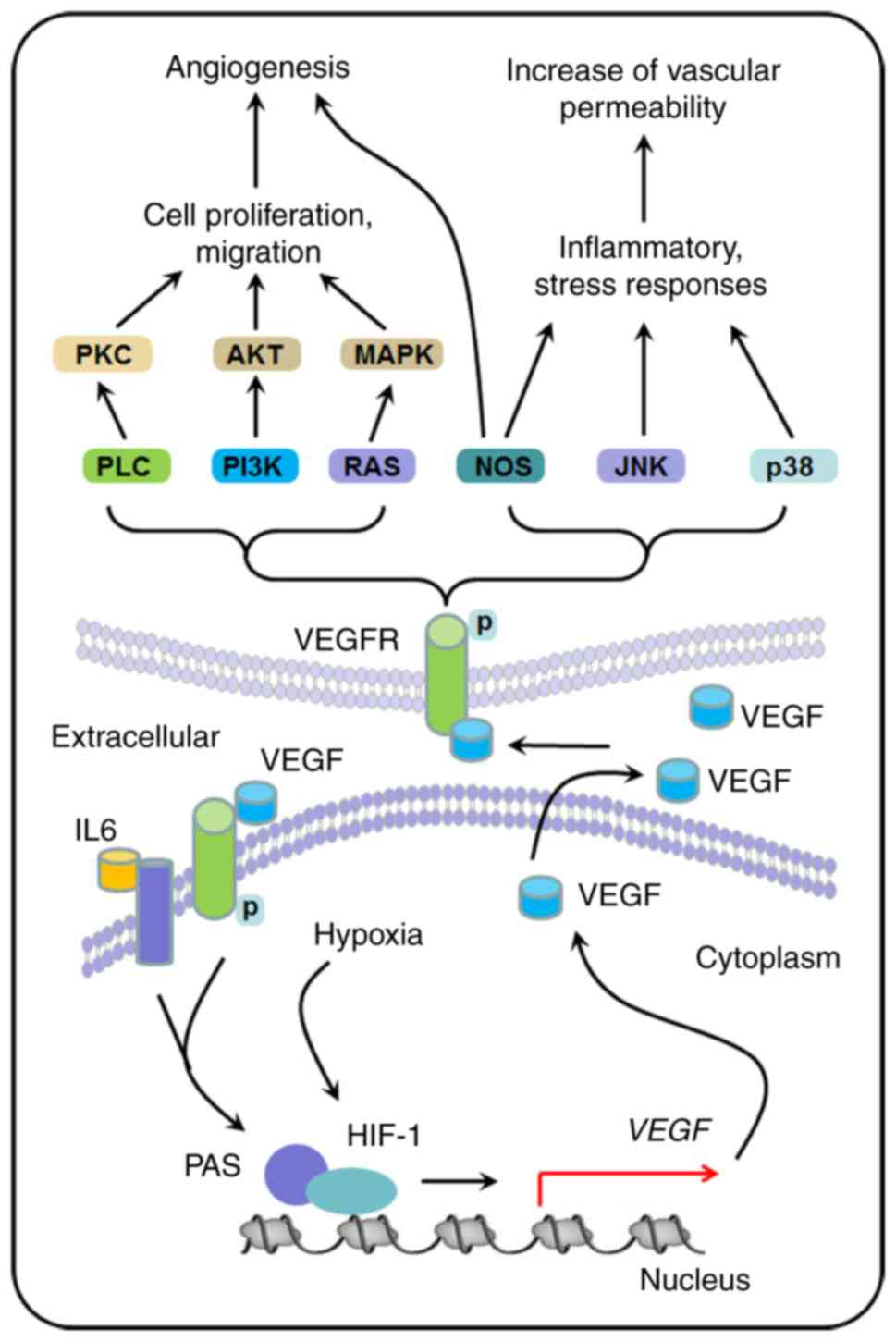 | Figure 2.VEGF signaling pathways and their
role in the pathogenesis of MPE. VEGF increases vascular
permeability and promotes tumor angiogenesis by binding to one of
its three receptors VEGFR1, −2 and −3 on vascular endothelial
cells. Upon activation, the VEGFR undergoes phosphorylation and
subsequently activates cell type-dependent signaling cascades,
including PLC, PI3K, NOS and MAPKs. Many factors of the local tumor
environment may contribute to the induction of VEGF expression in
tumor cells, including the occurrence of hypoxia and presence of
various growth factors (e.g., VEGF and IL-6). Hypoxia can activate
hypoxia-associated transcription factors, including HIF-1,
resulting in transcription of the VEGF gene. Through its role in
regulation of vascular permeability and angiogenesis, VEGF serves a
central role in the accumulation of pleural effusion in tumor
patients. HIF-1, hypoxia-inducible factor-1; IL, interleukin; JNK,
c-jun NH2-terminal kinase; MAPKs, mitogen-activated protein
kinases; MPE, malignant pleural effusion; NOS, nitric oxide
synthases; PAS, pathway activation signature; PI3K,
phosphoinositide 3-kinase; PKC, protein kinase C; PLC,
phosphoinositide phospholipase C; VEGF, vascular endothelial growth
factor; VEGFR, VEGF receptor. |
Elevated VEGF expression has been well demonstrated
in various tumor cells in humans, including pancreatic, stomach,
colorectal, breast and prostate cancers, melanoma, and cancers of a
number of other tissues (34).
Patients with various types of lung cancer, particularly those with
NSCLC, usually exhibit elevated VEGF expression levels in cancer
cells (17–19). Numerous factors in the local tumor
environment may contribute to the induction of VEGF expression in
tumor cells, including the occurrence of hypoxia and the presence
of various growth factors (including epidermal growth factor,
transforming growth factor and insulin-like growth factor) and
hormones (35,36) (Fig.
2). Among these factors, hypoxia can activate
hypoxia-associated transcription factors and trigger the
transcription of hypoxia-inducible genes (37). One hypoxia-inducible gene is
hypoxia-inducible factor-1 (HIF-1); transcription of the
HIF-1 gene can be induced by hypoxia via transcriptional
activation of the mechanistic target of rapamycin (33). Binding of HIF-1 to the regulatory
promoter region of the VEGF gene can enhance transcription
of the VEGF gene locus (38). Elevated levels of VEGF in the blood
and in malignant tissues of patients with solid tumors are often
associated with the expression levels of HIF-1 (33). Notably, increased levels of VEGF in
MPE may also be caused by interactions between VEGF and VEGFRs,
which further stimulate tumor cells and mesothelial cells alike to
secrete VEGF (12). IL-6 can
trigger transcription of the VEGF gene, whereas IL-6
itself appears to be a VEGF-targeted gene in numerous cancer cells
(39,40). This autocrine signaling-mediated
IL-6 production can further promote the expression of VEGF in
cancer, particularly in NSCLC cells (41) (Fig.
2).
VEGF-targeted strategies for the management
of MPE in patients with NSCLC
Current MPE management
Effective management of MPE remains a clinical
challenge and current methods for the treatment of MPE in patients
with NSCLC include diuretics, limited intake of salt, thoracic
puncture, long-term catheter drainage and intrathoracic
administration of chemotherapy drugs or biological agents (3,4).
There remains a lack of consensus on which approach is most
effective and what the dose, interval and course should be for the
administration of the drugs (3,4). In
addition, identification of the potential long-term effects and the
best combination of drugs requires large samples of patients and
randomized controlled clinical studies (3,4).
With an improved understanding of the pathogenesis of MPE,
particularly the appreciation of a role for angiogenesis in tumor
metastatic progression due to the development of vascular-dependent
tumor growth theory since 1971, significant advances in the
clinical management of MPE in patients with NSCLC have been made in
recent years (3,4,7).
Novel drugs targeting these effectors and signaling pathways for
angiogenesis, coupled with the development of more effective
anti-cell proliferation drugs, have provided new strategies for the
clinical management of MPE in patients with NSCLC (7). Of these novel drugs, recombinant
human endostatin (endostar) and the monoclonal VEGF antibody
bevacizumab have demonstrated promising therapeutic benefits for
patients with NSCLC and MPE (42).
Endostar to target vascular
endothelial cells
In 1997, an endogenous glycoprotein with 184 amino
acids (molecular mass, ~20 kDa) was isolated from mouse endosomes
and was named endostatin by O'Reilly et al (43). Endostatin has been demonstrated to
possess strong antivascular activity, with limited side effects,
and was able to almost completely inhibit tumor-induced
angiogenesis in murine models (44). Subsequently, a recombinant human
endostatin with an additional 9 amino acid sequence (MGGSHHHHH)
added to the N-terminal of the protein was engineered; the product
of this modification was named endostar (45). Such modification has been reported
to significantly enhance the purification, solubility and stability
of the protein (45). Endostar
also has a longer half-life than endostatin, with much improved
medicinal properties and efficacy, resulting in a significant
improvement of its biological function (45). It is now a common angiogenesis
antagonist used to treat lung cancer; in particular, it is used for
the management of relapse and metastasis in patients with NSCLC
(46), and has been widely used in
clinical practice to treat other tumors, including squamous cell
carcinoma (47).
Antiangiogenic mechanisms of
endostatin and endostar
The process of tumor angiogenesis is finely
regulated by a complex interaction between proangiogenic growth and
antiangiogenic factors (5,11,12).
Given the wide variety of antitumor growth effects of endostatin,
the following mechanistic themes have been speculated for the
antiangiogenic effects of endostatin/endostar. Firstly,
endostatin/endostar has been demonstrated to act on the endothelial
cells of newly formed blood vessels, inhibit endothelial cell
migration and induce endothelial cell apoptosis, thus resulting in
limited tumor growth (47).
Secondly, endostatin/endostar can downregulate the expression of
Neuropilin-1 and VEGFA, which are two potent proangiogenic growth
factors in numerous types of tumor due to their actions in
triggering endothelial cell proliferation (48). Thirdly, endostatin/endostar can
interact with the heparin sulfate proteoglycan receptors and block
the receptor binding of proangiogenic growth factors, including
VEGF and β fibroblast growth factor, thus resulting in inhibition
of endothelial cell proliferation and tumor angiogenesis (49). Fourthly, endostatin/endostar has
been reported to specifically recognize and bind to integrin α5β1
and αVβ3 on the endothelial cell surface (50). Such interactions may prevent the
adhesion of endothelial cells in extracellular matrix-mediated
migration, induce tyrosine phosphorylation of adhesion focal kinase
and paxillin to promote elastic fiber formation, and consequently
inhibit tumor cell adhesion and metastatic progression (50). Finally, endostatin/endostar can
inhibit the transcription of MMP2 and MMP9 genes
(51). In addition, recent studies
using murine models have revealed that overexpression of endostatin
may result in downregulation of the VEGFC gene, and
inhibition of lymphangiogenesis and tumor metastasis to nearby
lymph nodes (52). Therefore,
recombinant human endostatin/endostar is a potent antiangiogenic
drug with a wide range of potential research prospects.
Endostatin/endostar in the clinical
management of MPE in patients with NSCLC
Endostar has been used in clinical practice for
numerous years for the treatment of advanced NSCLC in combination
with vinorelbine and cisplatin (53,54).
Emerging evidence suggests that endostar in combination with
chemotherapy (vinorelbine plus cisplatin) can prolong the time to
progression (TTP), and improve the overall response rate (RR) and
clinical benefit rate (CBR) with a favorable toxic profile in
patients with advanced NSCLC (55). One phase III clinical trial was
carried out between April 2003 and July 2004 to investigate the
therapeutic effects of endostar in combination with chemotherapy
(vinorelbine plus cisplatin) on advanced NSCLC; a total of 486
patients were recruited for the study from 24 medical centers
across the country (55). The
results demonstrated that the RRs of the experimental group and the
control group (NP) were 35.4 and 19.5% (P<0.001), the CBRs were
73.3 and 64.0% (P<0.05), and the median TTPs were 6.3 and 3.6
months (P<0.001), respectively. For patients with relapsed
NSCLC, the RRs of the endostar in combination with chemotherapy
group and the control group were 23.9 and 8.5% (P<0.01), the
CBRs were 65.2 and 61.7% (P=0.68), and the median TTPs were 5.7 and
3.2 months (P<0.001), respectively. No significant difference
was identified in the incidence of moderate to severe adverse
reactions in patients between the endostar in combination with
chemotherapy group and the control group (55).
Endostar has been used via intracavity injection for
the treatment of patients with malignant serous effusions,
including MPE and malignant peritoneal effusion. Evidence indicates
that endostar alone or in combination with chemotherapy drugs is
safe and effective for the treatment of malignant serous effusions
in patients with cancer (56–69).
Only a few of these clinical studies will be discussed in the
present study and others are summarized in Table II. Results from a recent
randomized controlled clinical study (56), with participation from numerous
institutions, provided strong evidence that endostar is effective
for the treatment of MPE. In addition, Qin et al (56) compared the efficacy of endostar on
MPE between patients treated with endostar or cisplatin alone, or
with the combination of the two drugs. The analysis revealed that
either endostar alone or in combination with cisplatin is effective
and safe for the treatment of patients with malignant cavity
effusion, with objective response rates (ORRs) of 51, 49 and 36%,
for the combination-treated group, endostar-treated group and
cisplatin group, respectively. Specifically, for patients with MPE,
the ORRs were 62, 58 and 38% for the combination-treated group,
endostar-treated group and cisplatin group, respectively. For
patients with ascites, the ORRs were 39, 42 and 32%, respectively
(56). These effects of endostar
on MPE are supported by results of a similar clinical report by Hu
et al (57). Similarly,
63.6% of patients with MPE demonstrated improvement following
treatment with endostar in combination with cisplatin, which was
significantly better than cisplatin treatment alone (40.6%;
P=0.022). The average progression-free survival (PFS) time for
patients with MPE who received treatment with endostar in
combination with cisplatin was 95 days, which was significantly
longer than that of patients treated with cisplatin alone (PFS, 53
days; P=0.039) (57). Endostar has
also been demonstrated to inhibit ascites formation and prolong
survival in mouse models of malignant ascites established using
S180 and H22 tumor cells (58).
The tumor cells collected from the ascites in endostar-treated mice
demonstrated a decrease in the expression of VEGF mRNA (58). In addition, treatment of S180 and
H22 tumor cells with endostar revealed a significant inhibition of
VEGF protein secretion and VEGF mRNA expression, but no
effect on cellular proliferation (58). The inhibitory effects of
recombinant human endostatin/endostar on tumor growth have also
been reported in other cancer types, including ovarian cancer,
malignant melanoma and colon cancer, and in liver
transplantation-associated angiogenesis (59).
| Table II.Endostatin/endostar in the clinical
management of MPE in patients with NSCLC. |
Table II.
Endostatin/endostar in the clinical
management of MPE in patients with NSCLC.
| Author (year) | Groups | Cases | Efficacy (%) | Clinical
outcome | (Refs.) |
|---|
| Qin (2016) | I: Cisplatin (50
mg/m2) | 42 | I: 47.62 | Improved quality of
life: Group I, 47.62%; Group II, 42.86% | (62) |
|
| II: Cisplatin (50
mg/m2) + Endostar (60 mg) |
| II: 76.19 |
|
|
| Xu et al
(2014) | I: Nedaplatin (60
mg) + Endostar (60 mg) | 70 | I: 74.28 | Quality of life
improved significantly, no significant difference in adverse
reactions | (63) |
|
| II: Nedaplatin (60
mg) |
| II: 48.57 |
|
|
| Huang (2014) | I: Cisplatin (50
mg/m2) | 50 | I: 48 | Quality of life
improved significantly, no significant difference in adverse
reactions | (64) |
|
| II: Cisplatin (50
mg/m2) + Endostar (30 mg) |
| II: 78 |
|
|
| Yang et al
(2013) | I: Cisplatin (40
mg/m2) | 42 | I: 42.86 | Quality of life
improved significantly, no significant difference in adverse
reactions | (65) |
|
| II: Cisplatin (40
mg/m2) + Endostar (30 mg) |
| II: 80.95 |
|
|
| Liu et al
(2010) | I: Cisplatin (40
mg/m2) | 96 | I: 43.75 | Improved quality of
life: Group I, 40.63%; Group II, 36.38%; Group III, 56.25% | (66) |
|
| II: Endostar (60
mg/m2) |
| II: 40.63 |
|
|
|
| III:Cisplatin (40
mg/m2) + Endostar (60 mg) |
| III: 78.13 |
|
|
| Huang (2010) | I: Cisplatin (60
mg/m2) | 36 | I: 43.75 | Quality of life
improved significantly, no significant difference in adverse
reactions | (67) |
|
| II: Cisplatin (60
mg/m2) + Endostar (45 mg) |
| II: 77.8 |
|
|
| Li (2014) | I: Cisplatin (60
mg/m2) | 42 | I: 42.86 | Improved quality of
life: Group I, 52.38%; Group II, 76.19% | (68) |
|
| II: Cisplatin (60
mg/m2) + Endostar (45 mg) |
| II: 80.95 |
|
|
| Tu (2014) | I: Cisplatin (40
mg/m2) | 90 | I: 51.11 | Quality of life
improved significantly, no significant difference in adverse
reactions | (69) |
|
| II: Cisplatin (40
mg/m2) + Endostar (45 mg) |
| II: 82.22 |
|
|
Despite the general inhibitory effects of
endostatin/endostar on tumor progression, and on MPE in patients
with NSCLC, opinions on the best dosage and administration, and the
duration of treatment remain controversial. In most stage II–IV
clinical studies, endostar was administered at 30–60
mg/m2, intravenously for 3–4 h/day for 1–14 days.
Increasing evidence indicates that the antitumor effects of
endostar are time- and dose-dependent; prolongation of
administration time and a gradual increase in its blood level can
improve its antitumor effects (47). When it was administered
intraperitoneally (i.p.) in a single bolus dose to tumor-bearing
mice, endostatin was rapidly cleared in the tumor tissues within 2
h, whereas endostatin administered continuously via implanted
mini-osmotic pump maintained systemic concentrations of 200–300
ng/ml for the duration of administration (42). In addition, continuous i.p.
administration of endostatin resulted in more effective tumor
suppression at significantly reduced doses (5-fold) compared with
bolus administration (42).
Results of clinical studies have also demonstrated that the
antitumor effects of continuous i.p. administration of
endostatin/endostar are better compared with the same dose in
short-term intravenous administration (60,61).
Continuous administration via implanted osmotic pump may be able to
maintain a stable plasma concentration of endostatin/endostar, so
that it can persistently act on newly-formed vascular endothelial
cells, resulting in a sustained and constant treatment effect
(60). Therefore, continuous
administration via an implanted mini-osmotic pump provides a novel
method to further improve the therapeutic effects of
endostatin/endostar on MPE in patients with NSCLC.
Bevacizumab to target VEGFA
The identification and isolation of VEGFA in 1989
provided a novel avenue for the development of antiangiogenic
strategies (9). Consequently, a
recombinant humanized monoclonal anti-VEGFA antibody, termed
bevacizumab, was generated to block angiogenesis by inhibiting
VEGFA. In 2006, the USA Food and Drug Administration approved
bevacizumab for use in first-line treatment for advanced
nonsquamous NSCLC in combination with carboplatin/paclitaxel
chemotherapy. Results of the BEYOND study led by Qingcun Zhou at
Tongji University suggested that bevacizumab is safe and effective
for the treatment of patients with advanced or recurrent
nonsquamous NSCLC in China, including patients with the epidermal
growth factor receptor mutation (70). Bevacizumab was then approved by the
Chinese Food and Drug Administration for NSCLC in China on July 1,
2015, providing an additional choice for the treatment of MPE in
patients with NSCLC.
The antiangiogenic mechanism of
bevacizumab
Acting by promoting the formation of new blood
vessels and increasing vascular permeability, VEGFA is one of the
most important key mediators for the development of MPE, including
in patients with NSCLC (12).
VEGFA has been demonstrated to induce inflammatory responses and
disrupt cell-cell connections to increase vascular permeability
and, consequently, promote tumor cell migration. VEGFR1 and VEGFR2,
two receptor tyrosine kinases, are the receptors for VEGFA on
endothelial cells (71). VEGFR2
may be more important than VEGFR1 for VEGFA-mediated endothelial
cell proliferation, angiogenesis and vascular permeability
(71). Upon ligation by VEGFA,
VEGFR2 can be activated through receptor dimerization and
autophosphorylation, thus resulting in activation of various
downstream signal cascades (71).
Bevacizumab can block the binding of VEGFA to its receptors and
thus inhibit activation of the downstream signaling pathways
(72,73).
Bevacizumab in the clinical management
of MPE in patients with NSCLC
Preclinical evidence suggests that bevacizumab can
reduce vascular permeability and decrease the formation of pleural
effusion (12). The efficacy of
bevacizumab in combination with paclitaxel/carboplatin in the
treatment of advanced nonsquamous NSCLC with MPE without
chemotherapy was studied in Japan, in a multi-center, clinical
phase II prospective study (74).
After 2–6 cycles of treatment with bevacizumab in combination with
paclitaxel/carboplatin, it was demonstrated that patients exhibited
an ORR of 60.8% and a disease control rate of 87.0%; the disease
control rate of MPE was higher compared with in patients who
received paclitaxel/carboplatin chemotherapy alone (with a disease
control rate of 78.3%) (74). Data
from two retrospective studies also confirmed that bevacizumab
combined with chemotherapy drugs through intravenous injection can
effectively control MPE in patients with nonsquamous NSCLC, with a
MPE control rate of 92.3% and a MPE release rate of ≤71.4%
(4). The efficacy of bevacizumab
combined with platinum through local pleural administration in the
treatment of MPE was investigated by Hsu et al (75). The results indicated that local
pleural administration of bevacizumab plus cisplatin, alongside the
systemic administration of paclitaxel, resulted in a much higher
ORR (83.3 vs. 50.0%; P<0.05), compared with that in patients who
received systemic administration of paclitaxel plus local cisplatin
only (75). Patients in the first
group demonstrated a significant reduction in the amount of pleural
effusion, accompanied with a markedly improved quality of life, and
tolerated the treatment well (75). Substantial additional clinical
studies have all reported that bevacizumab is safe and effective
for the treatment of MPE in patients with NSCLC (Table III) (76–82).
Therefore, local administration of bevacizumab in the pleural
cavity plus systemic administration of chemotherapy drugs may
effectively control MPE in patients with advanced nonsquamous
NSCLC.
| Table III.Bevacizumab in the clinical
management of malignant pleural effusion in patients with non-small
cell lung cancer. |
Table III.
Bevacizumab in the clinical
management of malignant pleural effusion in patients with non-small
cell lung cancer.
| Author (year) | Groups | Cases | Efficacy (%) | Clinical
outcome | (Refs.) |
|---|
| Lin et al
(2010) | I: Cisplatin +
Bevacizumab | 94 | I: 70.2 | Significant
improvement of quality of life, with very mild side effects | (76) |
|
| II: Cisplatin |
| II: 44.7 |
|
|
| Chi et al
(2016) | I: Pemetrexed +
Carboplatin | 46 | I: 65.21 | Significant
improvement of quality of life, with very mild side effects | (77) |
|
| II: Pemetrexed +
Carboplatin + Bevacizumab |
| II: 86.96 |
|
|
| Chen and Xia
(2015) | I: Cisplatin +
Bevacizumab | 54 | I: 85.7 | Significant
improvement of quality of life, with very mild side effects | (78) |
|
| II: Cisplatin |
| II: 69.2 |
|
|
| Qu et al
(2015) | I: Cisplatin +
Bevacizumab | 63 | I: 84.3 | Significant
improvement of quality of life, with very mild side effects | (79) |
|
| II: Cisplatin |
| II: 61.3 |
|
|
| Huang (2016) | I: Cisplatin +
Bevacizumab | 73 | I: 81.08 | Significant
improvement of quality of life, with very mild side effects | (80) |
|
| II: Cisplatin |
| II: 58.33 |
|
|
| Han et al
(2013) | I: Pemetrexed +
Carboplatin + Bevacizumab | 42 | I: 85.0 | Significant
improvement of quality of life, with very mild side effects | (81) |
|
| II: Pemetrexed +
Carboplatin |
| II: 68.2 |
|
|
| Liu et al
(2016) | I: Bevacizumab (d1,
pleural cavity) + | 84 | I: 83.3 | Significant
improvement of quality of life, with very mild side effects | (82) |
|
| Cisplatin (d1,
d3)/Pemetrexed (d1) |
|
|
|
|
|
| II: Cisplatin (d1,
d3)/Pemetrexed (d1) |
| II: 64.29 |
|
|
VEGF in the prognosis of MPE in various
subtypes of NSCLC and in patients with advanced NSCLC
VEGF can promote the occurrence and development of
MPE in patients with NSCLC directly (via increasing vascular
permeability) and indirectly (via promoting angiogenesis and tumor
migration). Accordingly, the therapeutic efficiency of
VEGF-targeted strategies for the management of MPE in patients with
NSCLC depends on their inhibitory effects on vascular permeability
and tumor angiogenesis. The most common types of NSCLC are squamous
cell carcinoma, large cell carcinoma and adenocarcinoma, but there
are several other types that occur less frequently. All types can
occur in unusual histological variants and as mixed cell-type
combinations and thus vary in metastatic features and VEGF-VEGFR
functional signatures (47).
Therefore, various types of NSCLC may differ in their response to
VEGF-targeted strategies for the management of associated MPE.
Nevertheless, although detailed information is limited in the
current literature and more comprehensive clinical studies are
required, it appears that endostar and bevacizumab are effective
for the treatment of MPE in most NSCLC types, including squamous
cell carcinoma and adenocarcinoma (83–85).
It is increasingly clear that levels of VEGF in
pleural effusion may be one of the critical indicators of the
prognosis of MPE in patients with advanced NSCLC, and anti-VEGF
therapy is of important therapeutic value (47). Firstly, VEGF is overexpressed in
the majority of patients with advanced NSCLC and MPE, and VEGF
levels in pleural effusion are increased and associated with the
prognosis of patients with advanced NSCLC and MPE (17–20).
A higher concentration of VEGF in pleural effusion implies a higher
risk of distant metastasis for patients with NSCLC (75). Specifically, it has been
demonstrated that the levels of VEGF and endostatin in pleural
effusion, together with the serum levels of endostatin, are
prognostic parameters for patients with advanced NSCLC and MPE
(17–20,86,87).
Secondly, anti-VEGF therapy is safe and effective for patients with
advanced NSCLC (47). In a number
of phase II trials in patients with advanced metastatic NSCLC, the
addition of bevacizumab to standard carboplatin/paclitaxel
chemotherapy significantly increased the TTP and increased the RR
when compared with chemotherapy alone. This was particularly
impressive in the subset of patients with non-squamous histology.
Bevacizumab is generally well tolerated and does not appear to
increase the incidence or severity of nausea/vomiting, neuropathy
and renal toxicity, which are typically associated with
carboplatin/paclitaxel chemotherapy (74,88–91).
Nevertheless, although bevacizumab improves outcomes when added to
platinum-based chemotherapy in advanced-stage non-squamous NSCLC, a
recent phase III trial study demonstrated that the addition of
bevacizumab to adjuvant chemotherapy did not improve overall
survival for patients with surgically resected early-stage NSCLC
(92). In the future, bevacizumab
may be used alongside novel molecular therapies or immuno-oncology
drugs, in order to optimize RRs and overcome resistance in patients
with advanced NSCLC (93).
Conclusion and perspectives
VEGF is of great significance to the diagnosis and
clinical treatment of MPE in patients with NSCLC. With the recent
development of molecular biological technology, great advances have
been made in the diagnosis of MPE, including biochemical analysis,
cytopathology and imaging examination of pleural effusion. Notably,
the pathogenesis of MPE involves numerous factors and complex
molecular mechanisms. With an improved understanding of the role
for VEGF in the development of MPE, particularly in patients with
NSCLC, targeting VEGF has provided a novel strategy for the
diagnosis and treatment of patients with MPE. Since approval of the
clinical use of endostar and bevacizumab, substantial clinical
studies have been conducted worldwide. Results from these studies
have provided strong evidence to suggest that endostar and
bevacizumab are safe and effective for the treatment of MPE,
particularly in patients with NSCLC. With treatment, patients with
NSCLC and MPE not only exhibited an improved quality of life but
also, to a certain extent, an improved survival rate. It has been
speculated that additional clinical studies, particularly
well-controlled ones with a larger number of patient cases
currently ongoing, may provide additional comprehensive insights
for the enhanced judgment of the efficacy of targeting VEGF in
patients with NSCLC and MPE.
Acknowledgements
The authors would like to thank Dr. Xian-Ming Chen
(Creighton University, Omaha, NE, USA) for helpful and stimulating
discussions.
Funding
No funding was received.
Availability of data and materials
All data generated or analyzed during this study are
included in this published article.
Authors' contributions
YC, NWM and HL wrote the paper. All authors read and
approved the final manuscript.
Ethics approval and consent to
participate
Not applicable.
Consent for publication
Not applicable.
Competing interests
The authors declare that they have no competing
interests.
Glossary
Abbreviations
Abbreviations:
|
CEA
|
carcinoembryonic antigen
|
|
HIF-1
|
hypoxia-inducible factor-1
|
|
IFN
|
interferon
|
|
IL
|
interleukin
|
|
MAPK
|
mitogen-activated protein kinase
|
|
MMP
|
matrix metalloproteinase
|
|
MPE
|
malignant pleural effusion
|
|
NSCLC
|
non-small cell lung cancer
|
|
ORR
|
objective response rate
|
|
PFS
|
progression-free survival
|
|
PI3K
|
phosphoinositide 3-kinase
|
|
PLC
|
phosphoinositide phospholipase C
|
|
RR
|
overall response rate
|
|
STAT
|
signal transducer and activator of
transcription
|
|
TNF
|
tumor necrosis factor
|
|
TTF-1
|
thyroid transfection factor-1
|
|
TTP
|
time to progression
|
|
VEGF
|
vascular endothelial growth factor
|
|
VEGFR
|
vascular endothelial growth factor
receptor
|
References
|
1
|
Wong MCS, Lao XQ, Ho KF, Goggins WB and
Tse SLA: Incidence and mortality of lung cancer: Global trends and
association with socioeconomic status. Sci Rep. 7:143002017.
View Article : Google Scholar : PubMed/NCBI
|
|
2
|
Chen W, Zheng R, Baade PD, Zhang S, Zeng
H, Bray F, Jemal A, Yu XQ and He J: Cancer Statistics in China,
2015. CA Cancer J Clin. 66:115–132. 2016. View Article : Google Scholar : PubMed/NCBI
|
|
3
|
Penz E, Watt KN, Hergott CA, Rahman NM and
Psallidas I: Management of malignant pleural effusion: Challenges
and solutions. Cancer Manag Res. 9:229–241. 2017. View Article : Google Scholar : PubMed/NCBI
|
|
4
|
Shi H: Guidelines to the diagnosis and
treatment of malignant pleural effusion. Chin J Intern Med.
53:166–167. 2014.
|
|
5
|
Zachary I: Signaling mechanisms mediating
vascular protective actions of vascular endothelial growth factor.
Am J Physiol Cell Physiol. 280:C1375–C1386. 2001. View Article : Google Scholar : PubMed/NCBI
|
|
6
|
Ide AG, Baker NH and Warren SL:
Vascularization of the brown pearce rabbit epithelioma transplant
as seen in the transparent ear chamber. Am J Roentgenol.
42:891–899. 1939.
|
|
7
|
Folkman J: Tumor angiogenesis: Therapeutic
implications. N Engl J Med. 285:1182–1186. 1971. View Article : Google Scholar : PubMed/NCBI
|
|
8
|
Senger DR, Galli SJ, Dvorak AM, Perruzzi
CA, Harvey VS and Dvorak HF: Tumor cells secrete a vascular
permeability factor that promotes accumulation of ascites fluid.
Science. 219:983–985. 1983. View Article : Google Scholar : PubMed/NCBI
|
|
9
|
Ferrara N and Henzel WJ: Pituitary
follicular cells secrete a novel heparin-binding growth factor
specific for vascular endothelial cells. Biochem Biophys Res
Commun. 161:851–858. 1989. View Article : Google Scholar : PubMed/NCBI
|
|
10
|
de Vries C, Escobedo JA, Ueno H, Houck K,
Ferrara N and Williams LT: The fms-like tyrosine kinase, a receptor
for vascular endothelial growth factor. Science. 255:989–991. 1992.
View Article : Google Scholar : PubMed/NCBI
|
|
11
|
Harmey JH and Bouchier-Hayes D: Vascular
endothelial growth factor (VEGF), a survival factor for tumor
cells: Implications for anti-angiogenic therapy. Bioessays.
24:280–283. 2002. View Article : Google Scholar : PubMed/NCBI
|
|
12
|
Bradshaw M, Mansfield A and Peikert T: The
role of vascular endothelial growth factor in the pathogenesis,
diagnosis and treatment of malignant pleural effusion. Curr Oncol
Rep. 15:207–216. 2013. View Article : Google Scholar : PubMed/NCBI
|
|
13
|
Zang J, Hu Y, Xu X, Ni J, Yan D, Liu S, He
J, Xue J, Wu J and Feng J: Elevated serum levels of vascular
endothelial growth factor predict a poor prognosis of
platinum-based chemotherapy in non-small cell lung cancer. Onco
Targets Ther. 10:409–415. 2017. View Article : Google Scholar : PubMed/NCBI
|
|
14
|
Popper HH: Progression and metastasis of
lung cancer. Cancer Metastasis Rev. 35:75–91. 2016. View Article : Google Scholar : PubMed/NCBI
|
|
15
|
Hu CP: Interpretation of expert consensus
2014 on diagnosis and treatment of malignant pleural effusion. J
Transl Int Med. 3:1–2. 2015. View Article : Google Scholar : PubMed/NCBI
|
|
16
|
Dvorak HF: Vascular permeability
factor/vascular endothelial growth factor: A critical cytokine in
tumor angiogenesis and a potential target for diagnosis and
therapy. J Clin Oncol. 20:4368–4380. 2002. View Article : Google Scholar : PubMed/NCBI
|
|
17
|
Fafliora E, Hatzoglou C, Gourgoulianis KI
and Zarogiannis SG: Systematic review and meta-analysis of vascular
endothelial growth factor as a biomarker for malignant pleural
effusions. Physiol Rep. 4:e129782016. View Article : Google Scholar : PubMed/NCBI
|
|
18
|
Sriram KB, Relan V, Clarke BE, Duhig EE,
Yang IA, Bowman RV, Lee YC and Fong KM: Diagnostic molecular
biomarkers for malignant pleural effusions. Future Oncol.
7:737–752. 2011. View Article : Google Scholar : PubMed/NCBI
|
|
19
|
Gu Y, Zhang M, Li GH, Gao JZ, Guo L, Qiao
XJ, Wang LH, He L, Wang ML, Yan L and Fu XH: Diagnostic values of
vascular endothelial growth factor and epidermal growth factor
receptor for benign and malignant hydrothorax. Chin Med J (Engl).
128:305–309. 2015. View Article : Google Scholar : PubMed/NCBI
|
|
20
|
Zhou WB, Bai M and Jin Y: Diagnostic value
of vascular endothelial growth factor and endostatin in malignant
pleural effusions. Int J Tunerc Lung Dis. 13:381–386. 2009.
|
|
21
|
Nam HS: Malignant pleural effusion:
Medical approaches for diagnosis and management. Tuberc Respir Dis
(Seoul). 76:211–217. 2014. View Article : Google Scholar : PubMed/NCBI
|
|
22
|
Chen Y, Liang B, Zhao YJ, Wang SC, Fan YB
and Wu GP: Transcription expression and clinical significance of
vascular endothelial growth factor mRNA and endostatin mRNA in
pleural effusions of patients with lung cancer. Diagn Cytopathol.
40:287–291. 2012. View Article : Google Scholar : PubMed/NCBI
|
|
23
|
Jiang B, Wu GP, Zhao YJ and Wang SC:
Transcription expression and clinical significance of TTF-1 mRNA in
pleural effusion of patients with lung cancer. Diagn Cytopathol.
36:849–854. 2008. View Article : Google Scholar : PubMed/NCBI
|
|
24
|
Palaoro LA, Blanco AM, Gamboni M, Rocher
AE and Rotenberg RG: Usefulness of ploidy, AgNor and
immunocytochemistry for differentiating benign and malignant cells
in serous effusions. Cytopathology. 18:33–39. 2007. View Article : Google Scholar : PubMed/NCBI
|
|
25
|
Light RW: Pleural effusions. Med Clin
North Am. 95:1055–1070. 2011. View Article : Google Scholar : PubMed/NCBI
|
|
26
|
Liu QF: Clinical value of VEGF combined
with CEA in the treatment of malignant pleural effusion of advanced
NSCLC. Practical Oncol J (China). 27:156–159. 2013.
|
|
27
|
Stathopoulos GT and Kalomenidis I:
Malignant pleural effusion: Tumor-host interactions unleashed. Am J
Respir Crit Care Med. 186:487–492. 2012. View Article : Google Scholar : PubMed/NCBI
|
|
28
|
Hamed EA, El-Noweihi AM, Mohamed AZ and
Mahmoud A: Vasoactive mediators (VEGF and TNF-alpha) in patients
with malignant and tuberculous pleural effusions. Respirology.
9:81–86. 2004. View Article : Google Scholar : PubMed/NCBI
|
|
29
|
Gerber HP and Ferrara N: Pharmacology and
pharmacodynamics of bevacizumab as monotherapy or in combination
with cytotoxic therapy in preclinical studies. Cancer Res.
65:671–680. 2005.PubMed/NCBI
|
|
30
|
Numnum TM, Rocconi RP, Whitworth J and
Barnes MN: The use of bevacizumab to palliate symptomatic ascites
in patients with refractory ovarian carcinoma. Gynecol Oncol.
102:425–428. 2006. View Article : Google Scholar : PubMed/NCBI
|
|
31
|
Ferrara N, Gerber HP and LeCouter J: The
biology of VEGF and its receptors. Nat Med. 9:669–676. 2003.
View Article : Google Scholar : PubMed/NCBI
|
|
32
|
Hicklin DJ and Ellis LM: Role of the
vascular endothelial growth factor pathway in tumor growth and
angiogenesis. J Clin Oncol. 23:1011–1027. 2005. View Article : Google Scholar : PubMed/NCBI
|
|
33
|
Pritchard-Jones RO, Dunn DB, Qiu Y, Varey
AH, Orlando A, Rigby H, Harper SJ and Bates DO: Expression of VEGF
(XXX)b, the inhibitory isoforms of VEGF, in malignant melanoma. Br
J Cancer. 97:223–230. 2007. View Article : Google Scholar : PubMed/NCBI
|
|
34
|
Fontanini G, Lucchi M, Vignati S, Mussi A,
Ciardiello F, De Laurentiis M, De Placido S, Basolo F, Angeletti CA
and Bevilacqua G: Angiogenesis as a prognostic indicator of
survival in nonsmall-cell lung carcinoma: A prospective study. J
Natl Cancer Inst. 89:881–886. 1997. View Article : Google Scholar : PubMed/NCBI
|
|
35
|
Ferrara N: Vascular endothelial growth
factor: Basic science and clinical progress. Endocr Rev.
25:581–611. 2004. View Article : Google Scholar : PubMed/NCBI
|
|
36
|
Voelkel NF, Vandivier RW and Tuder RM:
Vascular endothelial growth factor in the lung. Am J Physiol Lung
Cell Moistiol. 290:L209–L221. 2006. View Article : Google Scholar
|
|
37
|
Semenza GL: Targeting HIF-1 for cancer
therapy. Nat Rev Cancer. 3:721–732. 2003. View Article : Google Scholar : PubMed/NCBI
|
|
38
|
Mayerhofer M, Valent P, Sperr WR, Griffin
JD and Sillaber C: BCR/ABL induces expression of vascular
endothelial growth factor and its transcriptional activator,
hypoxia induciblefactor-1alpha, through a pathway involving
phosphoinositide 3-kinase and the mammalian target of rapamycin.
Blood. 100:3767–3775. 2002. View Article : Google Scholar : PubMed/NCBI
|
|
39
|
Loeffler S, Fayard B, Weis J and
Weissenberger J: Interleukin-6 induces transcriptional activation
of vascular endothelial growth factor (VEGF) in astrocytes in vivo
and regulates VEGF promoter activity in glioblastoma cells via
direct interaction between STAT3 and Sp1. Int J Cancer.
115:202–213. 2005. View Article : Google Scholar : PubMed/NCBI
|
|
40
|
Cohen T, Nahari D, Cerem LW, Neufeld G and
Levi BZ: Interleukin 6 induces the expression of vascular
endothelial growth factor. J Biol Chem. 271:736–741. 1996.
View Article : Google Scholar : PubMed/NCBI
|
|
41
|
Wójcik E, Jakubowicz J, Skotnicki P,
Sas-Korzyńska B and Kulpa JK: IL-6 and VEGF in small cell lung
cancer patients. Anticancer Res. 30:1773–1778. 2010.PubMed/NCBI
|
|
42
|
Yan J, Jiang Y and Xu H: Endu treatment of
malignant pleural effusion. Practical J Cancer. 27:538–539.
2012.
|
|
43
|
O'Reilly MS, Boehm T, Shing Y, Fukai N,
Vasios G, Lane WS, Flynn E, Birkhead JR, Olsen BR and Folkman J:
Endostatin: An endogenous inhibitor of angiogenesis and tumor
growth. Cell. 88:277–285. 1997. View Article : Google Scholar : PubMed/NCBI
|
|
44
|
Hayes AJ, Li LY and Lippman ME: Science,
medicine, and the future. Antivascular therapy: A new approach to
cancer treatment. BMJ. 318:853–856. 1999. View Article : Google Scholar : PubMed/NCBI
|
|
45
|
Xu X, Mao W, Chen Q, Zhuang Q, Wang L, Dai
J, Wang H and Huang Z: Endostar, a modified recombinant human
endostatin, suppresses angiogenesis through inhibition of
Wnt/β-catenin signaling pathway. PLoS One. 9:e1074632014.
View Article : Google Scholar : PubMed/NCBI
|
|
46
|
Rong B, Yang S, Li W, Zhang W and Ming Z:
Systematic review and meta-analysis of Endostar (rh-endostatin)
combined with chemotherapy versus chemotherapy alone for treating
advanced non-small cell lung cancer. World J Surg Oncol.
10:1702012. View Article : Google Scholar : PubMed/NCBI
|
|
47
|
Mohajeri A, Sanaei S, Kiafar F, Fattahi A,
Khalili M and Zarghami N: The challenges of recombinant endostatin
in clinical application: Focus on the different expression systems
and molecular bioengineering. Adv Pharm Bull. 7:21–34. 2017.
View Article : Google Scholar : PubMed/NCBI
|
|
48
|
Shichiri M and Hirata Y: Antiangiogenesis
signals by endostatin. FASEB J. 15:1044–1053. 2001. View Article : Google Scholar : PubMed/NCBI
|
|
49
|
Hajitou A, Grignet C, Devy L, Berndt S,
Blacher S, Deroanne CF, Bajou K, Fong T, Chiang Y, Foidart JM and
Noël A: The antitumoral effect of endostatin and angiostatin is
associated with a down-regulation of vascular endothelial growth
factor expression in tumor cells. FASEB J. 16:1802–1804. 2002.
View Article : Google Scholar : PubMed/NCBI
|
|
50
|
Li DH, Liu QG, Wang HP, Yu M, Jiang J,
Tang CH and Ning YL: Study on recombinant human endostatin in the
treatment of malignant pleural effusion in the elderly. Shaanxi Med
J. 44:1138–1139. 2015.(In Chinese).
|
|
51
|
Murakami A, Tabata C, Tabata R, Okuwa H
and Nakano T: Clinical role of pleural effusion MMP-3 levels in
malignant pleural mesothelioma. Oncol Lett. 3:581–585. 2012.
View Article : Google Scholar : PubMed/NCBI
|
|
52
|
Ma X, Yao Y, Yuan D, Liu H, Wang S, Zhou C
and Song Y: Endostar suppresses angiogenesis and lymphangiogenesis
of malignant pleural effusion in mice. PLoS One. 7:e534492012.
View Article : Google Scholar : PubMed/NCBI
|
|
53
|
Ishii H, Yazawa T, Sato H, Suzuki T, Ikeda
M, Hayashi Y, Takanashi Y and Kitamura H: Enhancement of pleural
treatment and lymph node metastasis of intrathoracic lung cancer
cells by vascular endothelial growth factors (VEGFs). Lung Cancer.
45:325–337. 2004. View Article : Google Scholar : PubMed/NCBI
|
|
54
|
Prager GW, Lackner EM, Krauth MT, Unseld
M, Poettler M, Laffer S, Cerny-Reiterer S, Lamm W, Kornek GV, et
al: Targeting of VEGF dependent transendothelial migration of
cancer cells by bevacizumab. Mol Oncol. 4:150–160. 2010. View Article : Google Scholar : PubMed/NCBI
|
|
55
|
Wang J, Sun Y, Liu Y, Yu Q, Zhang Y, Li K,
Zhu Y, Zhou Q, Hou M, Guan Z, et al: Results of randomized,
multicenter, double-blind phase III trial of rh-endostatin (YH-16)
in treatment of advanced non-small cell lung cancer patients.
Zhongguo Fei Ai Za Zhi. 8:283–290. 2005.(In Chinese). PubMed/NCBI
|
|
56
|
Qin S, Yang L, Liang J, Cheng Y, Tan Q, Bi
J, Wang L, Hu B, Shi J, Sun G, et al: Prospective, randomized,
multicenter clinical study of endometrial and cisplatin in the
treatment of malignant pleural effusion. Chin J Clin Oncol.
22:193–202. 2017.(In Chinese).
|
|
57
|
Hu X, Shi Y, Wang H, Zhang C, Liu P, Wang
Y and Li J: A clinical study on intra-thoracic chemotherapy of
recombinant human endostatin combined with cisplatin for malignant
pleural effusion. Clin Med J. 3:23–27. 2015.
|
|
58
|
Wei H, Qin S, Yin X, Chen Y and Hua H:
Endostar inhibits ascites formation and prolonged survival in mouse
models of malignant ascites. Oncol Lett. 9:2694–2700. 2015.
View Article : Google Scholar : PubMed/NCBI
|
|
59
|
Qin S, Liu X, Wang L, Chen Y, Qian J, Hui
H, Gong X, Yang L and He Z: Clinical study of recombinant human
endostatin combined with chemotherapy in the treatment of advanced
malignant tumors of lung. Clin J Onol. 34:426–428. 2012.(In
Chinese).
|
|
60
|
Kisker O, Becker CM, Prox D, Fannon M,
D'Amato R, Flynn E, Fogler WE, Sim BK, Allred EN, Pirie-Shepherd SR
and Folkman J: Continuous administration of endostatin by
intraperitoneally implanted osmotic pump improves the efficacy and
potency of therapy in a mouse xenograft tumor model. Cancer Res.
61:7669–7674. 2011.
|
|
61
|
Shen H, Zhao J, Weng SS, Fang XF, Zhang YY
and Huang JJ: Continuous administration of endostar plus GP
chemotherapy in local advanced or metastatis lung squamous cell
carcinoma. Acta Medica Mediterr. 32:57–62. 2016.
|
|
62
|
Qin M: Clinical observation of cisplatin
plus endostar endovascular infusion in the treatment of advanced
non-small cell lung cancer with malignant pleural effusion. China
Pract Med. 11:228–229. 2016.
|
|
63
|
Xu J, Qi DL, Li XB and Wang RX:
Recombinant human endostatin combined with chemotherapy in the
treatment of malignant pleural effusion of non-small cell lung
cancer. Chin J Clin Oncol. 24:1573–1576. 2014.
|
|
64
|
Huang L: Clinical observation on treatment
of malignant pleural effusion of Non-small cell lung cancer with.
Jilin Med. 35:4308–4309. 2014.
|
|
65
|
Yang Y, Lin R and Cao G: Combined with
cisplatin in the treatment of malignant pleural effusion in
non-small cell lung cancer. Chin Med. 22:21–22. 2013.
|
|
66
|
Liu W, Ham M, Yin N and Li J: Clinical
study on the treatment of non-small cell lung cancer with malignant
pleural effusion by intrapleural injection with. Shandong
Pharmaceutical. 50:79–80. 2010.
|
|
67
|
Huang J: Observation on the therapeutic
effect of Endu combined with cisplatin intrapleural injection on
malignant pleural effusion. J Clin Med Practical. 13:63–64.
2010.
|
|
68
|
Li W: Observation of curative effect of
intrapleural perfusion of malignant pleural effusion. J Med Forum.
32:170–172. 2011.
|
|
69
|
Tu J: Clinical study of recombinant human
endostatin combined with intrapleural injection of cisplatin in the
treatment of malignant pleural effusion of non-small cell lung
cancer. Pract J Cancer. 12:1592–1594. 2014.(In Chinese).
|
|
70
|
Zhou C, Wu YL, Chen G, Liu X, Zhu Y, Lu S,
Feng J, He J, Han B, Wang J, et al: BEYOND: A randomized,
double-blind, placebo-controlled, multicenter, phase III study of
first-line carboplatin/paclitaxel plus bevacizumab or placebo in
Chinese patients with advanced or recurrent nonsquamous
non-small-cell lung cancer. J Clin Oncol. 33:2197–2204. 2015.
View Article : Google Scholar : PubMed/NCBI
|
|
71
|
Shibuya M: Vascular endothelial growth
factor (VEGF) and its receptor (VEGFR) signaling in angiogenesis: A
crucial target for anti- and pro-angiogenic therapies. Genes
Cancer. 2:1097–1105. 2011. View Article : Google Scholar : PubMed/NCBI
|
|
72
|
Wu Z, Wu Q, Sun D, Wang Z, Shi Y and Dai
G: Progress in response prediction for Bevacizumab in anti-tumor
therapy. Prog Mod Bio. 14:4570–4573. 2014.
|
|
73
|
Guan F, Li Z, Yuan S and Gao J: Current
status and clinical application of bevacizumab. Chin Sci.
12:302016.
|
|
74
|
Tamiya M, Tamiya A, Yamadori T, Nakao K,
Asami K, Yasue T, Otsuka T, Shiroyama T, Morishita N, Suzuki H, et
al: Phase 2 study of bevacizumab with carboplatin-paclitaxel for
non-small cell lung cancer with malignant pleural effusion. Med
Oncol. 30:6762013. View Article : Google Scholar : PubMed/NCBI
|
|
75
|
Hsu LH, Hsu PC, Liao TL, Feng AC, Chu NM
and Kao SH: Pleural fluid osteopontin, vascular endothelial growth
factor and urokinase-type plasminogen activator levels as
predictors of pleurodesis outcome and prognosticators in patients
with malignant pleural effusion: A prospective cohort study. BMC
Cancer. 16:4632016. View Article : Google Scholar : PubMed/NCBI
|
|
76
|
Lin FH, Su WP and Jin R: Clinical study of
bevacizumab combined with cisplatin in the treatment of malignant
pleural effusion of non-small cell lung cancer. Clin J Clin Med.
44:698–700. 2010.(In Chinese).
|
|
77
|
Chi J, Bai Y and Chen H: Clinical study of
bevacizumab combined with carboplatin in the treatment of malignant
pleural effusion of non-small cell lung cancer. Chin J Clin
Pharmacol. 13:1175–1180. 2016.(In Chinese).
|
|
78
|
Chen L and Xia SY: Effects and safety of
bevacizumab combined with cisplatin in the treatment of malignant
pleural effusion of non-small cell lung cancer. J Shanghai Jiaotong
Univ. 8:1194–1198. 2015.
|
|
79
|
Qu B, Jiang W and Zhou Z: Clinical study
of bevacizumab combined with cisplatin in the treatment of
malignant pleural effusion of non-small cell lung cancer. J Chin
Med Univ. 7:648–652. 2015.(In Chinese).
|
|
80
|
Huang B: Efficacy of bevacizumab combined
with cisplatin in the treatment of malignant pleural effusion in
non-small cell lung cancer. Intern J Resp Dis. 11:814–817.
2016.
|
|
81
|
Han N, Zhang MX, Yu SY and Cheng Y:
Clinical study of bevacizumab combined with cisplatin/pemetrexed in
the treatment of malignant pleural effusion of non-squamous cell
carcinoma of non-squamous cell carcinoma. J Huazhong Univ Sci Tech.
5:588–591. 2013.
|
|
82
|
Liu HP, Gong CP, Qu L, Li X, Hu HT and
Zhang YL: Efficacy of bevacizumab combined with
cisplatin/pemetrexed in the treatment of malignant pleural effusion
of non-small cell lung cancer. Med J Nat Defending Forces Southwest
Chin. 12:1148–1151. 2016.
|
|
83
|
Lu J, Xie Q, Chen Q, Sun WH, Zhong AH, Shi
Q, Liao SX and Zhu JX: The expression of BRMS1 in lung cancer and
its effect on invasion and metastasis of lung cancer cells. J Clin
Pulmonary Med. 9:1664–1667. 2016.
|
|
84
|
Usui K, Sugawara S, Nishitsuji M, Fujita
Y, Inoue A, Mouri A, Watanabe H, Sakai H, Kinoshita I, Ohhara Y, et
al: A phase II study of bevacizumab with carboplatin-pemetrexed in
non-squamous non-small cell lung carcinoma patients with malignant
pleural effusions: North East Japan Study Group Trial NEJ013A. Lung
Cancer. 99:131–136. 2016. View Article : Google Scholar : PubMed/NCBI
|
|
85
|
Masago K, Fujimoto D, Fujita S, Hata A,
Kaji R, Ohtsuka K, Okuda C, Takeshita J and Katakami N: Response to
bevacizumab combination chemotherapy of malignant pleural effusions
associated with non-squamous non-small-cell lung cancer. Mol Clin
Oncol. 3:415–419. 2015. View Article : Google Scholar : PubMed/NCBI
|
|
86
|
Tamiya M, Tamiya A, Yasue T, Nakao K,
Omachi N, Shiroyama T, Tani E, Hamaguchi M, Morishita N, Suzuki H,
et al: Vascular endothelial growth factor in plasma and pleural
effusion is a biomarker for outcome after bevacizumab plus
carboplatin-paclitaxel treatment for non-small cell lung cancer
with malignant pleural effusion. Anticancer Res. 36:2939–2944.
2016.PubMed/NCBI
|
|
87
|
Gkiozos I, Tsagouli S, Charpidou A, Grapsa
D, Kainis E, Gratziou C and Syrigos K: Levels of vascular
endothelial growth factor in serum and pleural fluid are
independent predictors of survival in advanced non-small cell lung
cancer: Results of a prospective study. Anticancer Res.
35:1129–1137. 2015.PubMed/NCBI
|
|
88
|
Zhang Y, Yu LK, Lu GJ, Xia N, Xie HY, Hu
W, Hao KK, Xu CH and Qian Q: Prognostic values of VEGF and
Endostatin with malignant pleural effusions in patients with lung
cancer. Asian Pac J Caner Prev. 15:8435–8440. 2014. View Article : Google Scholar
|
|
89
|
Hooper CE, Elvers KT, Welsh GI, Millar AB
and Maskell NA: VEGF and sVEGFR-1 in malignant pleural effusions:
Association with survival and pleurodesis outcomes. Lung Cancer.
77:443–449. 2012. View Article : Google Scholar : PubMed/NCBI
|
|
90
|
Sandler AB, Johnson DH and Herbst RS:
Anti-vascular endothelial growth factor monoclonals in non-small
cell lung cancer. Clin Cancer Res. 10:4258–4262. 2004. View Article : Google Scholar
|
|
91
|
Kitamura K, Kubota K, Ando M, Takahashi S,
Nishijima N, Sugano T, Toyokawa M, Miwa K, Kosaihira S, Noro R, et
al: Bevacizumab plus chemotherapy for advanced non-squamous
non-small-cell lung cancer with malignant pleural effusion. Cancer
Chemother Pharmacol. 71:457–461. 2013. View Article : Google Scholar : PubMed/NCBI
|
|
92
|
Wakelee HA, Dahlberg SE, Keller SM, Tester
WJ, Gandara DR, Graziano SL, Adjei AA, Leighl NB, Aisner SC,
Rothman JM, et al: Adjuvant chemotherapy with or without
bevacizumab in patients with resected non-small-cell lung cancer
(E1505): An open-label, multicentre, randomised, phase 3 trial.
Lancet Oncol. 18:1610–1623. 2017. View Article : Google Scholar : PubMed/NCBI
|
|
93
|
Assoun S, Brosseau S, Steinmetz C, Gounant
V and Zalcman G: Bevacizumab in advanced lung cancer: State of the
art. Future Oncol. 13:2515–2535. 2017. View Article : Google Scholar : PubMed/NCBI
|


