Introduction
Epithelial cell sheets, which overlap over the
external and internal surfaces of organs, function as a diffusion
barrier that regulates the selective permeation of substances and
ions (1–3). It has been revealed that tight
junctions (TJs) serve crucial functions in the formation of these
cell sheets (4). TJs, which are
composed of a network of strands that encircle the cells, form the
closest contacts along the apical border of the cell membrane
between epithelial cells (5,6). At
present, it is well accepted that TJs serve a crucial role in
epithelial and endothelial physiology (7,8).
Considering that the majority of cancer is derived from the
epithelium (9), the cellular
processes that mediate the acquisition of a tumorigenic phenotype
of epithelial cells have become an important area of scientific
research (10,11). Recently, the destruction of the
structure of TJs has become a well-accepted factor that endows
transformed epithelial cells with metastatic capability (12). Recent research has reported that
TJs have a vital effect on cell polarity, and may additionally
affect cell proliferation, metastasis and invasion. The destruction
of the structure of TJs was revealed to lead to the disruption of
epithelial cell cohesion and the promotion of epithelial cell
invasiveness (5,6,13).
The claudin proteins, which are the primary
molecules involved in TJs, include 27 integral membrane proteins
(8,14,15).
Claudin family members possess four transmembrane domains that form
two extracellular loops, and amino and carboxyl terminal tails that
extend into the cytoplasm (16).
The extracellular loops of claudin proteins were revealed to be
necessary for epithelial barrier integrity and for the maintenance
of TJ structure and function (17,18).
Additionally, the claudin proteins were reported to interact with
other TJ proteins and to be involved in cell signaling pathways via
a PDZ domain in the C-terminus within the cytoplasm (14,19–23).
These studies have suggested that the abnormal expression of
claudin proteins may serve a particular role in cancer progression
(24).
In the majority of tissues, the combination of
numerous claudin proteins leads to the formation of TJs via
homotypic or heterotypic interactions, or interactions with other
TJ proteins (25,26). Previously, it was suggested that
the expression profiles of claudin proteins may vary among
different tissue type (27). The
functions of claudin proteins may be highly tissue-specific and may
depend on the active molecular pathway in epithelial cells
(28). Alterations in claudin
expression are common phenomena that are associated with
tumorigenesis and cancer progression (29). Thus, the objective of the present
study was to investigate the expression profiles of claudins, which
are TJ molecules, in gastric carcinoma and non-neoplastic mucosal
tissues.
Patients and methods
Patients
Sections were collected from 100 patients with
gastric carcinoma who were treated at the First Hospital of Jilin
University (Changchun, China) during the period between October
2006 and September 2011. There were 61 males and 39 females and the
patients' age ranged from 41 to 86 years with a median of 63 years.
The cases were selected based on the following criteria:
Pathologically confirmed diagnosis of gastric carcinoma; no
previous malignant disease or second primary tumor; and no history
of radiotherapy and chemotherapy. All the patients with gastric
carcinoma were graded and classified according to the International
Union against Cancer staging system (30). Histologically normal gastric
tissues were additionally obtained from patients who were treated
at the First Hospital of Jilin University during the period between
October 2006 and September 2011 with inflamed or enlarged tonsils
that were identified to be histologically non-neoplastic. There
were 52 men and 48 women with average age of 43 years. The medical
records of the patients were reviewed to determine the clinical and
pathological characteristics. The follow-up durations were between
19 and 60 months. Among the patients, 46 were diagnosed with
metastatic disease, 29 had recurrent tumors and 24 succumbed to
mortality due to the disease. Prior patient consent was obtained
from patients. The present study was approved by the Ethics
Committee of Jilin University (no. 20100136) for the use of patient
samples for research purposes.
Materials
Rabbit polyclonal antibodies against claudin-2 (cat.
no. ab53032), claudin-5 (cat. no. ab15106), claudin-7 (cat. no.
ab183738), claudin-8 (cat. no. ab192398), zonula occludens-1 (cat.
no. ab96587), E-cadherin (cat. no. ab15148), GAPDH (cat. no.
ab9485) were purchased from Abcam (Cambridge, MA, USA), and a
UltraSensitive™ streptavidin-peroxidase immunohistochemistry
reagent kit (cat. no. KIT-9710) was purchased from Fuzhou Maixin
Biological Technology Development Company (Fujian, China).
Immunohistochemistry
An immunohistochemical analysis was performed
according to the manufacturer's protocol of UltraSensitiveTM SP
(Mouse/Rabbit) IHC kit (cat. no. KIT-9710, Maixin Biological
Technology Development Company, Fujian, China). Sections (1.5 mm
thick) were incubated at 4°C overnight with the rabbit anti-human
claudin-2 antibody, the rabbit anti-human claudin-5 antibody, the
rabbit anti-human claudin-7 antibody, the rabbit anti-human
claudin-8 antibody, the rabbit anti-human zonula occludens-1
antibody, the rabbit anti-human E-cadherin antibody diluted 1:450,
1:300, 1:350, 1:400 1:400 and 1:400 respectively. Subsequently, the
slides were incubated with goat anti-rabbit amplification reagent
(included in the IHC kit) for 30 min at room temperature and
followed by incubation with diaminobenzidine (DAB) for 5 min at
room temperature. For negative controls, the tissue sections were
incubated with isotype antibodies (diluted at same concentration
with primary antibodies) the at 4°C overnight. All sections were
scored by two pathologists using a light microscope (E100; Nikon
Instruments Inc, Japan; magnification, ×400).
Western blot analysis
Western blotting was used to detect the expression
of claudin proteins in 12 human gastric carcinoma tissues and
non-neoplastic mucosae, which were randomly selected from the 70
samples of gastric carcinoma tissues and homologous non-neoplastic
mucosal tissues. Tissue lysates were prepared from each gastric
carcinoma tissues and non-neoplastic mucosae, and protein
concentration was determined using BCA Protein Assay Kit (Pierce;
Thermo Fisher Scientific, Inc., Waltham, MA, USA). Western blotting
was performed as previously described (31). Twenty micrograms of total proteins
were separated on 10% SDS-PAGE and then transferred onto
nitrocellulose membrane (Thermo Fisher Scientific, Inc.). Briefly,
the membranes were probed overnight at 4°C with the aforementioned
primary antibodies (anti-claudin-2, 1: 1,000; anti-claudin-5,
1:1,000; anti-claudin-7, 1:1,000; anti-claudin-8, 1:1,000 and GAPDH
1:1,000), and incubated with HRP-conjugated secondary antibodies
(1:1,000 dilution; cat. no. 4414; Cell Signaling Technology, Inc.,
Danvers, MA, USA). Immunoreactive bands were detected using ECL
western blot reagents (GE Healthcare, Chicago, IL, USA) and
analyzed with Image Lab 6.0.1 Software (Bio-Rad Laboratories, Inc.
Hercules, CA, USA).
Reverse transcription-quantitative
polymerase chain reaction (RT-qPCR) for the detection of claudin
mRNA
RNA was isolated from frozen specimens using TRIzol
reagent (Thermo Fisher Scientific, Inc., Waltham, MA, USA)
according to the manufacturer's protocol. RT-qPCR reactions were
performed as previously described (32). The relative expression was based on
the expression ratio of a target gene compared with that of GAPDH.
The primers used were as follows: Claudin-2 forward,
5′-CCAACCTCAGCCAGAGAGAGG-3′ and reverse,
5′-TCCCCAAACCCACTAATCACA-3′; claudin-5 forward,
5′-CCTTCATCGGCAACAGCATC-3′ and reverse, 5′-CGTACACCTTGCACTGCATC-3′;
claudin-7 forward, 5′-ATGGCCAACTCGGGCCTGCAACTG-3′ and reverse,
5′-TCACACGTATTCCTTGGAGGAATT-3′; claudin-8 forward,
5′-CGTCTTGGCTTTCTTGGCTTTCATG-3′ and reverse,
5′-GGCAACCCAGCTGACAGG-3′; and GAPDH forward,
5′-AACGTGTCAGTCGTGGACCTG-3′ and reverse,
5′-AGTGGGTGTCGCTGTFGAAGT-3′.
Criteria for the positive expression
of claudin proteins in gastric tissues
The staining and scoring of the claudin protein
expression levels were classified semi-quantitatively based on the
total combined scores of the percentage of positively stained tumor
cells together with the staining intensity (33). A tumor was scored ‘0’ if <5% of
tumor cells stained positive, ‘1’ if 5–30% of cells were positive,
‘2’ if 30–50% of cells were positive and ‘3’ if >50% of cells
were positively stained. The staining intensity was scored as ‘0’
if no cells were stained or if only weak staining was present, ‘1’
if moderate staining was present, and ‘2-3’ in cases of strong
staining. The final score of the claudin protein expression was
defined as ‘low claudin expression’ if the sum of the positivity
score and the staining intensity score was 0–1, and ‘high claudin
expression’ if the sum was 2–3. In each case, ≥5 different areas of
the tumor were examined and the mean of the results was used as the
final expression score.
Follow-up
Patients were followed-up to 60 months to evaluate
metastasis and to determine survival. Survival time was calculated
as the time from the beginning of diagnosis to the time of
mortality or loss to follow-up. By the end of September 2016, all
patients had received follow-up either on an outpatient basis or by
telephone interview. The mortality status of each patient was
confirmed.
Statistical analysis
All data are presented as the mean ± standard
deviation. The χ2 test/χ2 goodness-of-fit
test was used to determine the prognostic significance and value. A
Student's t-test was used to analyze the significance of the
differences between two groups. Origin 7.5 laboratory data analysis
software (OriginLab, Northampton, MA, USA) and image processing
software (Image-Pro Plus 6.0, Media Cybernetics, Inc., Rockville,
MD, USA) were utilized to quantify the data. Survival was analyzed
by the Kaplan-Meier method following by log-rank tests.
Results
Claudin-2 expression in gastric cancer
is not notably different compared with that in non-neoplastic
mucosa
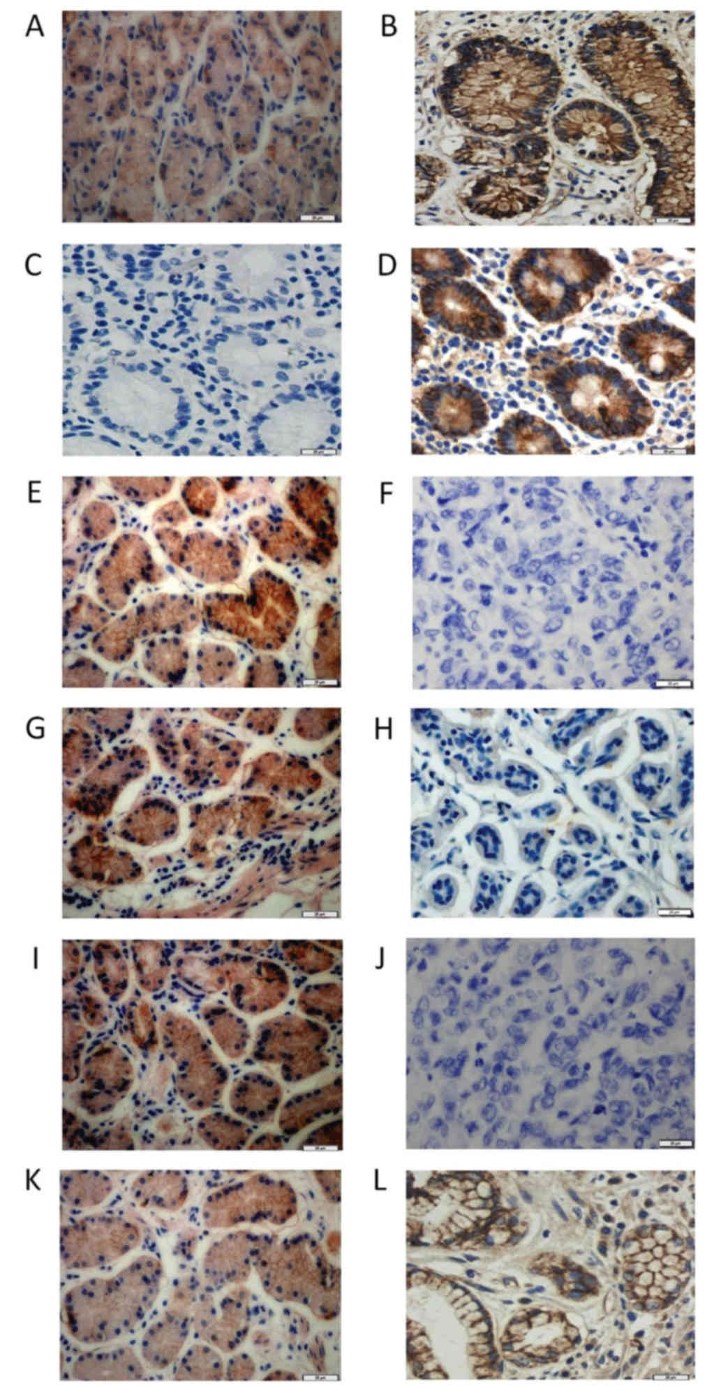
The expression of claudin proteins was investigated
in the membranes of gastric cancer tissues and non-neoplastic
mucosal tissues. Claudin-2 was highly expressed in 37.0% (37/100)
of gastric cancer tissues and 41.0% (41/100) of non-neoplastic
tissues. The results of the present study indicated that claudin-2
expression in gastric cancer samples exhibited no notable
difference compared with histologically normal gastric tissues
(χ2 test/χ2 goodness-of-fit test; P=0.374;
Fig. 1A and B). The expression of
claudin-2 was not associated with age (P=1.000), histological grade
(P=1.000), Ki67 (P=0.232), clinical staging (P=1.000) and lymph
node metastasis (P=0.368; Table
I). The expression levels of claudin-5, −7 and −8 varied
between gastric cancer tissues and in non-neoplastic mucosa.
 | Table I.Expression of claudin-2 and
claudin-5, and the clinicopathological characteristics in patients
with gastric carcinoma. |
Table I.
Expression of claudin-2 and
claudin-5, and the clinicopathological characteristics in patients
with gastric carcinoma.
| Factor | n | Claudin-2
(high) | Claudin-2
(low) | P-value | n | Claudin-5
(high) | Claudin-5
(low) | P-value |
|---|
| Gastric carcinoma
tissue | 100 | 37 | 63 | 0.374 | 100 | 47 | 53 | <0.01 |
| Non-neoplastic
tissue | 100 | 41 | 59 |
| 100 | 19 | 81 |
|
| Age, years |
|
|
|
|
|
|
|
|
|
≤60 | 29 | 10 | 19 | 1.000a | 27 | 15 | 12 | 0.261a |
|
>60 | 71 | 27 | 44 |
| 73 | 32 | 41 |
|
| Histological
grade |
|
|
|
|
|
|
|
|
|
Well-differentiated | 35 | 12 | 23 | 1.000a | 32 | 16 | 16 | 0.614a |
|
Moderately and
poorly-differentiated | 65 | 25 | 40 |
| 68 | 31 | 37 |
|
| Lymph node
metastasis |
|
|
|
|
|
|
|
|
| + | 46 | 16 | 30 | 0.368 | 39 | 15 | 24 | <0.01 |
| − | 64 | 21 | 43 |
| 61 | 32 | 29 |
|
| Ki67 |
|
|
|
|
|
|
|
|
| + | 29 | 11 | 18 | 0.232a | 27 | 12 | 15 | 0.443a |
| − | 71 | 26 | 45 |
| 73 | 35 | 38 |
|
| Clinical stage |
|
|
|
|
|
|
|
|
|
I–II | 27 | 11 | 16 | 1.000a | 27 | 15 | 12 | 0.863a |
|
III–IV | 73 | 26 | 57 |
| 73 | 32 | 41 |
|
Membrane expression of claudin-5 was high in 47.0%
(47/100) of gastric cancer tissues and 19.0% (19/100) of
non-neoplastic tissues (Fig. 1C and
D). Claudin-7 expression was detected to be high expressed in
22.0% (22/100) of gastric cancer tissues and 54.0% (54/100) in
non-neoplastic tissues (Fig. 1E and
F). High expression of claudin-8 protein was detected in 31.0%
(31/100) of gastric cancer tissues and 53.0% (53/100) of
non-neoplastic tissues (Fig. 1G and
H). A basolateral membrane marker (E-cadherin) and TJ (zonula
occludens-1) were used as the positive control, to make the results
of claudin localization to the membrane more visible (Fig. 1I-L).
As mentioned previously, the results of the present
study suggested that membrane expression of claudin-5 was
significantly increased in gastric cancer samples compared with
histologically normal gastric tissue (P<0.01). Additionally, the
membrane expression of claudin-7 and claudin-8 in gastric cancer
tissues was significantly decreased compared with non-neoplastic
tissues (P<0.01).
As presented in Table
I, claudin-5 expression was associated with lymph node
metastasis (P<0.01), although it had no association with age
(P=0.261), histological grade (P=0.614), clinical staging (P=0.863)
or expression of Ki67 (P=0.443). In addition, as presented in
Table II, the expression of
claudin-7 was associated with clinical staging (P<0.01) and
lymph node metastasis (P<0.01), although it had no association
with age (P=0.782), expression of Ki67 (P=0.382) or histological
grade (P=1.000). In addition, the expression of claudin-8 was
associated with lymph node metastasis (P<0.01), although it had
no association with age (P=1.000), clinical staging (P=1.000),
histological grade (P=0.524) or expression of Ki67 (P=0.746;
Table II).
| Table II.Expression of claudin-7 and claudin-8
and the clinicopathological characteristics in patients with
gastric carcinoma. |
Table II.
Expression of claudin-7 and claudin-8
and the clinicopathological characteristics in patients with
gastric carcinoma.
| Factor | n | Claudin-7
(high) | Claudin-7
(low) | P-value | n | Claudin-8
(high) | Claudin-8
(low) | P-value |
|---|
| Gastric carcinoma
tissue | 100 | 22 | 78 | <0.01 | 100 | 31 | 69 | <0.01 |
| Non-neoplastic
tissue | 100 | 54 | 46 |
| 100 | 53 | 47 |
|
| Age, years |
|
|
|
|
|
|
|
|
|
≤60 | 29 | 6 | 23 | 0.782a | 29 | 8 | 21 | 1.000a |
|
>60 | 71 | 16 | 55 |
| 71 | 23 | 48 |
|
| Histological
grade |
|
|
|
|
|
|
|
|
|
Well-differentiated | 35 | 7 | 28 | 1.000a | 35 | 8 | 27 | 0.524a |
|
Moderately- and
poorly-differentiated | 65 | 15 | 50 |
| 65 | 23 | 42 |
|
| Lymph node
metastasis |
|
|
|
|
|
|
|
|
| + | 46 | 6 | 40 | <0.01 | 46 | 7 | 39 | <0.01 |
| − | 64 | 16 | 48 |
| 64 | 24 | 40 |
|
| Ki67 |
|
|
|
|
|
|
|
|
| + | 29 | 5 | 24 | 0.382a | 29 | 7 | 22 | 0.746a |
| − | 71 | 17 | 54 |
| 71 | 24 | 47 |
|
| Clinical stage |
|
|
|
|
|
|
|
|
|
I–II | 27 | 12 | 15 | <0.01 | 27 | 10 | 17 | 1.000a |
|
III–IV | 73 | 10 | 63 |
| 73 | 21 | 52 |
|
Expression of claudin proteins in
non-neoplastic mucosa and gastric cancer tissues
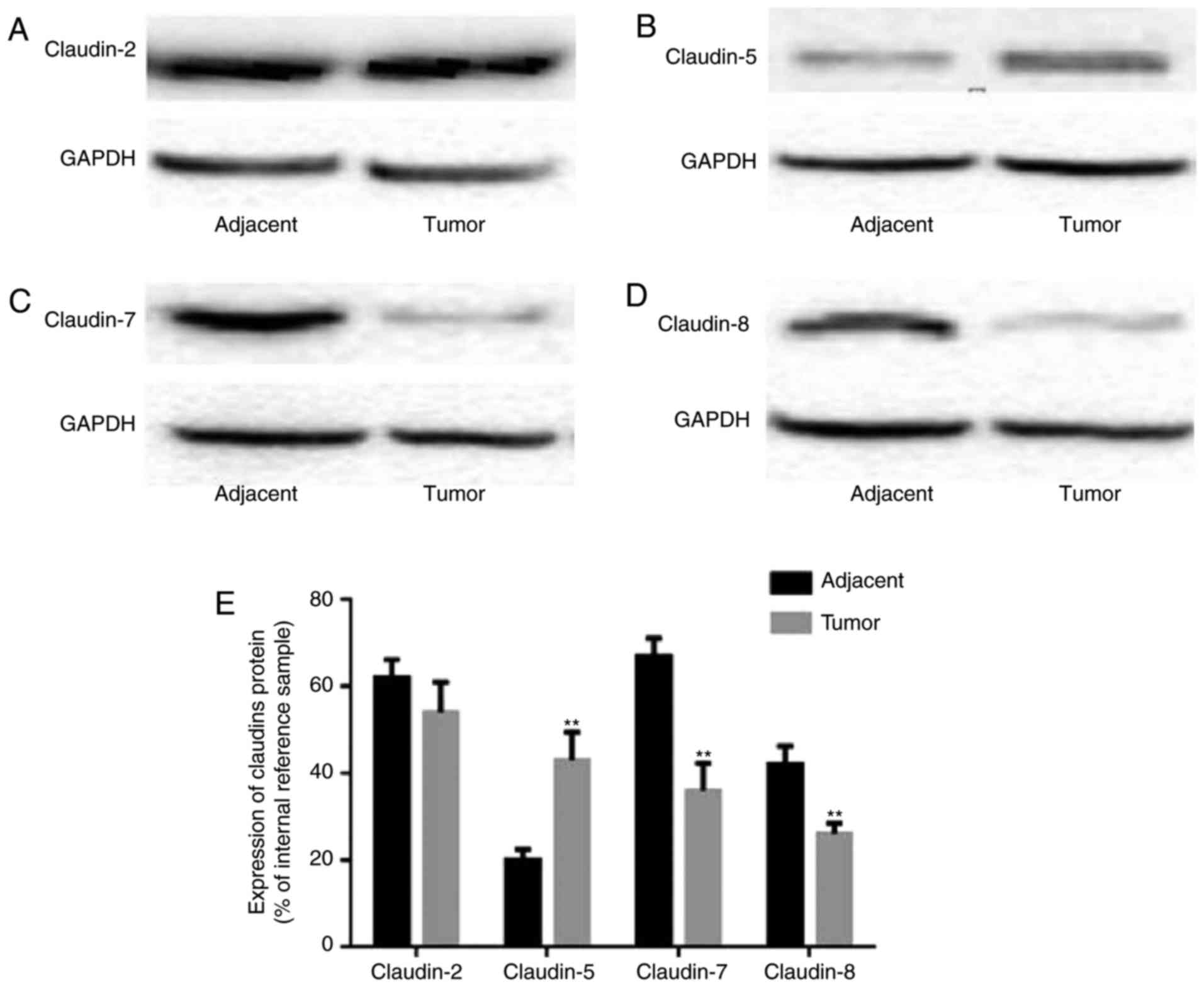
Semi-quantitative immunoblotting was utilized to
investigate statistical differences in claudin expression between
non-neoplastic mucosa and gastric cancer tissues. Claudin-2, −5, −7
and −8 were detected at approximately 22 kDa in all tissues.
According to the scanning results, claudin-2 expression levels did
not exhibit notable differences between gastric cancer tissues and
non-neoplastic tissues (Fig. 2A).
The expression of claudin-5 was upregulated in gastric cancer
tissues compared with in non-neoplastic tissues (Fig. 2B). In addition, the expression of
claudin-7 and claudin-8 was significantly downregulated in gastric
cancer tissues compared with non-neoplastic tissues (Fig. 2C and D, respectively). The
statistical differences in the expression of claudins are presented
in Fig. 2E.
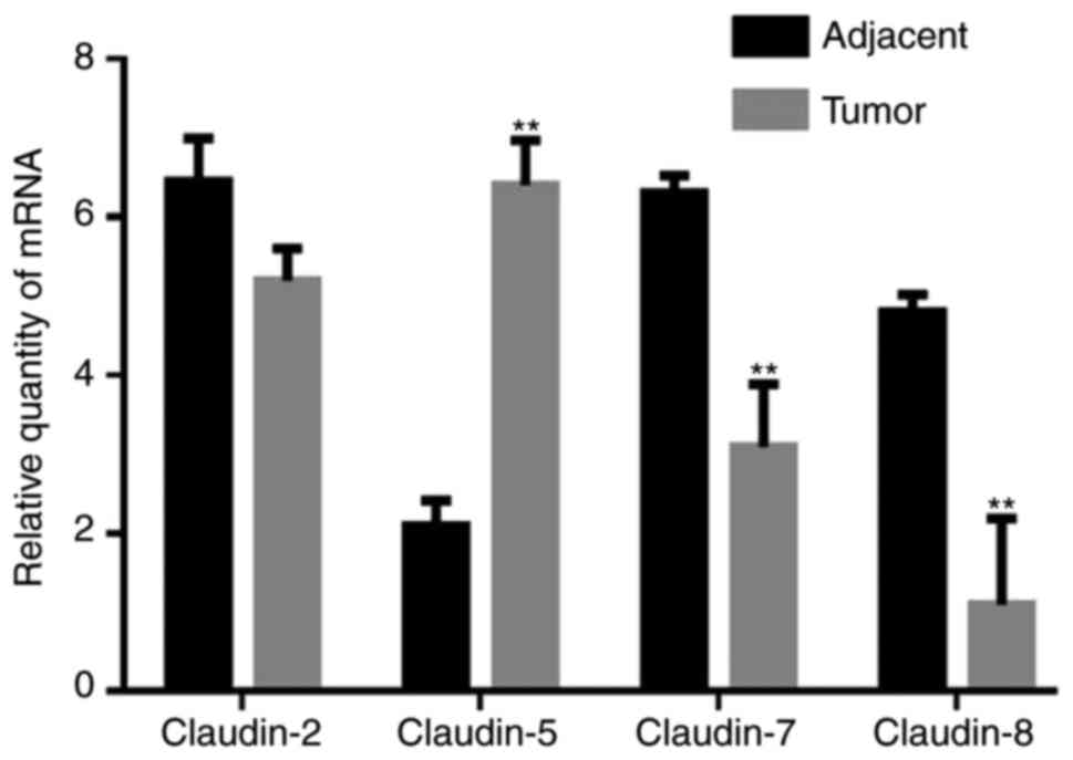
Expression of claudin mRNA in gastric
cancer and non-neoplastic mucosa
qPCR was used to determine the statistical
differences in the mRNA expression of claudins between
non-neoplastic mucosa and gastric cancer tissues. As presented in
Fig. 3, the difference in
claudin-2 mRNA expression levels between gastric cancer tissues and
non-neoplastic mucosa was not statistically significant. Compared
with non-neoplastic tissues, the mRNA expression levels of
claudin-5 were significantly upregulated in gastric cancer tissues,
while the mRNA expression levels of claudin-7 and claudin-8 were
significantly downregulated in gastric cancer tissues. This
corresponded with the results obtained by immunoblotting.
Claudin-7 and claudin-8 are
concurrently expressed in gastric cancer tissues
In addition, the association between claudin-7 and
claudin-8 expression in gastric cancers tissues and non-neoplastic
mucosa was investigated using the χ2 test/χ2
goodness-of-fit test. As presented in Tables III and IV, the association between claudin-7 and
claudin-8 was reported.
| Table III.Association between the expression of
claudin-7 and claudin-8 in gastric carcinoma tissues. |
Table III.
Association between the expression of
claudin-7 and claudin-8 in gastric carcinoma tissues.
|
| Claudin-8
(high) | Claudin-8
(low) | φ* | P-value |
|---|
| Claudin-7
(high) | 33 | 21 | 0.754 | <0.01 |
| Claudin-7
(low) | 20 | 26 |
|
|
| Table IV.Association between the expression of
claudin-7 and claudin-8 in non-neoplastic mucosae. |
Table IV.
Association between the expression of
claudin-7 and claudin-8 in non-neoplastic mucosae.
|
| Claudin-8
(high) | Claudin-8
(low) | φ* | P-value |
|---|
| Claudin-7
(high) | 15 | 7 | 0.782 | <0.01 |
| Claudin-7
(low) | 16 | 62 |
|
|
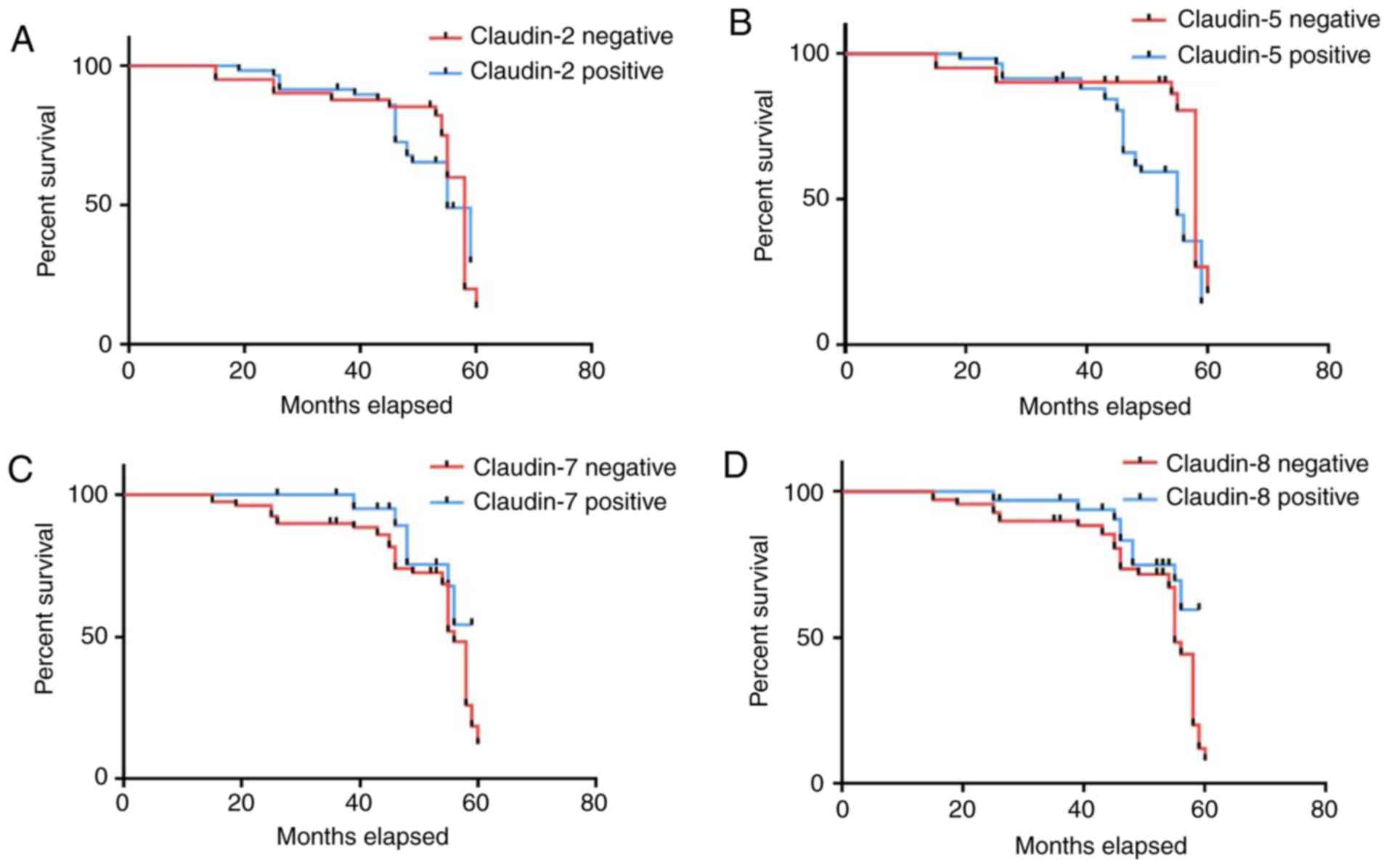
Clinical correlations and
survival
Follow-up ranged between 19 and 60 months. Patients
with tumors that were positive for claudin-7 and claudin-8 proteins
had a significantly longer survival compared with those whose
tumors were negative for these two proteins (P=0.002 and P=0.011,
respectively); patients with tumors that were positive for
claudin-5 protein expression had a significantly shorter survival
duration compared with those whose tumors were negative for this
protein (P=0.004; Fig. 4).
Discussion
The loss of TJ structure caused by aberrant
expression of claudin proteins has been suggested to be of extreme
importance in the promotion of the diffusion of nutrients and other
factors that are necessary for the survival and proliferation of
cancer cells (34,35). Previous research has revealed that
the expression of TJ claudin proteins is frequently altered in
various cancers (36). This was
demonstrated in a study that suggested that the expression of
claudin-1 was downregulated in pancreatic cancer cells and that
re-expression of claudin-1 reduced the invasive ability of these
cells (11,36). Similarly, it was revealed that the
expression of claudin-8 was downregulated in head and neck cancer,
and invasive breast cancer (37).
These reports of downregulated claudin protein expression in a
variety of human cancer cells are in agreement with the generally
accepted idea that tumorigenesis occurs along with the loss of TJ
integrity caused by the downregulation of claudin proteins
(38–40). Conversely, several studies have
suggested that the expression levels of numerous claudin proteins
are increased in cancer (14,41).
For instance, a number of reports have revealed the upregulation of
claudin-3 and claudin-4 in breast cancers (42). Additionally, a majority of the
studies published thus far have reported increased expression of
claudin proteins in various human cancer types; the differential
regulation of claudin proteins in various types of cancer
represents an opportunity to determine the mechanism of different
therapeutic responses (34,43–45).
In view of the specificity of claudin expression
profiles in human cancer, it has been revealed that claudin
proteins may serve as useful molecular markers for cancer
diagnostics. There are numerous publications on the expression
profiles of claudin proteins in gastric cancer. For instance, it
has been demonstrated that claudin-1 may be a biomarker for
intestinal-type gastric cancer with reduced survival (46). Claudin-4 was significantly
correlated with tumor T stage and with intestinal type
classification in gastric cancer (47). In addition, the expression of
claudin-10 and claudin-17 was downregulated, while the expression
of claudin-14 was upregulated, in gastric cancer tissues compared
with in tumor adjacent tissues (48). The expression of claudin-11 and −23
was greatly increased in paracancerous gastric tissue compared with
cancerous tissue (49). These
results suggested that the functions of claudin proteins may be
highly tissue-specific and may be the candidate biomarkers of
gastric cancer progression. Additionally, Lin et al
(34) revealed that the expression
of the TJ proteins claudin-2, −6 and −11 varies between human
gastric cancer and adjacent non-neoplastic tissues. The expression
of claudin-2 and claudin-6 was downregulated while the expression
of claudin-11 was upregulated in gastric cancer tissue. Differing
from the results of the present study, the localizations of claudin
proteins in research of Lin et al (34) were distributed in the cytoplasm and
the expression of claudin-2 was downregulated in gastric cancer
tissues compared with adjacent non-neoplastic tissues. There were
certain notable deficiencies in research of Lin et al
(34); for example, the
localizations of claudin proteins were distributed in cytoplasm,
and they did not appear to be TJ-associated; only 28 samples of
non-neoplastic tissues adjacent to the tumors, rather than
non-neoplastic mucosal tissues, were examined for the expression of
claudin proteins. In summary, testing the application of claudins
as cancer biomarkers in gastric cancer is not novel; however, the
expression profiles of the TJ proteins claudin-2, −5, −7 and −8 in
gastric cancer and gastric non-neoplastic mucosal tissues requires
further investigation.
In the present study, it was revealed that the
expression levels of claudin-7 and claudin-8 were downregulated,
while the expression levels of claudin-5 were upregulated in
gastric carcinoma compared with non-neoplastic mucosa. The
correlations between the expression of claudin-5, −7 and −8 and
lymph node metastasis were additionally observed, which revealed
that the expression levels of these claudins may have the potential
to be established as prognostic indicators in patients with gastric
carcinoma. In addition, claudin-7 and claudin-8 were concurrently
expressed in the mucosae and in gastric carcinoma tissues, which
suggested that claudin-7 and claudin-8 may participate in the
composition of TJ structure in gastric tissues. In addition,
patients with tumors that were positive for claudin-7 and claudin-8
protein expression had a significantly longer survival time
compared with those with negative tumors, while those with tumors
that were positive for claudin-5 protein expression had a
significantly shorter survival time compared with those with
negative tumors.
In view of the observations made in the present
study, patients may be screened following surgery for the
expression of these claudin proteins. Due to the association
between the expression of these claudin proteins and survival,
these proteins may represent novel tumor markers and therapeutic
targets. In the future, the specific mechanism that is responsible
for the observations in the present study, on how alterations in
claudin protein expression affect the malignant and oncogenic
phenotype of gastric carcinoma, may be investigated.
In summary, the results of the present study
inferred that the expression of claudin-5, −7 and −8 was altered
between human non-neoplastic mucosa and gastric carcinoma tissues,
and that their expression was associated with lymph node
metastasis. Additionally, claudin-7 and claudin-8 were concurrently
expressed in non-neoplastic mucosal and gastric carcinoma
tissues.
Acknowledgements
The authors would like to thank Dr William Orr,
Department of Pathology, University of Manitoba, Canada, for help
with this manuscript.
Funding
No funding was received.
Availability of data and materials
The datasets used and/or analyzed during the present
study are available from the corresponding author on reasonable
request.
Authors' contributions
LY performed the experiments and analyzed the data.
XS contributed to the conception and design of the study. XM
designed the present study and revised the manuscript. All authors
read and approved the final manuscript.
Ethics approval and consent to
participate
All procedures performed in the present study
involving human participants were in accordance with the ethical
standards of the institutional and/or national research committee
and with the 1964 Helsinki declaration and its later amendments or
comparable ethical standards. The present study (no. JLU01354) was
approved by the Ethics Committee of Jilin University. This article
does not contain any studies with animals performed by any of the
authors.
Consent for publication
Not applicable.
Competing interest
The authors declare that they have no competing
interests.
References
|
1
|
Dejana E: Endothelial cell-cell junctions:
Happy together. Nat Rev Mol Cell Biol. 5:261–270. 2004. View Article : Google Scholar : PubMed/NCBI
|
|
2
|
Brandner JM, Haftek M and Niessen CM:
Adherens junctions, desmosomes and tight junctions in epidermal
barrier function. Open Dermatol J. 4:14–20. 2010. View Article : Google Scholar
|
|
3
|
Förster C: Tight junctions and the
modulation of barrier function in disease. Histochem Cell Biol.
130:55–70. 2008. View Article : Google Scholar : PubMed/NCBI
|
|
4
|
Niessen CM: Tight junctions/adherens
junctions: Basic structure and function. J Invest Dermatol.
127:2525–2532. 2007. View Article : Google Scholar : PubMed/NCBI
|
|
5
|
Martin TA and Jiang WG: Loss of tight
junction barrier function and its role in cancer metastasis.
Biochim Biophys Acta. 1788:872–891. 2009. View Article : Google Scholar : PubMed/NCBI
|
|
6
|
Schneeberger EE and Lynch RD: The tight
junction: A multifunctional complex. Am J Physiol Cell Physiol.
286:C1213–C1228. 2004. View Article : Google Scholar : PubMed/NCBI
|
|
7
|
Zhang GH, Wu L and Yu GY: Tight junctions
and paracellular fluid and ion transport in salivary glands. Chin J
Dent Res. 16:13–46. 2013.PubMed/NCBI
|
|
8
|
Van Itallie CM and Anderson JM: The
molecular physiology of tight junction pores. Physiology
(Bethesda). 19:331–338. 2004.PubMed/NCBI
|
|
9
|
Coradini D, Casarsa C and Oriana S:
Epithelial cell polarity and tumorigenesis: New perspectives for
cancer detection and treatment. Acta Pharmacol Sin. 32:552–564.
2011. View Article : Google Scholar : PubMed/NCBI
|
|
10
|
Lee M and Vasioukhin V: Cell polarity and
cancer-cell and tissue polarity as a non-canonical tumor
suppressor. J Cell Sci. 121:1141–1150. 2008. View Article : Google Scholar : PubMed/NCBI
|
|
11
|
Morin PJ: Claudin proteins in human
cancer: Promising new targets for diagnosis and therapy. Cancer
Res. 65:9603–9606. 2005. View Article : Google Scholar : PubMed/NCBI
|
|
12
|
Tabariès S and Siegel PM: The role of
claudins in cancer metastasis. Oncogene. 36:1176–1190. 2017.
View Article : Google Scholar : PubMed/NCBI
|
|
13
|
González-Mariscal L, Tapia R and Chamorro
D: Crosstalk of tight junction components with signaling pathways.
Biochim Biophys Acta. 1778:729–756. 2008. View Article : Google Scholar : PubMed/NCBI
|
|
14
|
Oliveira S and Morgado-Díaz JA: Claudins:
Multifunctional players in epithelial tight junctions and their
role in cancer. Cell Mol Life Sci. 64:17–28. 2007. View Article : Google Scholar : PubMed/NCBI
|
|
15
|
Morita K, Furuse M, Fujimoto K and Tsukita
S: Claudin multigene family encoding four-transmembrane domain
protein components of tight junction strands. Proc Natl Acad Sci.
96:511–516. 1999. View Article : Google Scholar : PubMed/NCBI
|
|
16
|
Krause G, Winkler L, Mueller SL, Haseloff
RF, Piontek J and Blasig IE: Structure and function of claudins.
Biochim Biophy Acta. 1778:631–645. 2008. View Article : Google Scholar
|
|
17
|
Elkouby-Naor L and Ben-Yosef T: Functions
of claudin tight junction proteins and their complex interactions
in various physiological systems. Int Rev Cell Mol Biol. 279:1–32.
2010. View Article : Google Scholar : PubMed/NCBI
|
|
18
|
Findley MK and Koval M: Regulation and
roles for claudin-family tight junction proteins. IUBMB Life.
61:431–437. 2009. View
Article : Google Scholar : PubMed/NCBI
|
|
19
|
Lal-Nag M and Morin PJ: The claudins.
Genome Biol. 10:2352009. View Article : Google Scholar : PubMed/NCBI
|
|
20
|
Ikari A, Sato T, Watanabe R, Yamazaki Y
and Sugatani J: Increase in claudin-2 expression by an
EGFR/MEK/ERK/c-Fos pathway in lung adenocarcinoma A549 cells.
Biochim Biophy Acta. 1823:1110–1118. 2012. View Article : Google Scholar
|
|
21
|
Itoh M, Furuse M, Morita K, Kubota K,
Saitou M and Tsukita S: Direct binding of three tight
junction-associated MAGUKs, ZO-1, ZO-2, and ZO-3, with the COOH
termini of claudins. J Cell Biol. 147:1351–1363. 1999. View Article : Google Scholar : PubMed/NCBI
|
|
22
|
D'Souza T, Agarwal R and Morin PJ:
Phosphorylation of claudin-3 at threonine 192 by cAMP-dependent
protein kinase regulates tight junction barrier function in ovarian
cancer cells. J Biol Chem. 280:26233–26240. 2005. View Article : Google Scholar : PubMed/NCBI
|
|
23
|
D'Souza T, Indig FE and Morin PJ:
Phosphorylation of claudin-4 by PKCepsilon regulates tight junction
barrier function in ovarian cancer cells. Exp Cell Res.
313:3364–3375. 2007. View Article : Google Scholar : PubMed/NCBI
|
|
24
|
Kominsky SL: Claudins: Emerging targets
for cancer therapy. Expert Rev Mol Med. 8:1–11. 2006. View Article : Google Scholar : PubMed/NCBI
|
|
25
|
Furuse M and Tsukita S: Claudins in
occluding junctions of humans and flies. Trends Cell Biol.
16:181–188. 2006. View Article : Google Scholar : PubMed/NCBI
|
|
26
|
Günzel D and Alan S: Claudins and the
modulation of tight junction permeability. Physiol Rev. 93:525–569.
2013. View Article : Google Scholar : PubMed/NCBI
|
|
27
|
Ouban A and Ahmed AA: Claudins in human
cancer: A review. Histol Histopathol. 25:83–90. 2010.PubMed/NCBI
|
|
28
|
Osanai M, Takasawa A, Murata M and Sawada
N: Claudins in cancer: Bench to bedside. Pflugers Arch. 469:55–67.
2017. View Article : Google Scholar : PubMed/NCBI
|
|
29
|
Kwon MJ: Emerging roles of claudins in
human cancer. Int J Mol Sci. 14:18148–18180. 2013. View Article : Google Scholar : PubMed/NCBI
|
|
30
|
Katai H, Yoshimura K, Maruyama K, Sasako M
and Sano T: Evaluation of the New International Union Against
Cancer TNM staging for gastric carcinoma. Cancer. 88:1796–1800.
2000. View Article : Google Scholar : PubMed/NCBI
|
|
31
|
Pan XY, Wang B, Che YC, Weng ZP, Dai HY
and Peng W: Expression of claudin-3 and claudin-4 in normal,
hyperplastic, and malignant endometrial tissue. Int J Gynecol
Cancer. 17:233–241. 2007. View Article : Google Scholar : PubMed/NCBI
|
|
32
|
Livak KJ and Schmittgen TD: Analysis of
relative gene expression data using real-time quantitative PCR and
the 2(-delta delta C(T)) method. Methods. 25:402–408. 2001.
View Article : Google Scholar : PubMed/NCBI
|
|
33
|
Yang Y, Yang H, McNutt MA, Xiong F, Nie X,
Li L and Zhou R: LAPTM4B overexpression is an independent
prognostic marker in ovarian carcinoma. Oncol Rep. 20:1077–1083.
2008.PubMed/NCBI
|
|
34
|
Lin Z, Zhang X, Liu Z, Liu Q, Wang L, Lu
Y, Liu Y, Wang M, Yang M, Jin X and Quan C: The distinct expression
patterns of claudin-2, −6, and −11 between human gastric neoplasms
and adjacent non-neoplastic tissues. Diagn Pathol. 8:1332013.
View Article : Google Scholar : PubMed/NCBI
|
|
35
|
Lu Z: Functions of claudin-7 in human lung
cancer. 2012.
|
|
36
|
Hewitt KJ, Agarwal R and Morin PJ: The
claudin gene family: Expression in normal and neoplastic tissues.
BMC Cancer. 6:1862006. View Article : Google Scholar : PubMed/NCBI
|
|
37
|
Kominsky SL, Argani P, Korz D, Evron E,
Raman V, Garrett E, Rein A, Sauter G, Kallioniemi OP and Sukumar S:
Loss of the tight junction protein claudin-7 correlates with
histological grade in both ductal carcinoma in situ and invasive
ductal carcinoma of the breast. Oncogene. 22:2021–2033. 2003.
View Article : Google Scholar : PubMed/NCBI
|
|
38
|
Singh AB, Sharma A and Dhawan P: Claudin
family of proteins and cancer: An overview. J Oncol.
2010:5419572010. View Article : Google Scholar : PubMed/NCBI
|
|
39
|
Swisshelm K, Macek R and Kubbies M: Role
of claudins in tumorigenesis. Adv Drug Deliv Rev. 57:919–928. 2005.
View Article : Google Scholar : PubMed/NCBI
|
|
40
|
Webb PG, Spillman MA and Baumgartner HK:
Claudins play a role in normal and tumor cell motility. BMC Cell
Biol. 14:192013. View Article : Google Scholar : PubMed/NCBI
|
|
41
|
Rangel LB, Agarwal R, D'Souza T, Pizer ES,
Alò PL, Lancaster WD, Gregoire L, Schwartz DR, Cho KR and Morin PJ:
Tight junction proteins claudin-3 and claudin-4 are frequently
overexpressed in ovarian cancer but not in ovarian cystadenomas.
Clin Cancer Res. 9:2567–2575. 2003.PubMed/NCBI
|
|
42
|
Szász MA: Claudins as prognostic factors
of breast cancer. Magy Onkol. 56:209–212. 2012.(In Hungarian).
PubMed/NCBI
|
|
43
|
Zhang X, Ruan Y, Li Y, Lin D, Liu Z and
Quan C: Expression of apoptosis signal-regulating kinase 1 is
associated with tight junction protein claudin-6 in cervical
carcinoma. Int J Clin Exp Pathol. 8:5535–5541. 2015.PubMed/NCBI
|
|
44
|
Wang H and Yang X: The expression patterns
of tight junction protein claudin-1, −3, and −4 in human gastric
neoplasms and adjacent non-neoplastic tissues. Int J Clin Exp
Pathol. 8:881–887. 2015.PubMed/NCBI
|
|
45
|
Zhu J and Wang R, Cao H, Zhang H, Xu S,
Wang A, Liu B, Wang Y and Wang R: Expression of claudin-5, −7, −8
and −9 in cervical carcinoma tissues and adjacent non-neoplastic
tissues. Int J Clin Exp Pathol. 8:9479–9486. 2015.PubMed/NCBI
|
|
46
|
Huang J, Li J, Qu Y, Zhang J, Zhang L,
Chen X, Liu B and Zhu Z: The expression of claudin 1 correlates
with β-catenin and is a prognostic factor of poor outcome in
gastric cancer. Int J Oncol. 44:1293–1301. 2014. View Article : Google Scholar : PubMed/NCBI
|
|
47
|
Zhu JL, Gao P, Wang ZN, Song YX, Li AL, Xu
YY, Wang MX and Xu HM: Clinicopathological significance of
claudin-4 in gastric carcinoma. World J Surg Oncol. 11:1502013.
View Article : Google Scholar : PubMed/NCBI
|
|
48
|
Gao M, Li W, Wang H and Wang G: The
distinct expression patterns of claudin-10, −14, −17 and E-cadherin
between adjacent non-neoplastic tissues and gastric cancer tissues.
Diagn Pathol. 8:2052013. View Article : Google Scholar : PubMed/NCBI
|
|
49
|
Lu Y, Jing J, Sun L, Gong Y, Chen M, Wang
Z, Sun M and Yuan Y: Expression of claudin-11, −23 in different
gastric tissues and its relationship with the risk and prognosis of
gastric cancer. PLoS One. 12:e01744762017. View Article : Google Scholar : PubMed/NCBI
|