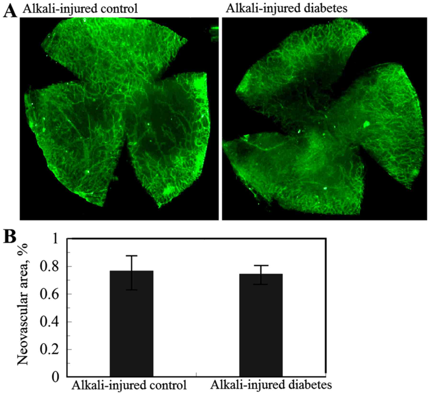
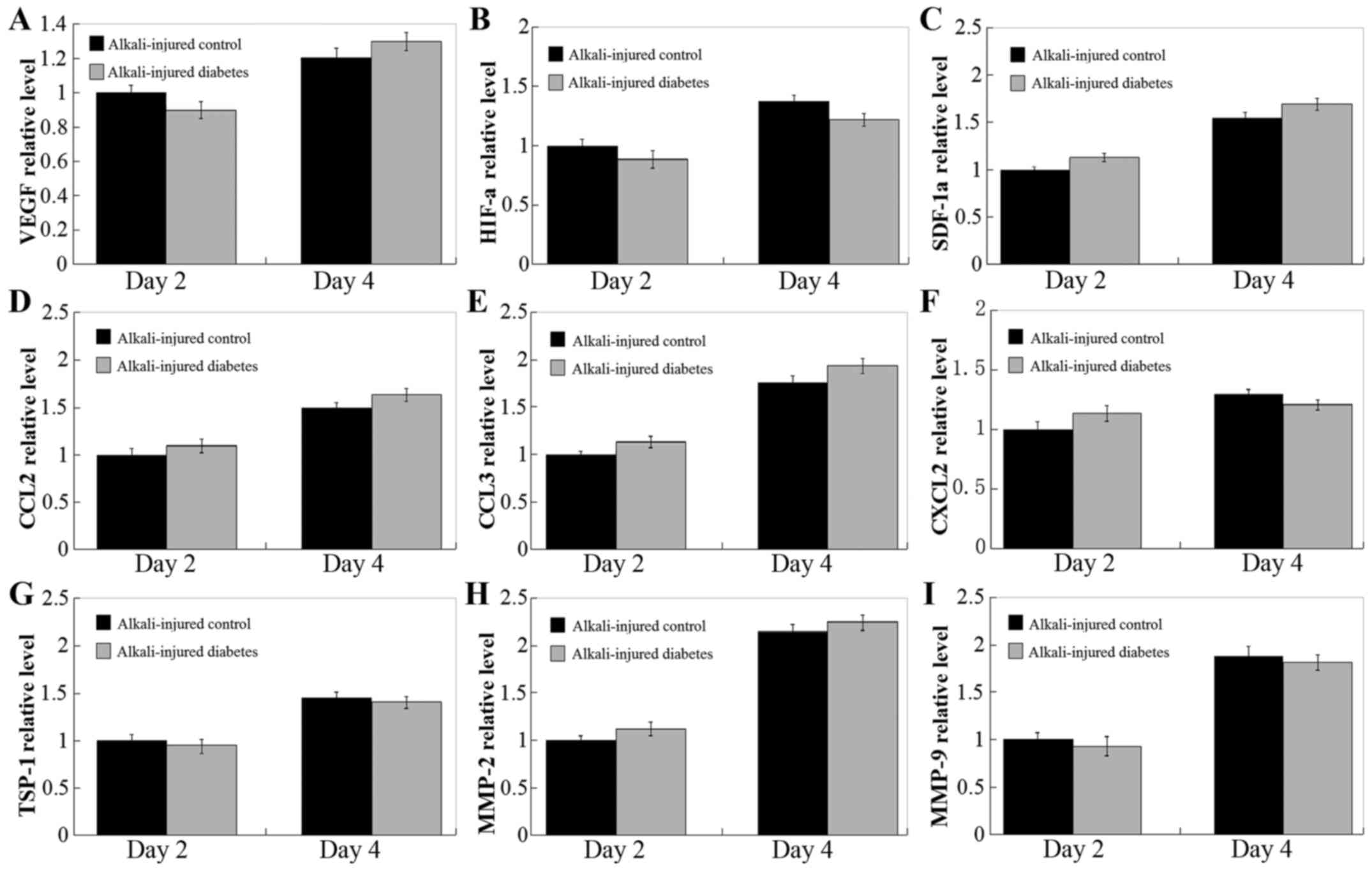
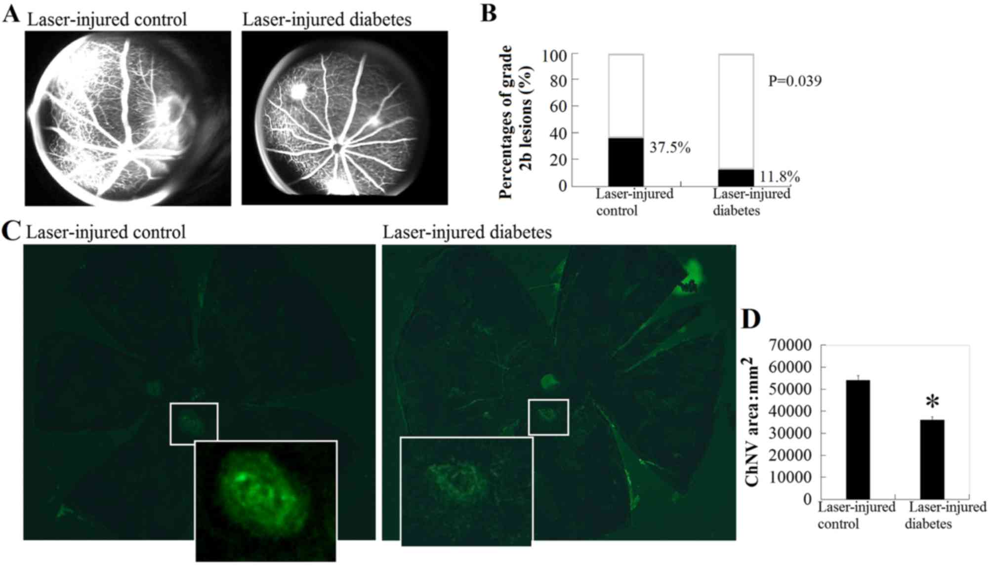
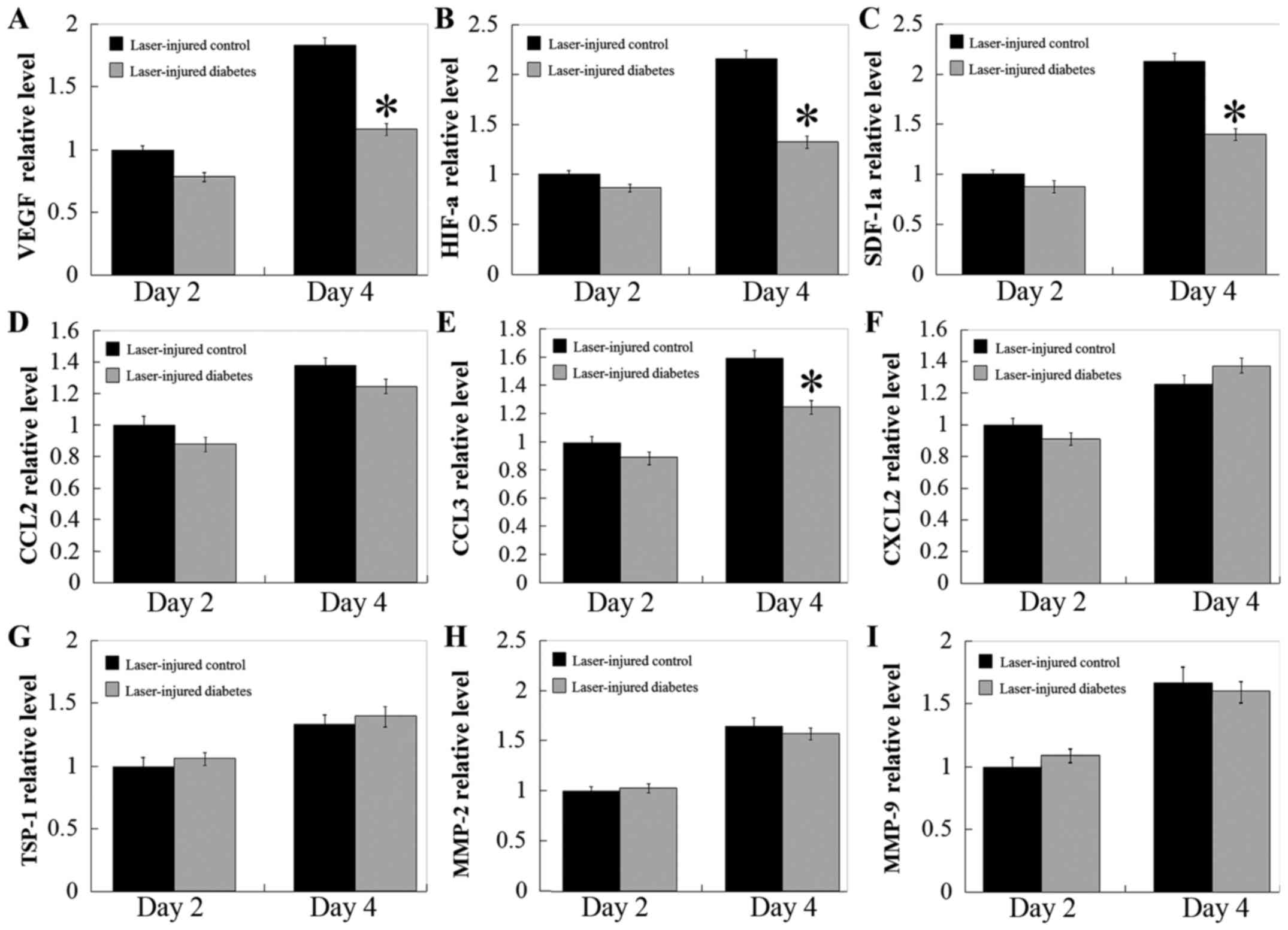
|
1
|
Wild S, Roglic G, Green A, Sicree R and
King H: Global prevalence of diabetes: Estimates for 2000 and
projections for 2030. Diabetes Care. 27:1047–1053. 2004. View Article : Google Scholar : PubMed/NCBI
|
|
2
|
Smith SC, Lamping DL and Maclaine GD:
Measuring health-related quality of life in diabetic peripheral
neuropathy: A systematic review. Diabetes Res Clin Pract.
96:261–270. 2012. View Article : Google Scholar : PubMed/NCBI
|
|
3
|
Laiteerapong N, Karter AJ, Liu JY, Moffet
HH, Sudore R, Schillinger D, John PM and Huang ES: Correlates of
quality of life in older adults with diabetes: The diabetes &
aging study. Diabetes Care. 34:1749–1753. 2011. View Article : Google Scholar : PubMed/NCBI
|
|
4
|
Keymel S, Heinen Y, Balzer J, Rassaf T,
Kelm M, Lauer T and Heiss C: Characterization of macro-and
microvascular function and structure in patients with type 2
diabetes mellitus. Am J Cardiovasc Dis. 1:68–75. 2011.PubMed/NCBI
|
|
5
|
Leiva A, Pardo F, Ramírez MA, Farías M,
Casanello P and Sobrevia L: Fetoplacental vascular endothelial
dysfunction as an early phenomenon in the programming of human
adult diseases in subjects born from gestational diabetes mellitus
or obesity in pregnancy. Exp Diabetes Res. 2011:3492862011.
View Article : Google Scholar : PubMed/NCBI
|
|
6
|
Ambati BK, Nozaki M, Singh N, Takeda A,
Jani PD, Suthar T, Albuquerque RJ, Richter E, Sakurai E, Newcomb
MT, et al: Corneal avascularity is due to soluble VEGF receptor-1.
Nature. 443:993–997. 2006. View Article : Google Scholar : PubMed/NCBI
|
|
7
|
Cursiefen C, Masli S, Ng TF, Dana MR,
Bornstein P, Lawler J and Streilein JW: Roles of thrombospondin-1
and −2 in regulating corneal and iris angiogenesis. Invest
Ophthalmol Vis Sci. 45:1117–1124. 2004. View Article : Google Scholar : PubMed/NCBI
|
|
8
|
Gao G and Ma J: Tipping the balance for
angiogenic disorders. Drug Discov Today. 7:171–172. 2002.
View Article : Google Scholar : PubMed/NCBI
|
|
9
|
Zhang SX and Ma JX: Ocular
neovascularization: Implication of endogenous angiogenic inhibitors
and potential therapy. Prog Retin Eye Res. 26:1–37. 2007.
View Article : Google Scholar : PubMed/NCBI
|
|
10
|
Lu P, Li L, Kuno K, Wu Y, Baba T, Li YY,
Zhang X and Mukaida N: Protective roles of the
fractalkine/CX3CL1-CX3CR1 interactions in alkali-induced corneal
neovascularization through enhanced antiangiogenic factor
expression. J Immunol. 108:4283–4291. 2008. View Article : Google Scholar
|
|
11
|
Liu G, Lu P, Li L, Jin H, He X, Mukaida N
and Zhang X: Critical role of SDF-1α-induced progenitor cell
recruitment and macrophage VEGF production in the experimental
corneal neovascularization. Mol Vis. 18:2128–2137. 2011.
|
|
12
|
Lu P, Li L, Liu G, van Rooijen N, Mukaida
N and Zhang X: Opposite roles of CCR2 and CX3CR1 macrophages in
alkali-induced corneal neovascularization. Cornea. 28:562–569.
2009. View Article : Google Scholar : PubMed/NCBI
|
|
13
|
Lu P, Li L, Liu G, Zhang X and Mukaida N:
Enhanced experimental corneal neovascularization along with
aberrant angiogenic factor expression in the absence of IL-1
receptor antagonist. Invest Ophthalmol Vis Sci. 50:4761–4768. 2009.
View Article : Google Scholar : PubMed/NCBI
|
|
14
|
Hoerster R, Muether PS, Vierkotten S,
Schröder S, Kirchhof B and Fauser S: In-vivo and ex-vivo
characterization of laser-induced choroidal neovascularization
variability in mice. Graefes Arch Clin Exp Ophthalmol.
250:1579–1586. 2012. View Article : Google Scholar : PubMed/NCBI
|
|
15
|
Fujimura S, Takahashi H, Yuda K, Ueta T,
Iriyama A, Inoue T, Kaburaki T, Tamaki Y, Matsushima K and Yanagi
Y: Angiostatic effect of CXCR3 expressed on choroidal
neovascularization. Invest Ophthalmol Vis Sci. 53:1999–2006. 2012.
View Article : Google Scholar : PubMed/NCBI
|
|
16
|
Badr G, Badr BM, Mahmoud MH, Mohany M,
Rabah DM and Garraud O: Treatment of diabetic mice with undenatured
whey protein accelerates the wound healing process by enhancing the
expression of MIP-1α, MIP-2, KC, CX3CL1 and TGF-β in wounded
tissue. BMC Immunol. 13:322012. View Article : Google Scholar : PubMed/NCBI
|
|
17
|
Towner RA, Smith N, Saunders D, Henderson
M, Downum K, Lupu F, Silasi-Mansat R, Ramirez DC, Gomez-Mejiba SE,
Bonini MG, et al: In vivo imaging of immuno-spin trapped radicals
with molecular magnetic resonance imaging in a diabetic mouse
model. Diabetes. 61:2405–2413. 2012. View Article : Google Scholar : PubMed/NCBI
|
|
18
|
Yang Y, Mao D, Chen X, Zhao L, Tian Q, Liu
C and Zhou BL: Decrease in retinal neuronal cells in
streptozotocin-induced diabetic mice. Mol Vis. 18:1411–1420.
2012.PubMed/NCBI
|
|
19
|
Semkova I, Fauser S, Lappas A, Smyth N,
Kociok N, Kirchhof B, Paulsson M, Poulaki V and Joussen AM:
Overexpression of FasL in retinal pigment epithelial cells reduces
choroidal neovascularization. FASEB J. 20:1689–1691. 2006.
View Article : Google Scholar : PubMed/NCBI
|
|
20
|
Livak KJ and Schmittgen TD: Analysis of
relative gene expression data using real-time quantitative PCR and
the 2(-Delta Delta C(T)) method. Methods. 25:402–408. 2001.
View Article : Google Scholar : PubMed/NCBI
|
|
21
|
Evans JR and Lawrenson JG: Antioxidant
vitamin and mineral supplements for slowing the progression of
age-related macular degeneration. Cochrane Database Syst Rev.
7:CD0002542017.PubMed/NCBI
|
|
22
|
Cheung LK and Eaton A: Age-related macular
degeneration. Pharmacotherapy. 33:838–855. 2013. View Article : Google Scholar : PubMed/NCBI
|
|
23
|
Sin HP, Liu DT and Lam DS: Lifestyle
modification, nutritional and vitamins supplements for age-related
macular degeneration. Acta Ophthalmol. 91:6–11. 2013. View Article : Google Scholar : PubMed/NCBI
|
|
24
|
McCusker MM, Durrani K, Payette MJ and
Suchecki J: An eye on nutrition: The role of vitamins, essential
fatty acids, and antioxidants in age-related macular degeneration,
dry eye syndrome, and cataract. Clin Dermatol. 34:276–285. 2016.
View Article : Google Scholar : PubMed/NCBI
|
|
25
|
Stevens RJ, Coleman RL, Adler AI, Stratton
IM, Matthews DR and Holman RR: Risk factors for myocardial
infarction case fatality and stroke case fatality in type 2
diabetes: UKPDS 66. Diabetes Care. 27:201–207. 2004. View Article : Google Scholar : PubMed/NCBI
|
|
26
|
Chung SS, Ho EC, Lam KS and Chung SK:
Contribution of polyol pathway to diabetes-induced oxidative
stress. J Am Soc Nephrol. 14(8 Suppl 3): S233–S236. 2003.
View Article : Google Scholar : PubMed/NCBI
|
|
27
|
Lee TS, Saltsman KA, Ohashi H and King GL:
Activation of protein kinase C by elevation of glucose
concentration: Proposal for a mechanism in the development of
diabetic vascular complications. Proc Natl Acad Sci USA.
86:5141–5145. 1989. View Article : Google Scholar : PubMed/NCBI
|
|
28
|
Giacco F and Brownlee M: Oxidative stress
and diabetic complications. Circ Res. 107:1058–1070. 2010.
View Article : Google Scholar : PubMed/NCBI
|
|
29
|
Schmidt AM, Yan SD, Wautier JL and Stern
D: Activation of receptor for advanced glycation end products: A
mechanism for chronic vascular dysfunction in diabetic vasculopathy
and atherosclerosis. Circ Res. 84:489–497. 1999. View Article : Google Scholar : PubMed/NCBI
|
|
30
|
Kolluru GK, Bir SC and Kevil CG:
Endothelial dysfunction and diabetes: Effects on angiogenesis,
vascular remodeling, and wound healing. Int J Vasc Med.
2012:9182672012.PubMed/NCBI
|
|
31
|
da Costa Pinto FA and Malucelli BE:
Inflammatory infiltrate, VEGF and FGF-2 contents during corneal
angiogenesis in STZ-diabetic rats. Angiogenesis. 5:67–74. 2002.
View Article : Google Scholar : PubMed/NCBI
|
|
32
|
Zagon IS, Klocek MS, Griffith JW, Sassani
JW, Komáromy AM and McLaughlin PJ: Prevention of exuberant
granulation tissue and neovascularization in the rat cornea by
naltrexone. Arch Ophthalmol. 126:501–506. 2008. View Article : Google Scholar : PubMed/NCBI
|
|
33
|
Waltenberger J, Lange J and Kranz A:
Vascular endothelial growth factor-A-induced chemotaxis of
monocytes is attenuated in patients with diabetes mellitus: A
potential predictor for the individual capacity to develop
collaterals. Circulation. 102:185–190. 2000. View Article : Google Scholar : PubMed/NCBI
|
|
34
|
Tchaikovski V, Olieslagers S, Böhmer FD
and Waltenberger J: Diabetes mellitus activates signal transduction
pathways resulting in vascular endothelial growth factor resistance
of human monocytes. Circulation. 120:150–159. 2009. View Article : Google Scholar : PubMed/NCBI
|
|
35
|
Grossniklaus HE, Ling JX, Wallace TM,
Dithmar S, Lawson DH, Cohen C, Elner VM, Elner SG and Sternberg P
Jr: Macrophage and retinal pigment epithelium expression of
angiogenic cytokines in choroidal neovascularization. Mol Vis.
8:119–126. 2002.PubMed/NCBI
|
|
36
|
Greenberg JI, Shields DJ, Barillas SG,
Acevedo LM, Murphy E, Huang J, Scheppke L, Stockmann C, Johnson RS,
Angle N and Cheresh DA: A role for VEGF as a negative regulator of
pericytes function and vessel maturation. Nature. 456:809–813.
2008. View Article : Google Scholar : PubMed/NCBI
|
|
37
|
Elner VM, Strieter RM, Elner SG,
Baggiolini M, Lindley I and Kunkel SL: Neutrophil chemotactic
factor (IL-8) gene expression by cytokine-treated retinal pigment
epithelial cells. Am J Pathol. 136:745–750. 1990.PubMed/NCBI
|
|
38
|
Losso JN, Truax RE and Richard G:
Trans-resveratrol inhibits hyperglycemia-induced inflammation and
connexin downregulation in retinal pigment epithelial cells. J
Agric Food Chem. 58:8246–8252. 2010. View Article : Google Scholar : PubMed/NCBI
|
|
39
|
Yuan Z, Feng W, Hong J, Zheng Q, Shuai J
and Ge Y: p38MAPK and ERK promote nitric oxide production in
cultured human retinal pigmented epithelial cells induced by high
concentration glucose. Nitric Oxide. 20:9–15. 2009. View Article : Google Scholar : PubMed/NCBI
|
|
40
|
Bento CF, Fernandes R, Matafome P, Sena C,
Seiça R and Pereira P: Methylglyoxal-induced imbalance in the ratio
of vascular endothelial growth factor to angiopoietin 2 secreted by
retinal pigment epithelial cells leads to endothelial dysfunction.
Exp Physiol. 95:955–970. 2010. View Article : Google Scholar : PubMed/NCBI
|
|
41
|
Villarroel M, García-Ramírez M, Corraliza
L, Hernández C and Simó R: High glucose concentration leads to
differential expression of tight junction proteins in human retinal
pigment epithelial cells. Endocrinol Nutr. 56:53–58. 2009.
View Article : Google Scholar : PubMed/NCBI
|
|
42
|
Tepper OM, Carr J, Allen RJ Jr, Chang CC,
Lin CD, Tanaka R, Gupta SM, Levine JP, Saadeh PB and Warren SM:
Decreased circulating progenitor cell number and failed mechanisms
of stromal cell-derived factor-1alpha mediated bone marrow
mobilization impair diabetic tissue repair. Diabetes. 59:1974–1983.
2010. View Article : Google Scholar : PubMed/NCBI
|
|
43
|
Gallagher KA, Liu ZJ, Xiao M, Chen H,
Goldstein LJ, Buerk DG, Nedeau A, Thom SR and Velazquez OC:
Diabetic impairments in NO-mediated endothelial progenitor cell
mobilization and homing are reversed by hyperoxia and SDF-1 alpha.
J Clin Invest. 117:1249–1259. 2007. View Article : Google Scholar : PubMed/NCBI
|
|
44
|
Jiraritthamrong C, Kheolamai P, U-Pratya
Y, Chayosumrit M, Supokawej A, Manochantr S, Tantrawatpan C,
Sritanaudomchai H and Issaragrisil S: In vitro vessel-forming
capacity of endothelial progenitor cells in high glucose
conditions. Ann Hematol. 91:311–320. 2012. View Article : Google Scholar : PubMed/NCBI
|
|
45
|
Cao C, Zhang H, Gong L, He Y and Zhang N:
High glucose conditions suppress the function of bone
marrow-derived endothelial progenitor cells via inhibition of the
eNOS-caveolin-1 complex. Mol Med Report. 5:341–346. 2012.
|
|
46
|
Klein R, Klein BE and Moss SE: Diabetes,
hyperglycemia, and age-related maculopathy. The beaver Dam Eye
study. Ophthalmology. 99:1527–1534. 1992. View Article : Google Scholar : PubMed/NCBI
|
|
47
|
Song SJ, Youm DJ, Chang Y and Yu HG:
Age-related macular degeneration in a screened South Korean
population: Prevalence, risk factors, and subtypes. Ophthalmic
Epidemiol. 16:304–310. 2009. View Article : Google Scholar : PubMed/NCBI
|
|
48
|
Choi JK, Lym YL, Moon JW, Shin HJ and Cho
B: Diabetes mellitus and early age-related macular degeneration.
Arch Ophthalmol. 129:196–199. 2011. View Article : Google Scholar : PubMed/NCBI
|
|
49
|
Topouzis F, Anastasopoulos E, Augood C,
Bentham GC, Chakravarthy U, de Jong PT, Rahu M, Seland J, Soubrane
G, Tomazzoli L, et al: Association of diabetes with age-related
macular degeneration in the EUREYE study. Br J Ophthalmol.
93:1037–1041. 2009. View Article : Google Scholar : PubMed/NCBI
|


