Introduction
Atherosclerosis (AS) is one of the most prevalent
diseases in elderly people. AS is characterized by the accumulation
of lipids in the intima of large- and medium-sized arteries
(1) (including coronary or carotid
arteries); this leads to a compromised flow to target organs,
resulting in hypoxic and ischemic cardiovascular events, including
myocardial infarction and stroke, which are two leading causes of
mortality worldwide (2,3). Therefore, the development of
treatments to prevent the formation and control the progression of
atherosclerotic lesions has attracted considerable attention.
Accumulating evidence has demonstrated that oxidized
low-density lipoprotein-(ox-LDL)-induced endothelial dysfunction is
the initial step in the development of AS (4). By binding to the lectin-like
endothelial ox-LDL receptor, ox-LDL promotes the production of
reactive oxygen species and activates the endoplasmic reticulum
stress response, which sequentially phosphorylates the
mitogen-activated protein kinase (MAPK) cascade and mediates
nuclear factor (NF)-κB activation to upregulate the expression of
pro-apoptotic proteins, including caspase-3, caspase-9, caspase-12
and Bcl-2 associated X, apoptosis regulator (Bax), ultimately
leading to apoptosis of endothelial cells (4–6).
Endothelial cell apoptosis may destroy the integrity of endothelium
and increase vascular permeability, which facilitates the
infiltration and deposition of local lipids within the arterial
wall and finally results in atherogenesis (7). Furthermore, disruption of the
endothelial lining may additionally cause the plaque instability
and rupture that triggers the coagulation cascade and platelet
aggregation, eventually increasing the formation of
atherothrombosis and resulting in sudden mortality (8–10).
Therefore, inhibition of endothelial dysfunction may represent a
promising approach for the treatment of AS (11–13).
In addition to platelets, a previous study suggested
that P2Y purinoreceptor 12 (P2Y12) receptors may additionally be
highly expressed in endothelial cells of culprit coronary plaques
(14). The use of the P2Y12
receptor antagonists clopidogrel or ticagrelor may improve
endothelial dysfunction in patients with acute (15), and stable coronary artery disease
(16,17) or pig models with coronary stent
restenosis (18), suggesting that
P2Y12 receptor blockers may be potential drugs for the treatment of
AS. This hypothesis has been preliminarily verified by Preusch
et al (19), which
documented that treatment with ticagrelor induces a reduction in
lesion size in the necrotic core area within the aortic sinus of
apolipoprotein-E-deficient (ApoE−/−) mice (an AS animal
model). In vitro studies additionally demonstrated that
ox-LDL uptake and induced apoptosis of RAW264.7 macrophages are
decreased following incubation with ticagrelor (19). Furthermore, Ren et al
(20) identified that clopidogrel
inhibits the progression of AS in a rabbit model by reducing the
ratio of Bcl-2/Bax in the vascular wall. However, the anti-AS
effect of P2Y12 receptor blockers remains rarely investigated. In
addition, whether a reduction in endothelial cell apoptosis is
involved and the mechanism of action have not been examined.
The aim of the present study was to further
investigate the anti-AS effects in ApoE−/− mice and
analyze the anti-apoptotic effects in the endothelial cell line
EAhy926 following treatment with ticagrelor, a novel P2Y12 receptor
inhibitor. In a previous study (21), it was observed that increased
expression of proprotein convertase subtilisin/kexin type 9
(PCSK9), encoding a neural apoptosis-regulated convertase 1, is
associated with ox-LDL-induced apoptosis in EAhy926 cells.
Furthermore, downregulation of PCSK9 by short hairpin (sh)RNA
inhibits apoptosis of EAhy926 cells and decreases levels of
apoptosis-associated proteins (Bax and caspase-3) and the
mitogen-activated protein kinase (MAPK) signaling pathway (21). Accordingly, it was hypothesized
that the inhibition of PCSK9 expression may be a potential
mechanism for ticagrelor to exert anti-AS effects. This has not
been reported previously, to the best of the authors' knowledge,
and was investigated in the present study.
Materials and methods
Cell culture and grouping
The human umbilical vein endothelial cell line
EAhy926 was provided by the Pathology Laboratory of Tianjin Medical
University (Tianjin, China). EAhy926 cells were maintained in
Dulbecco's modified Eagle's medium (HyClone; GE Healthcare Life
Sciences, Logan, UT, USA) supplemented with 10% fetal bovine serum
(HyClone; GE Healthcare Life Sciences, Logan, UT, USA), 100 U/ml
penicillin G and 100 µg/ml streptomycin and cultured at 37°C in a
humidified incubator with 5% CO2.
EAhy926 cells were divided into four groups;
control, ox-LDL, ox-LDL + 40 µmol/l ticagrelor (T40) and ox-LDL +
60 µmol/l ticagrelor (T60). Oxidized LDL (50 µg/ml; Beijing Xinyuan
Jiahe Biotech Co., Ltd., Beijing, China) was used to induce
endothelial dysfunction of cells, as previously described (20). Cells in each group were treated for
24 h.
Apoptosis assay
The cultured cells were detached by trypsinization
and stained with Annexin V labeled with fluorescein isothiocyanate
(BD Pharmingen; BD Biosciences, San Jose, CA, USA) and propidium
iodide (1 mg/ml; Sigma-Aldrich; Merck KGaA, Darmstadt, Germany) in
the dark for 15 min at room temperature. Cells were subsequently
analyzed using a flow cytometer (FACSCalibur; BD Biosciences) with
CellQuest software (Version 6.0; BD Biosciences).
Reverse transcription-quantitative
polymerase chain reaction (RT-qPCR)
Total RNA was extracted from EAhy926 cells using
TRIzol® reagent (Invitrogen; Thermo Fisher Scientific,
Inc., Waltham, MA, USA), followed by RT into cDNA using an M-MLV RT
system (Takara Biotechnology Co., Ltd., Dalian, China) with the
reaction parameters of incubation at 65°C for 10 min, 42°C for 30
min and 70°C for 10 min. qPCR was performed using an ABI-7500
Real-Time PCR System (Applied Biosystems; Thermo Fisher Scientific,
Inc.) with a SYBR-Green PCR kit [Biocentury Transgene (China) Co.,
Ltd., Shenzhen, China]. The primers used are listed in Table I. The PCR reaction parameters were
incubation at 95°C for 10 sec, then 40 cycles of 95°C for 10 sec,
58°C for 30 sec and 72 for 30 sec. The relative expression of
genes, using a housekeeping gene (GAPDH) as an internal standard,
was calculated by the 2−∆∆Cq method (22). All measurements were performed in
triplicate.
 | Table I.Primers used in the present
study. |
Table I.
Primers used in the present
study.
| Gene | Primer | Annealing
temperature, °C | Product size,
bp |
|---|
| GAPDH |
| 58 | 163 |
| F |
5′-CACATGGCCTCCAAGGAGTA-3 |
|
|
| R |
5′-TCCCCTCTTCAAGGGGTCTA-3′ | 58 |
|
| PCSK9 |
| 58 | 140 |
| F |
5′-TGGAACTCACTCACTCTGGG-3′ |
|
|
| R |
5′-AAGAATCCTGCCTCCTTGGT-3′ | 58 |
|
| Bax |
| 58 | 119 |
| F |
5′-TGATCAGAACCATCATGGGC-3′ |
|
|
| R |
5′-GGACATCAGTCGCTTCAGTG-3′ | 58 |
|
| Bcl-2 |
| 58 | 156 |
| F |
5′-GAAGAAGCCACCCTCAAGC-3′ | 58 |
|
| R |
5′-AGCAAGGACACCCGCACTC-3′ |
|
|
| Caspase-3 |
| 58 | 106 |
| F |
5′-GAGGCCGACTTCTTGTATGC-3′ |
|
|
| R |
5′-GTTTCAGCATGGCACAAAGC-3′ | 58 |
|
| NF-κB |
| 58 | 186 |
| F |
5′-AGACAAATGGGCTACACCGA-3′ |
|
|
| R |
5′-AAAGCTGAGTTTGCGGAAGG-3′ | 58 |
|
Western blotting
Proteins were extracted from EAhy926 cells using a
radioimmunoprecipitation assay buffer (Beyotime Institute of
Biotechnology, Haimen, China). Protein concentrations were measured
using a 2-D Quant kit (GE Healthcare Life Sciences, Little
Chalfont, UK) and equal amounts of protein (30 µg) from each group
were separated by SDS-PAGE on 10% gels prior to being transferred
to polyvinylidene difluoride membranes (EMD Millipore, Billerica,
MA, USA). Subsequent to blocking with 5% non-fat milk at 4°C for 1
h, membranes were probed with anti-PCSK9, caspase-3, Bax, Bcl-2,
p38, phosphorylated (p)-p38, extracellular signal-regulated kinase
(ERK), p-ERK, c-Jun N-terminal kinases (JNK), p-JNK or β-actin
primary antibodies at 4°C overnight. Membranes were subsequently
incubated with horseradish peroxidase-conjugated secondary
antibodies at room temperature for 1 h. The protein bands were
visualized with an enhanced chemiluminescence reagent (EMD
Millipore). β-actin was used as an internal control. Detailed
information regarding the antibodies used is listed in Table II. Densitometry was performed
using Image-Pro Plus software (Version 6.0; Media Cybernetics,
Rockville, MD, USA).
| Table II.Antibody details. |
Table II.
Antibody details.
|
|
| Primary
antibody | Secondary
antibody |
|
|---|
|
|
|
|
|
|
|---|
| Antibody | Supplier | Catalog no. | Dilution | Species raised | Supplier | Catalog no. | Dilution | Molecular weight,
kDa |
|---|
| PCSK9 | Sigma-Aldrich | SAB1302902 | 1:500 | Rabbit | Abcam | ab191866 | 1:2,000 | 72 |
| Caspase-3 | Abcam | ab32042 | 1:100 | Rabbit | Abcam | ab191866 | 1:5,000 | 32 |
| Bax | Abcam | ab32503 | 1:5,000 | Rabbit | Abcam | ab191866 | 1:10,000 | 21 |
| Bcl-2 | Abcam | ab32124 | 1:1,000 | Rabbit | Abcam | ab191866 | 1:5,000 | 26 |
| p38 | Abcam | ab27986 | 1:1,000 | Rabbit | Abcam | ab191866 | 1:10,000 | 38 |
| p-p38 | Abcam | ab4822 | 1:1,000 | Rabbit | Abcam | ab191866 | 1:10,000 | 38 |
| ERK | Abcam | ab17942 | 1:1,000 | Rabbit | Abcam | ab191866 | 1:5,000 | 42-44 |
| p-ERK | Abcam | ab214362 | 1:1,000 | Rabbit | Abcam | ab191866 | 1:5,000 | 42-44 |
| JNK | Abcam | ab179461 | 1:1,000 | Rabbit | Abcam | ab191866 | 1:5,000 | 46 |
| p-JNK | Abcam | ab124956 | 1:1,000 | Rabbit | Abcam | ab191866 | 1:5,000 | 46 |
| β-actin | Abcam | ab8226 | 1:10,000 | Mouse | Abcam | ab131368 | 1:10,000 | 43 |
Animal model
All animal experiments conformed with the Regulation
of Animal Care Management of the Ministry of Public Health,
People's Republic of China and were approved by the Ethical
Committee of Second Hospital of Tianjin Medical University
(Tianjin, China).
Six-week-old male weighing 20–25 g C57BL/6 mice
(n=10) and homozygous ApoE−/− mice (n=10) were purchased
from Beijing University (Beijing, China). The mice were housed in a
specific pathogen-free facility under controlled conditions
(temperature, 22±2°C; relative humidity, 55±15%; noise, <60 dB;
light/dark cycle, 12/12 h) and were used following one week of
accommodation. All animals were provided with free access to
water/food. ApoE−/− mice were fed with a high-fat diet
containing 0.25% cholesterol and 15% cocoa butter (Hunan Huakang
Biotech Inc., Hunan, China) to induce the formation of the
atherosclerotic plaques. Normal C57BL/6 mice were given standard
chow throughout the experiment to serve as a control. After 20
weeks of feeding, ApoE−/− mice were randomly divided
into the model group (AS, n=5) and treatment group (ticagrelor,
n=5). Mice in the treatment group received ticagrelor (100 mg/kg,
corresponding to concentration T60 in vitro) through
intragastric administration for 10 consecutive days, while an equal
amount of saline was intragastrically administered to AS and
control mice in the same period.
Biochemical analysis
Mice in each group were anesthetized with 5%
pentobarbital sodium (50 mg/kg) by intraperitoneal injection and
blood was collected through direct cardiac puncture. The samples
were subsequently centrifuged at 1,200 × g for 10 min at room
temperature to obtain the plasma for biochemical measurements. The
plasma levels of triglyceride (TG), cholesterol (TC), low-density
lipoprotein cholesterol (LDL-C) and high-density lipoprotein
cholesterol (HDL-C) were determined using an automatic biochemical
analyzer (Olympus AU5400; Olympus Corporation, Tokyo, Japan).
Histological analysis
Following cardiac exsanguination, the aorta were
quickly harvested, fixed in 4% paraformaldehyde at 4°C for 24 h,
dehydrated in a graded series of alcohol (70, 80, 90, 95 and 100%,
each 90 min), embedded in 5-µm-thick paraffin and stained with
hematoxylin (5 min) & eosin (5 min) at room temperature. Images
were captured using a light microscope (OLYMPUS X81; Olympus
Corporation) at magnification, ×10 and ×40.
Immunohistochemical staining
The embedded tissue sections were incubated
overnight at 4°C with a primary antibody against PCSK9 (1:200;
Sigma-Aldrich; Merck KGaA) following a rehydration in a graded
series of alcohol (100, 95, 80, and 70%, each 5 min), hydrogen
peroxide-induced endogenous peroxidase activity inhibition,
microwave-based antigen retrieval (121°C for 10 min) and
non-specific binding blocked with 10% normal goat serum
(Invitrogen; Thermo Fisher Scientific, Inc.; at room temperature,
10 min). Following three washes in sterile PBS, the sections were
incubated with the secondary antibody (1:500; Sigma-Aldrich; Merck
KGaA) at room temperature for 30 min. The immunohistochemical
reaction color was developed with diaminobenzidine (Vector
Laboratories, Inc., Burlingame, CA, USA; 2–8 min) and
counterstained with hematoxylin (2 min) at room temperature. The
expression of PCSK9 in the tissues was viewed under a light
microscope (Olympus X81; Olympus Corporation) at magnification, ×10
and ×40 and semi-quantitatively measured using Image Pro Plus
software (Version 6.0; Media Cybernetics, Inc.).
Statistical analysis
Statistical analyses were performed using SPSS
software (Version 18.0; SPSS Inc., Chicago, IL, USA). Data are
expressed as the mean ± standard error. of three independent
repeats. The significant difference between two experimental groups
was analyzed using Student's t-test; however, statistical
significance of differences among three groups were assessed using
one-way analysis of variance followed by the Least Significant
Difference post hoc test for multiple comparisons. P<0.05 was
considered to indicate a statistically significant difference.
Results
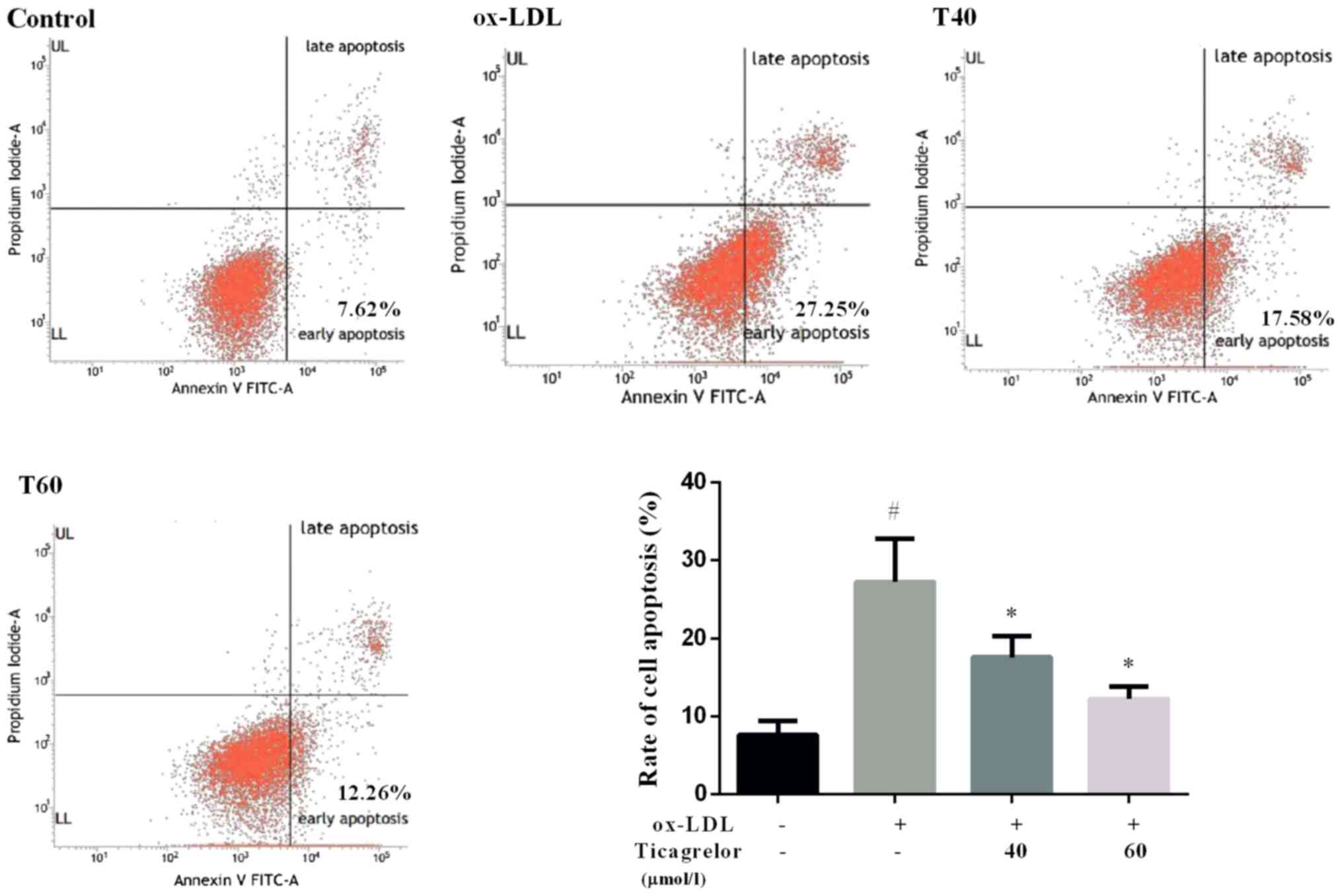
Ticagrelor inhibits ox-LDL-induced
apoptosis in EAhy926 cells
Damage in the vascular endothelium has been
suggested as an initial step in the pathogenesis of AS (4). To determine whether ticagrelor
alleviates AS, the effect of ticagrelor on ox-LDL-induced
endothelial dysfunction was evaluated. The present data
demonstrated that treatment with 50 µg/ml ox-LDL resulted in a
significant increase in apoptosis of EAhy926 cells compared with
the control (27.25±5.54 vs. 7.62±1.76%; P<0.05; Fig. 1). However, the addition of
ticagrelor was able to decrease ox-LDL-induced apoptosis,
particularly at a higher concentration (T40, 17.58±2.66%; T60,
12.26±1.54%; Fig. 1). Therefore,
60 µmol/l ticagrelor was used for subsequent experimentation.
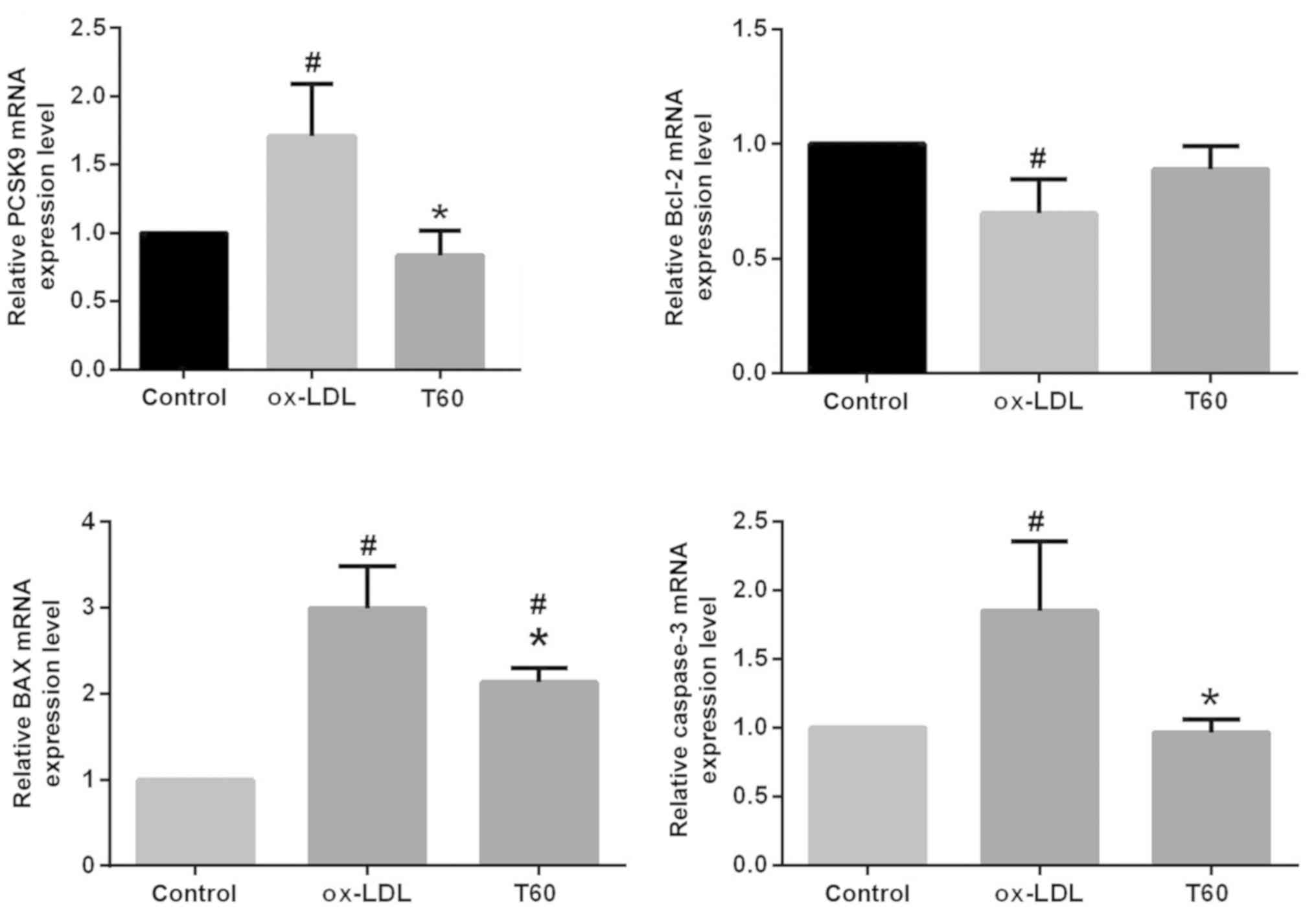
Ticagrelor downregulates PCSK9
expression and downstream apoptosis pathways in EAhy926 cells
To elucidate the mechanisms by which ticagrelor
inhibits endothelial apoptosis, the expression of PCSK9 and
downstream apoptosis pathway genes were additionally evaluated. As
expected, the mRNA expression level of PCSK9 determined by RT-qPCR
was significantly decreased following treatment with ticagrelor
(P<0.05; Fig. 2). The use of
ticagrelor additionally upregulated mRNA expression of the
anti-apoptotic factor Bcl-2 and significantly downregulated the
expression of the pro-apoptotic factors Bax and caspase-3 (Fig. 2; P<0.05). This result was
similarly observed in the analysis of protein expression levels by
western blotting (Fig. 3).
Furthermore, the expression levels of apoptosis pathway proteins,
including p-p38, p-JNK, JNK and p-ERK, were significantly
suppressed following treatment with ticagrelor; the JNK pathway may
be of particular importance, as difference between the p-JNK/JNK
ratios was statistically significant (P<0.05; Fig. 3). These results suggested that
ticagrelor reduced ox-LDL-induced endothelial apoptosis by
downregulating PCSK9 and downstream apoptosis pathways.
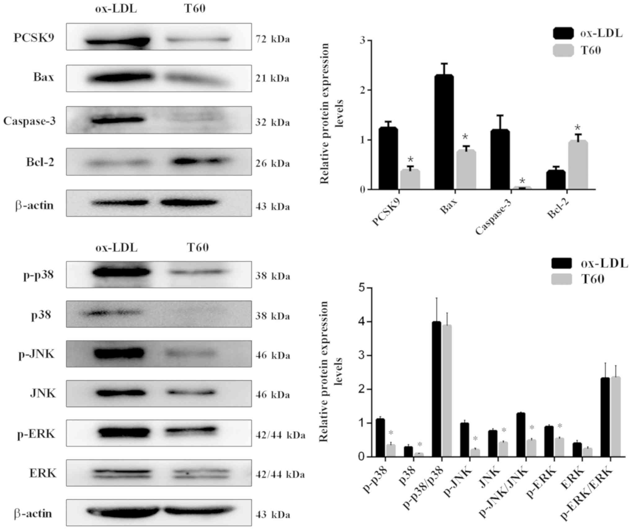 | Figure 3.Protein expression levels of PCSK9,
apoptosis-associated genes (Bax, Bcl-2 and caspase-3) and pathways
(JNK, p38 and ERK) detected by western blotting. All results are
expressed as the mean ± standard error. *P<0.05 vs. respective
ox-LDL. Ox-LDL, oxidized low-density lipoprotein; Bcl-2, B-cell
lymphoma 2; Bax, Bcl-2 associated X, apoptosis regulator; PCSK9,
proprotein convertase subtilisin/kexin type 9; T60, 60 µmol/l
ticagrelor; p, phosphorylated; JNK, c-Jun N-terminal kinases; ERK,
extracellular signal-regulated kinases. |
Intragastric administration of
ticagrelor reduces AS development in ApoE−/− mice
To further confirm the anti-atherosclerotic role and
mechanisms of action of ticagrelor in vivo, an AS animal
model, of ApoE−/− mice fed a high-fat diet, was
constructed. The health condition of the majority of mice was
excellent during the modeling, with no mortality observed in any
group. Neither treatment affected food intake, and body weight
increased in all groups after 20 weeks, particularly the AS and
ticagrelor groups due to the high-fat diet provided (Table III). Unexpectedly, there was no
significant difference in body weight between the AS and ticagrelor
groups (Table III). Likewise,
plasma concentrations of TC, HDL and LDL were equivalent in
ApoE−/− mice irrespective of ticagrelor exposure
(Table IV). These results
suggested that ticagrelor had no effect on lipid metabolism.
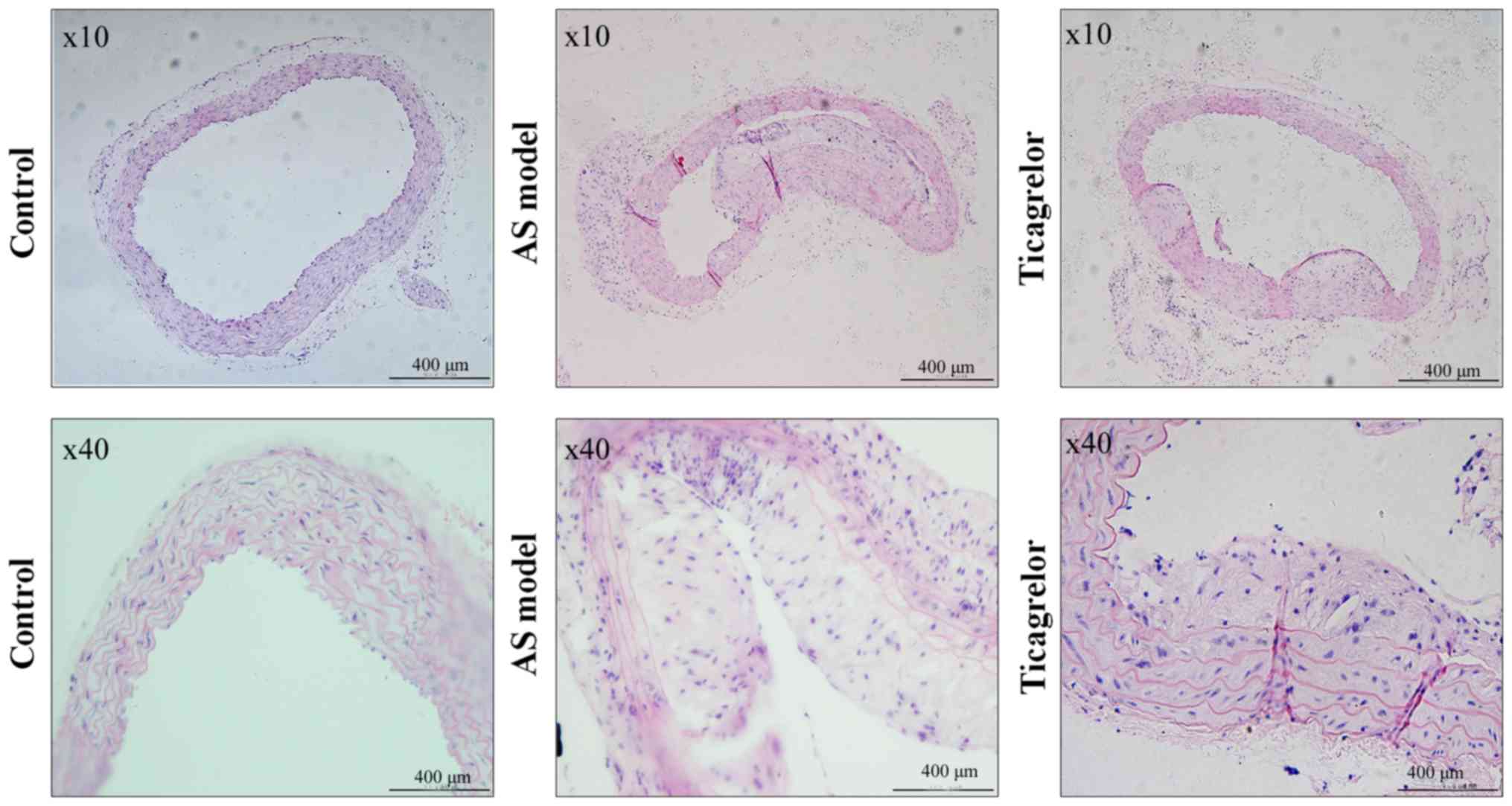
Histological analysis in the control group revealed that the
vascular endothelium of the aorta was complete, smooth and arranged
in order, without an increase in aortic arch inner membrane
thickness and formation of plaques in the arteries (Fig. 4). However, the vascular endothelium
was not intact and was even partly falling off in the AS model
group. Additionally, abundant plaque formation and lipid deposition
were observed in the lumen, accompanied by infiltration of numerous
inflammatory cells (Fig. 4).
Ticagrelor markedly decreased the atherosclerotic plaque area and
increased the lumen area (Fig. 4),
demonstrating the anti-AS effect of this drug.
| Table III.Body weight of control and
apolipoprotein E-deficient mice. |
Table III.
Body weight of control and
apolipoprotein E-deficient mice.
| Group | 0 weeks, g | 20 weeks, g |
|---|
| Control (n=10) | 14.46±0.3779 |
29.19±0.5523a |
| AS model (n=5) | 14.84±0.6562 |
31.72±0.8672a,b |
| Ticagrelor
(n=5) | 14.68±0.6160 |
31.04±0.4442a |
| Table IV.Plasma lipids profile of control and
apolipoprotein E-deficient mice. |
Table IV.
Plasma lipids profile of control and
apolipoprotein E-deficient mice.
| Group | TG, mmol/l | TC, mmol/l | HDL-C, mmol/l | HDL-L, mmol/l |
|---|
| Control (n=10) | 0.556±0.033 | 1.740±0.071 | 1.242±0.057 | 0.164±0.018 |
| AS model (n=5) |
1.220±0.129a |
19.56±1.892a |
2.860±0.246a |
6.210±0.560a |
| Ticagrelor
(n=5) |
1.550±0.338a |
16.44±1.032a |
2.554±0.292a |
4.782±0.329a |
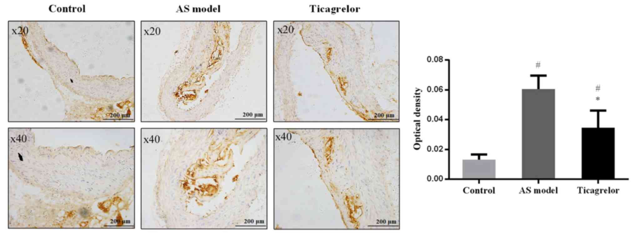
The aortas of the mice were additionally collected
to analyze the expression of PCSK9 by immunohistochemistry to
determine the association between PCSK9 and AS and the mechanism of
action of ticagrelor. As anticipated, there were no brown-yellow
particles (representing PCSK9 expression) around the blue nuclei in
the control group. In contrast, PCSK9 was abundantly expressed in
the aortic plaque of the AS group. Compared with the AS group, the
expression level of PCSK9 was significantly decreased following
treatment with ticagrelor (P<0.05; Fig. 5). These results further illustrated
that downregulation of PCSK9 may be an important mechanism for
ticagrelor to alleviate the formation and development of AS.
Discussion
Increasing evidence demonstrates that P2Y12 receptor
antagonists may improve endothelial dysfunction in patients with
acute (15), stable coronary
artery disease (16,17) or pig models with coronary stent
restenosis (18); however, the
endothelial function in these studies was determined by measuring
the vasomotor responses. The effects of P2Y12 receptor antagonists
on endothelial cell apoptosis, a key event in the pathogenesis of
AS, have not been investigated. In the present study, for the first
time to the best of the authors' knowledge, the influence of
ticagrelor, a relatively novel P2Y12 receptor inhibitor (23), on ox-LDL-induced apoptosis of the
endothelial cell line EAhy926, was examined. As expected, the
present results demonstrated that treatment with ticagrelor was
able to decrease ox-LDL-induced apoptosis, particularly at higher
doses (40 µmol/l vs. ox-LDL, 17.58±2.66 vs. 27.25±5.54%; 60 µmol/l
vs. ox-LDL, 12.26±1.54 vs. 27.25±5.54%). This is in agreement with
a previous study that demonstrated that clopidogrel reduces
palmitic acid-induced apoptosis of human vascular endothelial cells
(24). However, this finding may
be attributed to the P2Y12 receptor expressed on endothelial cells
(14) and to the interaction
between the endothelium and platelets (25), as the P2Y12 receptor is primarily
expressed in blood platelets (23). Ticagrelor may promote the
degradation of adenine nucleotides released from platelets to
adenosine (26) and thus inhibit
ADP/ATP binding to endothelial P2Y receptors (13), blocking the production of
pro-inflammatory factors (including nitric oxide and PGI2)
(27,28) and the downregulation of nucleolin
(29), which eventually suppresses
cell cycle arrest in S phase and cell apoptosis and stimulates cell
proliferation (29,30).
PCSK9 is a protein that was initially identified to
be upregulated in apoptotic neural cells, and for this reason it
was additionally termed neural apoptosis regulated convertase 1
(31,32). However, previous studies documented
that PCSK9 is additionally involved in endothelial cell apoptosis.
Knock-out of PCSK9 by small interfering RNA or shRNA inhibits
ox-LDL-induced endothelial cell apoptosis and alleviates the
formation of atherosclerotic plaques (21,33).
Therefore, it was speculated that downregulation of PCSK9 may be an
underlying mechanism for ticagrelor to inhibit endothelial cell
apoptosis. This was demonstrated, for the first time to the best of
the authors' knowledge, by the present results at the mRNA and
protein expression level. Although the way in which ticagrelor
regulates PCSK9 expression remains to be further elucidated, it is
hypothesized it may serve as a link between PCSK9 and platelets
(34). It has been demonstrated
that increased PCSK9 serum levels are positively associated with
the platelet count (r=0.218), plateletcrit (r=0.250) (35) and platelet reactivity (r=0.30) in
patients with acute coronary syndrome (36). Therefore, the anti-platelet agent
ticagrelor may influence PCSK9 expression by reducing
platelets.
Caspase-3 and Bax are common genes that serve
important roles in the process of apoptosis; whereas Bcl-2 exerts
an anti-apoptotic effect via a mitochondria-dependent caspase-9
pathway. MAPKs are a group of associated serine-threonine protein
kinases, which participate in the mediation of cell growth,
apoptosis, proliferation and function. Therefore, these genes were
predicted to be involved in ox-LDL-induced endothelial cell
apoptosis and AS. This postulation has been confirmed by numerous
studies. Chen et al (37)
demonstrated that ox-LDL is able to induce endothelial cell
apoptosis via a Bax-mitochondria-caspase protease pathway.
Furthermore, Yu et al (38)
demonstrated that ox-LDL activates the cell MAPKs, particularly JNK
and p38. Therefore, inhibition of the expression of caspase-3, Bax
and blocking of MAPK activation may protect endothelial cells
against apoptosis. In our previous study, it was observed that
shRNA-PCSK9 markedly inhibits the expression of pro-apoptotic
proteins and promotes anti-apoptotic proteins accompanied with
alteration in the phosphorylation of p38 and c-Jun N-terminal
kinases (21). This was
additionally concluded in the study conducted by Wu et al
(33). Accordingly, it was
hypothesized that suppression of these PCSK9-mediated apoptosis
signaling pathways may be the inhibitory mechanism of ticagrelor
for endothelial cell apoptosis and AS. Therefore, the expression
levels of caspase-3, Bcl-2, Bax and MAPK were additionally analyzed
following treatment with ticagrelor. In agreement with previous
studies on anti-apoptosis drugs (6,38,39),
the present results demonstrated that ticagrelor suppressed
ox-LDL-induced p38MAPK, JNK, ERK activation (particularly JNK),
caspase-3 and Bcl-2; however, upregulated Bax in EAhy926 cells.
In addition to in vitro experiments, an in
vivo study was conducted. In accordance with previous studies,
the present results demonstrated that the use of ticagrelor
inhibited the progression of AS (20) and reduced the lesion size (19). Compared with the study of Preusch
et al (19) who
additionally used ApoE−/− mice, the present results may
be of increased use to researchers as a high-fat diet was fed to
the mice.
There are some limitations in the present study.
First, further studies with quantitative assessment are still
required to confirm the therapeutic effects of ticagrelor on the
atherosclerotic plaque. Second, the MAPK agonists should be used to
further validate the mechanisms of ticagrelor in AS.
In conclusion, the present results preliminarily
demonstrated that ticagrelor may suppress ox-LDL-induced
endothelial cell apoptosis and alleviate AS by downregulating PCSK9
and the downstream apoptotic MAPK signaling pathway. These results
may provide a theoretical basis for the use of ticagrelor to treat
patients with AS in a clinical setting.
Acknowledgements
Not applicable.
Funding
The present study was supported by the National
Natural Science Foundation of China (grant nos. 81570298 and
81700304), the Research Fund for Key Laboratory of Second Hospital
of Tianjin Medical University (grant no. 2017ZDSYS02), and Youth
Research Fund for Central Laboratory of Second Hospital of Tianjin
Medical University (grant no. 2016ydey03).
Availability of data and materials
The datasets from current study are available from
the corresponding author on reasonable request.
Authors' contributions
XX, XL and GL designed the study. XX and JL
performed the experiments. SZ and JYL were involved in the
statistical analyses. TL and MA participated in the interpretation
of the data. XX, XL and GL wrote and revised the manuscript. All
authors read and approved the final manuscript.
Ethics approval and consent to
participate
All animal experiments conformed with the Regulation
of Animal Care Management of the Ministry of Public Health,
People's Republic of China and were approved by the Ethical
Committee of Second Hospital of Tianjin Medical University
(Tianjin, China).
Patient consent for publication
Not applicable.
Competing interests
The authors declare that they have no competing
interests.
References
|
1
|
Clarke R, Du H, Kurmi O, Parish S, Yang M,
Arnold M, Guo Y, Bian Z, Wang L, Chen Y, et al: Burden of carotid
artery atherosclerosis in Chinese adults: Implications for future
risk of cardiovascular diseases. Eur J Prev Cardiol. 24:647–656.
2017. View Article : Google Scholar : PubMed/NCBI
|
|
2
|
Barquera S, Pedroza-Tobías A, Medina C,
Hernández-Barrera L, Bibbins-Domingo K, Lozano R and Moran AE:
Global overview of the epidemiology of atherosclerotic
cardiovascular disease. Arch Med Res. 46:328–338. 2015. View Article : Google Scholar : PubMed/NCBI
|
|
3
|
Mathers CD and Loncar D: Projections of
global mortality and burden of disease from 2002 to 2030. PLoS Med.
3:e4422006. View Article : Google Scholar : PubMed/NCBI
|
|
4
|
Chen M, Masaki T and Sawamura T: LOX-1,
the receptor for oxidized low-density lipoprotein identified from
endothelial cells: Implications in endothelial dysfunction and
atherosclerosis. Pharmacol Ther. 95:89–100. 2002. View Article : Google Scholar : PubMed/NCBI
|
|
5
|
Hong D, Bai YP, Gao HC, Wang X, Li LF,
Zhang GG and Hu CP: Ox-LDL induces endothelial cell apoptosis via
the LOX-1-dependent endoplasmic reticulum stress pathway.
Atherosclerosis. 235:310–317. 2014. View Article : Google Scholar : PubMed/NCBI
|
|
6
|
Bao MH, Zhang YW and Zhou HH: Paeonol
suppresses oxidized low-density lipoprotein induced endothelial
cell apoptosis via activation of LOX-1/p38MAPK/NF-κB pathway. J
Ethnopharmacol. 146:543–551. 2013. View Article : Google Scholar : PubMed/NCBI
|
|
7
|
Xu F, Sun Y, Chen Y, Sun Y, Li R, Liu C,
Zhang C, Wang R and Zhang Y: Endothelial cell apoptosis is
responsible for the formation of coronary thrombotic
atherosclerotic plaques. Tohoku J Exp Med. 218:25–33. 2009.
View Article : Google Scholar : PubMed/NCBI
|
|
8
|
Durand E, Scoazec A, Lafont A, Boddaert J,
Al Hajzen A, Addad F, Mirshahi M, Desnos M, Tedgui A and Mallat Z:
In vivo induction of endothelial apoptosis leads to vessel
thrombosis and endothelial denudation a clue to the understanding
of the mechanisms of thrombotic plaque erosion. Circulation.
109:2503–2506. 2004. View Article : Google Scholar : PubMed/NCBI
|
|
9
|
Badimon L and Vilahur G: Thrombosis
formation on atherosclerotic lesions and plaque rupture. J Intern
Med. 276:618–632. 2015. View Article : Google Scholar
|
|
10
|
Raskob GE, Angchaisuksiri P, Blanco AN,
Büller H, Gallus A, Hunt BJ, Hylek EM, Kakkar TL, Konstantinides
SV, Mccumber M, et al: Thrombosis: A major contributor to global
disease burden. Semin Thromb Hemost. 40:724–735. 2014. View Article : Google Scholar : PubMed/NCBI
|
|
11
|
Zhang Y, Mu Q, Zhou Z, Song H, Zhang Y, Wu
F, Jiang M, Wang F, Zhang W, Li L, et al: Protective effect of
irisin on atherosclerosis via suppressing oxidized low density
lipoprotein induced vascular inflammation and endothelial
dysfunction. PLoS One. 11:e01580382016. View Article : Google Scholar : PubMed/NCBI
|
|
12
|
Wang L, Hao Q, Wang YD, Wang WJ and Li DJ:
Protective effects of dehydroepiandrosterone on atherosclerosis in
ovariectomized rabbits via alleviating inflammatory injury in
endothelial cells. Atherosclerosis. 214:47–57. 2011. View Article : Google Scholar : PubMed/NCBI
|
|
13
|
Wang GF, Shi CG, Sun MZ, Wang L, Wu SX,
Wang HF, Xu ZQ and Chen DM: Tetramethylpyrazine attenuates
atherosclerosis development and protects endothelial cells from
ox-LDL. Cardiovasc Drugs Ther. 27:199–210. 2013. View Article : Google Scholar : PubMed/NCBI
|
|
14
|
Lee CW, Hwang I, Park CS, Lee H, Park DW,
Kang SJ, Lee SW, Kim YH Park SW and Park SJ: Comparison of
differential expression of P2Y12 receptor in culprit
coronary plaques in patients with acute myocardial infarction
versus stable angina pectoris. Am J Cardiol. 108:799–803. 2011.
View Article : Google Scholar : PubMed/NCBI
|
|
15
|
Jeong HS, Hong SJ, Cho SA, Kim JH, Cho JY,
Lee SH, Joo HJ, Park JH, Yu CW and Lim DS: Comparison of ticagrelor
versus prasugrel for inflammation, vascular function, and
circulating endothelial progenitor cells in diabetic patients with
non-ST-segment elevation acute coronary syndrome requiring coronary
stenting. JACC Cardiovasc Interv. 10:1646–1658. 2017. View Article : Google Scholar : PubMed/NCBI
|
|
16
|
Warnholtz A, Ostad MA, Velich N, Trautmann
C, Schinzel R, Walter U and Munzel T: A single loading dose of
clopidogrel causes dose-dependent improvement of endothelial
dysfunction in patients with stable coronary artery disease:
Results of a double-blind, randomized study. Atherosclerosis.
196:689–695. 2008. View Article : Google Scholar : PubMed/NCBI
|
|
17
|
Willoughby SR, Luu LJ, Cameron JD, Nelson
AJ, Schultz CD, Worthley SG and Worthley MI: Clopidogrel improves
microvascular endothelial function in subjects with stable coronary
artery disease. Heart Lung Circ. 23:534–541. 2014. View Article : Google Scholar : PubMed/NCBI
|
|
18
|
Kim HK, Jeong MH, Lim KS, Kim JH, Lim HC,
Kim MC, Hong YJ, Kim SS, Park KH and Chang KS: Effects of
ticagrelor on neointimal hyperplasia and endothelial function,
compared with clopidogrel and prasugrel, in a porcine coronary
stent restenosis model. Int J Cardiol. 240:326–331. 2017.
View Article : Google Scholar : PubMed/NCBI
|
|
19
|
Preusch MR, Rusnak J, Staudacher K, Mogler
C, Uhlmann L, Sievers P, Bea F, Katus HA, Blessing E and Staudacher
I: Ticagrelor promotes atherosclerotic plaque stability in a mouse
model of advanced atherosclerosis. Drug Des Devel Ther.
10:2691–2699. 2016. View Article : Google Scholar : PubMed/NCBI
|
|
20
|
Ren H, Li ML, Jiang J, Zhang Y, Zhang Y
and Zhu X: Effects of clopidogrel on vascular proliferation and
apoptosis in an atherosclerotic rabbit model. J Cardiovasc
Pharmacol. 55:617–624. 2010. View Article : Google Scholar : PubMed/NCBI
|
|
21
|
Li J, Liang X, Wang Y, Xu Z and Li G:
Investigation of highly expressed PCSK9 in atherosclerotic plaques
and ox-LDL-induced endothelial cell apoptosis. Mol Med Rep.
16:1817–1825. 2017. View Article : Google Scholar : PubMed/NCBI
|
|
22
|
Livak KJ and Schmittgen TD: Analysis of
relative gene expression data using real-time quantitative PCR and
the 2(-Delta Delta C(T)) method. Methods. 25:402–408. 2001.
View Article : Google Scholar : PubMed/NCBI
|
|
23
|
Capodanno D, Dharmashankar K and
Angiolillo DJ: Mechanism of action and clinical development of
ticagrelor, a novel platelet ADP P2Yreceptor antagonist. Expert Rev
Cardiovasc Ther. 8:151–158. 2010. View Article : Google Scholar : PubMed/NCBI
|
|
24
|
Wang J, Chen L, Li H, Yang J, Gong Z, Wang
B and Zhao X: Clopidogrel reduces apoptosis and promotes
proliferation of human vascular endothelial cells induced by
palmitic acid via suppression of the long non-coding RNA HIF1A-AS1
in vitro. Mol Cell Biochem. 404:203–210. 2015. View Article : Google Scholar : PubMed/NCBI
|
|
25
|
Sachais BS: Platelet-endothelial
interactions in atherosclerosis. Curr Atheroscler Rep. 3:412–416.
2001. View Article : Google Scholar : PubMed/NCBI
|
|
26
|
Nylander S, Femia EA, Scavone M, Berntsson
P, Asztély AK, Nelander K, Löfgren L, Nilsson RG and Cattaneo M:
Ticagrelor inhibits human platelet aggregation via adenosine in
addition to P2Y12 antagonism. J Thromb Haemost. 11:1867–1876.
2013.PubMed/NCBI
|
|
27
|
Heitzer T, Rudolph V, Schwedhelm E,
Karstens M, Sydow K, Ortak M, Tschentscher P, Meinertz T, Böger R
and Baldus S: Clopidogrel improves systemic endothelial nitric
oxide bioavailability in patients with coronary artery disease:
Evidence for antioxidant and antiinflammatory effects. Arterioscler
Thromb Vasc Biol. 26:1648–1652. 2006. View Article : Google Scholar : PubMed/NCBI
|
|
28
|
Gwozdz P, Csanyi G, Luzak B, Gajda M,
Mateuszuk L, Chmura-Skirlinska A, Watala C and Chlopicki S:
Endothelial dysfunction and circulating platelet activation in
apoE/LDLR/-mice along the development of atherosclerosis. In:
Conference on Frontiers in Cardiovascular Biology. Cardiovasc Res.
87 Suppl:S94. 2010.
|
|
29
|
Wang W, Luo J, Xiang F, Liu X, Jiang M,
Liao L and Hu J: Nucleolin down-regulation is involved in
ADP-induced cell cycle arrest in S phase and cell apoptosis in
vascular endothelial cells. PLoS One. 9:e1101012014. View Article : Google Scholar : PubMed/NCBI
|
|
30
|
Coutinho-Silva R, Stahl L, Cheung KK, de
Campos NE, de Oliveira Souza C, Ojcius DM and Burnstock G: P2X and
P2Y purinergic receptors on human intestinal epithelial carcinoma
cells: Effects of extracellular nucleotides on apoptosis and cell
proliferation. Am J Physiol Gastrointest Liver Physiol.
288:G1024–G1035. 2005. View Article : Google Scholar : PubMed/NCBI
|
|
31
|
Seidah NG, Benjannet S, Wickham L,
Marcinkiewicz J, Jasmin SB, Stifani S, Basak A, Prat A and Chrétien
M: The secretory proprotein convertase neural apoptosis-regulated
convertase 1 (NARC-1): Liver regeneration and neuronal
differentiation. Proc Natl Acad Sci USA. 100:928–933. 2003.
View Article : Google Scholar : PubMed/NCBI
|
|
32
|
Kysenius K, Muggalla P, Mätlik K, Arumäe U
and Huttunen HJ: PCSK9 regulates neuronal apoptosis by adjusting
ApoER2 levels and signaling. Cell Mol Life Sci. 69:1903–1906. 2012.
View Article : Google Scholar : PubMed/NCBI
|
|
33
|
Wu CY, Tang ZH, Jiang L, Li XF, Jiang ZS
and Liu LS: PCSK9 siRNA inhibits HUVEC apoptosis induced by ox-LDL
via Bcl/Bax-caspase9-caspase3 pathway. Mol Cell Biochem.
359:347–358. 2012. View Article : Google Scholar : PubMed/NCBI
|
|
34
|
Gurbel PA, Navarese EP and Tantry US:
Exploration of PCSK9 as a cardiovascular risk factor: Is there a
link to the platelet? J Am Coll Cardiol. 70:1463–1466. 2017.
View Article : Google Scholar : PubMed/NCBI
|
|
35
|
Li S, Zhu CG, Guo YL, Xu RX, Zhang Y, Sun
J and Li JJ: The relationship between the plasma PCSK9 levels and
platelet indices in patients with stable coronary artery disease. J
Atheroscler Thromb. 22:76–84. 2015. View Article : Google Scholar : PubMed/NCBI
|
|
36
|
Navarese EP, Kolodziejczak M, Winter MP,
Alimohammadi A, Lang IM, Buffon A, Lip GY and Siller-Matula JM:
Association of PCSK9 with platelet reactivity in patients with
acute coronary syndrome treated with prasugrel or ticagrelor: The
PCSK9-REACT study. Int J Cardiol. 227:644–649. 2017. View Article : Google Scholar : PubMed/NCBI
|
|
37
|
Chen TG, Chen TL, Chang HC, Tai YT, Cherng
YG, Chang YT and Chen RM: Oxidized low-density lipoprotein induces
apoptotic insults to mouse cerebral endothelial cells via a
Bax-mitochondria-caspase protease pathway. Toxicol Appl Pharmacol.
219:42–53. 2007. View Article : Google Scholar : PubMed/NCBI
|
|
38
|
Yu W, Ying H, Tong F, Zhang C, Quan Y and
Zhang Y: Protective effect of the silkworm protein 30Kc6 on human
vascular endothelial cells damaged by oxidized low density
lipoprotein (Ox-LDL). PLoS One. 8:e687462013. View Article : Google Scholar : PubMed/NCBI
|
|
39
|
Zhang L, Jia YH, Zhao XS, Zhou FH, Pan YY,
Wan Q, Cui XB, Sun XG, Chen YY, Zhang Y and Cheng SB:
Trichosanatine alleviates oxidized low-density lipoprotein induced
endothelial cells injury via inhibiting the LOX-1/p38 MAPK pathway.
Am J Transl Res. 8:5455–5464. 2016.PubMed/NCBI
|