Introduction
Allergic diseases, including allergic asthma,
allergic rhinitis and allergic dermatitis, affect 30–40% of the
world population; furthermore, incidence and mortality rates
associated with allergic diseases are increasing, particularly
within young populations (1). At
present, >50% of allergic diseases are induced by house dust
mites (HDMs), a common source of inhaled allergens (2). A total of 37 HDM allergen groups have
been denominated (http://www.allergen.org); of these, the allergen
groups 1 and 2 are the most clinically relevant, with >80 and
>90% of patients with HDM allergies exhibiting an immunoglobulin
(Ig)E response to groups 1 and 2 HDM allergens, respectively
(3).
IgE is an important pathogenic mediator of allergic
immune responses. The detection of allergen-specific IgE is an
effective diagnostic method and anti-IgE therapy is used to treat
IgE-mediated allergic diseases (4). The indirect IgE-ELISA method is
frequently used to detect allergen-specific IgE in serum samples,
due to its simplicity and low cost compared with automated
ImmunoCAP® systems; however, there are certain
limitations (5). The normal range
of the human serum levels of IgE is 50–300 ng/ml, which is notably
low compared with that of IgG (~10 mg/ml) (6). In addition, the levels of HDM
allergen-specific IgE, even in sera from patients with HDM
allergies, are markedly decreased compared with the normal range of
total IgE (7). Thus, the
sensitivity of the indirect ELISA method is reduced by high titers
of IgG that compete to bind with coated antigens (8). Furthermore, indirect ELISA frequently
uses an HDM extract mixture as a coated antigen; the sensitivity of
this method is reduced by the low amounts of effective allergen
components in these mixtures.
Recombinant allergens are increasingly used in the
diagnosis and treatment of allergic diseases, due to their high
purity and consistency (9). Howard
et al (10) defined the
evolution of IgE responses to 112 recombinant or native allergen
components during childhood, which may aid the identification of
better diagnostic and prognostic biomarkers of allergic diseases.
Mas et al (11) reported
the use of the recombinant protein Salsola Kali in the diagnosis of
allergic disease induced by Salsola kali. The combined
expression of various antigens or major antigen epitopes as a
fusion protein may increase the sensitivity of detection of
antibodies targeted against allergens from a particular organism.
He et al (12) engineered a
recombinant antigen with epitopes from four hepatitis C viral
fragments to aid the detection of anti-hepatitis C antibodies. Dai
et al (13) constructed and
overexpressed a fusion gene comprising three Mycobacterium
tuberculosis antigen proteins; using the fusion polyprotein as
an immunogen, multi-target antibodies were produced that exhibited
significantly increased sensitivity for the clinical diagnosis of
tuberculosis than mono-target antibodies reactive to the three
respective antigens.
Omalizumab is a recombinant DNA-derived humanized
IgG monoclonal antibody that suppresses allergic symptoms by
binding to human IgE (14); thus,
free IgE levels are reduced, preventing interactions between IgE
and immune cells and decreasing the serum levels of inflammatory
mediators (15). The detection of
allergen-specific IgE is required for the diagnosis and management
of IgE-mediated allergic disease. In the present study, two fusion
allergens derived from the major allergenic HDM species
Dermatophagoides farina (Der f), Der f 1 and Der f 2, were cloned
and expressed. Subsequently, a novel capture IgE-ELISA was
developed using a recombinant Der f 1/2 fusion protein (rDer f
1/2), which was designed to improve the sensitivity of HDM
allergen-specific IgE detection. The capture ELISA method involved
the coating of wells with omalizumab to enrich serum IgE and reduce
interference from IgG. Preliminary experiments were conducted using
the assay to determine its reliability for accurate anti-allergen
detection and test the potential of rDer f 1/2 fusion protein-based
ELISAs for the diagnosis of HDM-allergic disease.
Materials and methods
Serum samples, reagents and
antibodies
All serum samples from HDM-sensitive individuals
(HDM-allergic sera) and non-allergic individuals (control sera)
were provided by The First Affiliated Hospital of Guangzhou Medical
College (Guangzhou, China) between March 2013 and July 2015. A
total of 28 subjects (13 males and 15 females, 18–55 years old)
were enrolled in the present study. The patients were subjected to
a skin prick test using dust mite allergen extract, serum samples
were separated by centrifugation at 500 × g for 15 min at room
temperature for the detection of IgE using the ImmunoCAP allergen
detection system (Phadia AB; Thermo Fisher Scientific, Inc.,
Waltham, MA, USA). IgE levels were defined as follows: Level 3,
3.5–17.5 IU/ml; level 4, 17.5–50.0 IU/ml; level 5, 50.0–100 IU/ml
and level 6, >100 IU/ml. The clinicopathological characteristics
of the patients are presented in Table
I. A random cohort of 71 HDM-allergic serum samples (37 males
and 34 females, 18–55 years old) were used to determine the
sensitivity of capture IgE-ELISA and indirect IgE-ELISA. In total,
20 non-allergic individuals (8 males and 12 females, 18–55 years
old) were used as negative controls. Ethical approval was obtained
from The First Affiliated Hospital of Guangzhou Medical College and
patients provided informed consent.
 | Table I.Clinicopathological factors of the
patients. |
Table I.
Clinicopathological factors of the
patients.
| Patient no. | Gender/age | ImmunoCAP
HDM-specific IgE testa | Clinical
history |
|---|
| 1 | F/45 | 3+ | AD |
| 2 | M/32 | 3+ | AR |
| 3 | F/18 | 3+ | – |
| 4 | F/50 | 3+ | AD |
| 5 | M/53 | 3+ | BA |
| 6 | M/34 | 3+ | – |
| 7 | F/31 | 3+ | BA |
| 8 | F/55 | 4+ | AR + BA |
| 9 | M/42 | 4+ | AR |
| 10 | F/24 | 4+ | AD |
| 11 | F/37 | 4+ | BA |
| 12 | F/52 | 4+ | BA |
| 13 | M/18 | 4+ | – |
| 14 | M/29 | 4+ | AR |
| 15 | F/35 | 5+ | – |
| 16 | M/37 | 5+ | AD |
| 17 | F/28 | 5+ | AR |
| 18 | M/53 | 5+ | AD + AR |
| 19 | F/48 | 5+ | AR |
| 20 | F/21 | 5+ | AD |
| 21 | M/46 | 5+ | BA |
| 22 | F/33 | 6+ | −b |
| 23 | M/41 | 6+ | AR |
| 24 | F/54 | 6+ | AD + BA |
| 25 | M/34 | 6+ | AR |
| 26 | M/21 | 6+ | AD |
| 27 | F/44 | 6+ | BA |
| 28 | F/27 | 6+ | AD + AR |
The restriction enzymes XhoI, NdeI and
pET28 plasmid were purchased from Takara Biotechnology Co., Ltd.
(Dalian, China). E. coli BL21(DE3)plysS cells were purchased
from Invitrogen (Thermo Fisher Scientific, Inc.).
Isopropyl-β-D-thiogalactopyranoside (IPTG) was purchased from
Sigma-Aldrich (Merck KGaA, Darmstadt, Germany). A Pierce™
3′-diaminobenzidine (DAB) kit was purchased from Pierce (Thermo
Fisher Scientific, Inc.). Biotinylated mouse anti-human IgE
antibody (cat. no. 9160-08), streptavidin-labeled horseradish
peroxidase (HRP; cat. no. 7100-05) and mouse anti-human IgE
HRP-labeled antibody (cat. no. 9160-05) were purchased from
SouthernBiotech (Birmingham, AL, USA).
Construction of the rDer f 1/2 fusion
expression vector
The pET28-Der f 1 and pET28-Der f 2 (Takara
Biotechnology Co., Ltd.) expression vectors were constructed as
previously described (16,17). Overlapping polymerase chain
reactions were performed to splice the Der f 1 and Der f 2 genes.
The construction of pET28-rDer f 1/2 plasmid was confirmed by DNA
Sanger sequencing (Sangon Biotech Co., Ltd., Shanghai, China). The
pET28-Der f 1/2 expression vector was engineered using NdeI and
XhoI restriction sites. The fusion sequence encoding Der f 1/2 was
reported in GenBank (accession no. MF074325.1; http://www.ncbi.nlm.nih.gov/nuccore/MF074325.1). Der f
1 amino acid residues 19–321 were linked to Der f 2 residues 18–146
via a GGGSS linker. The resultant recombinant protein contained a
hexahistidine (6×His) tag in its C-terminus.
Expression and purification of rDer f
1, rDer f 2 and rDer f 1/2 proteins
In total, 5 µl recombinant plasmid pET28-Der f 1/2
was transformed into 50 µl Escherichia coli BL21(DE3) pLysS cells
using heat shock method (18).
Cells were inoculated in Luria-Bertani medium (Merck KGaA,
Darmstadt, Germany) containing 0.01 mg/ml kanamycin at 37°C for
overnight. IPTG (1 mM)-induced expression was observed following
growth for 3 h at 37°C, at which time cells were harvested by
centrifugation at 8,000 × g for 2 min at 4°C. Pellets were
resuspended in buffer (20 mM Tris-HCl, 150 mM NaCl, pH 8.0) and the
cells were lysed by ultrasonic homogenization. The supernatant and
precipitate were collected and analyzed via SDS-PAGE (described
below). Gels were stained with Coomassie G-250 Brilliant Blue for
protein analysis as previously described (19). rDer f 1, rDer f 2 and rDer f 1/2 in
inclusion body fractions were solubilized with 6 M guanidine
hydrochloride in 100 mM Tris (pH 8.0) for 2 h at room temperature
(20). The proteins were purified
by nickel affinity chromatography (Ni Sepharose 6 Fast Flow; cat.
no. 17531801; GE Healthcare, Chicago, IL, USA) under denaturing
conditions as previously described (21). Inclusion body surfaces frequently
contain DNA, endotoxins and heteroproteins; washing with low
concentrations of denaturing agent reduces impurities, thereby
improving inclusion body protein purity (22). Deagglomeration of inclusion bodies
into free loose structures with high concentrations of guanidine
hydrochloride enables the material to be stored in buffer in a
soluble state. The solution was treated with 20 mM
β-mercaptoethanol for 30 min prior to diluting renaturation.
Denatured proteins were refolded and diluted 10-fold with 0.5 M
L-arginine (pH 8.0) (23).
Arginine buffer was added to the denatured protein solution slowly
(flow rate of 0.5 ml/min) and incubated overnight at 4°C. The
solution was then dialyzed for 24 h using buffer containing 20 mM
Tris and 150 mM NaCl. Following renaturation, protein
concentrations were determined by the Bradford method.
IgE-western blotting and IgE-dot
blotting
In total, 20 µg of rDer f 1/2 fusion protein was
loaded in each well. Proteins were separated by 12% SDS-PAGE and
then transferred to polyvinylidene difluoride membranes for western
blotting. For dot blotting, 2 µl of the allergens (rDer f 1, rDer f
2 and rDer f 1/2) was separately spotted onto nitrocellulose
membranes at a concentration of 1 µg/µl. The membranes were blocked
with 5% Difco™ Skim Milk (DSM; BD Biosciences, San Jose, CA, USA)
diluted in TBS containing 0.05% Tween 20 (TBST) at 4°C overnight.
Serum samples from 15 HDM-allergic patients and 15 control subjects
were also analyzed and incubated for 2 h at 37°C. Subsequently,
mouse anti-human IgE Fc-HRP antibody was applied (1:2,000 in TBST
with 1% DSM) for 1 h at 37°C. Antigen-antibody complexes on
membranes were visualized using a Pierce™ DAB kit.
Preparation of biotinylated Der f 1/2
fusion protein
rDer f 1/2 contained a 6×His tag attached to its
C-terminus. It was biotinylated using EZ-Link Sulfo-NHS-Biotin
reagents (Thermo Fisher Scientific, Inc.), which enable simple and
efficient molecular labeling (24). Fusion protein dissolved in PBS (pH
7.2) was incubated with 10 mM biotin reagent solution at room
temperature for 30 min. Upon demonstration of protein labeling, the
labeled fusion protein was purified by desalting column (HiTrap
Desalting; GE Healthcare) in preparation for capture ELISA.
Indirect and capture IgE-ELISA
The IgE binding activities of recombinant proteins
were detected by indirect and capture IgE-ELISAs. For indirect
IgE-ELISA, 96-well plates were coated with recombinant antigen (100
ng/well, diluted in carbonate buffer, pH 9.6) at 4°C overnight. The
plates were then blocked with 5% (w/v) DSM in PBS containing 0.05%
Tween 20 (PBST) for 3 h at 37°C. Following washing, the plates were
incubated with serum from HDM-allergic patients (1:5) for 2 h at
37°C, followed by incubation with mouse anti-human IgE
biotin-labeled (1:2,000) for 1.5 h at 37°C. The plates were washed
and subsequently incubated with streptavidin-HRP (1:4,000) for 30
min at 37°C. The plates were washed with PBST. Bound
biotinylated-labeled antibody was detected by adding 100 µl of
3,3′,5,5′-tetramethylbenzidine (TMB; 1 mM); the reaction was
stopped with 50 µl of H2SO4 (2 M). For
capture IgE-ELISA, wells were coated with omalizumab (500 ng/well)
overnight at 4°C. The wells were blocked with 5% DSM for 3 h at
37°C and HDM-allergic human serum (1:5) was subsequently added to
the wells prior to incubation for 2 h at 37°C. To select an optimal
concentration, a standard serial dilution (50, 100, 200 and 400
ng/well) of biotinylated rDer f 1/2 was initially employed for 1 h
at 37°C; 50 ng/well was selected for subsequent experiments. Each
protein solution was incubated in wells with streptavidin-HRP for
0.5 h at 37°C, prior to addition of TMB. The TMB reaction was
conducted for 10 min at 37°C prior to the application of
H2SO4. For indirect and capture IgE-ELISAs,
the absorbance was measured at 450 nm by a microplate reader
(Bio-Rad Laboratories, Inc., Hercules, CA, USA).
Statistical analysis
The experimental data were presented as the mean ±
standard error of the mean. Data were analyzed using GraphPad Prism
7 (GraphPad Software, Inc., La Jolla, CA). Differences between
groups were determined by analysis of one-way variance followed by
Dunnett's t-test for multiple comparisons. P<0.05 was considered
to indicate a statistically significant difference.
Results
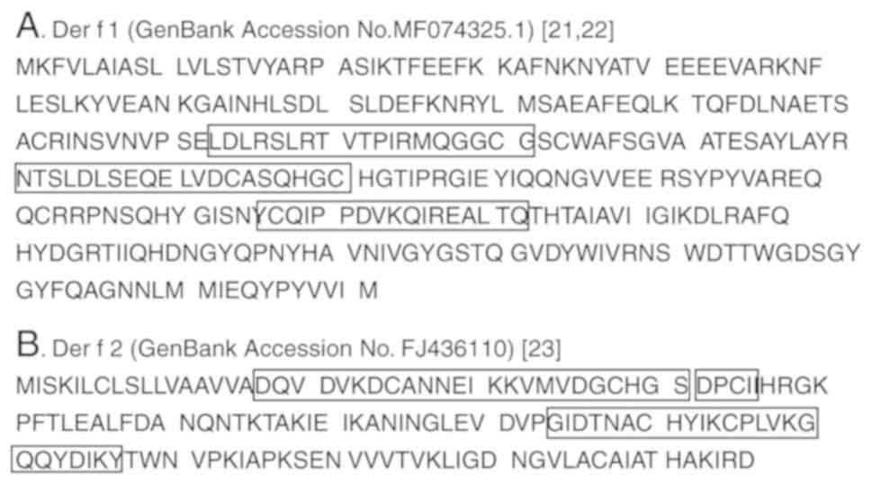
Linear IgE B cell epitopes of Der f 1
and Der f 2
B cell epitopes are antigenic regions in proteins
recognized by B cells and are considered to be important for the
diagnosis of allergies via the serum (25). The literature was investigated to
identify linear IgE B cell epitopes in Der f 1 and Der f 2; it was
revealed that Der f 1 (GenBank accession no. EF139428.1) and Der f
2 (GenBank accession no. FJ436110) separately possess three B cell
epitopes. The amino acid sequences of the epitopes in Der f 1 and
Der f 2 (26–28) are presented in Fig. 1. It was hypothesized that Der f
1/2, a fusion protein comprising these B cell epitopes, would bind
with a broader range of specific IgEs, rendering it suitable for
the diagnosis of HDM allergy.
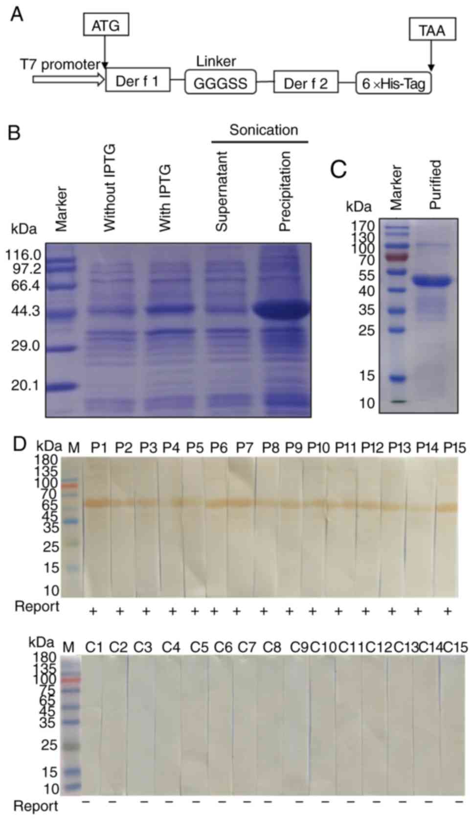
Expression, purification and IgE
binding activity of rDer f 1/2
A schematic diagram of the rDer f 1/2 construct is
presented in Fig. 2A. The rDer f
1/2 protein was expressed in the form of inclusion bodies,
solubilized with guanidine hydrochloride and identified by SDS-PAGE
(Fig. 2B). Purification of rDer f
1/2 by nickel affinity chromatography resulted in the presence of a
single band migrating at a theoretical molecular weight of 46 kDa
(Fig. 2C). IgE western blot
analysis revealed that rDer f 1/2 bound to IgEs when incubated with
serum samples from 15 patients with diagnosed HDM allergies, but
did not bind to IgEs in sera obtained from the 15 non-allergic
control subjects (Fig. 2D). These
results indicated that rDer f 1/2 exhibited a selective, strong
binding affinity for IgE in HDM-allergic sera.
Analysis of the binding of specific
IgE to rDer f 1, rDer f 2 and rDer f 1/2
SDS-PAGE of purified E. coli-expressed rDer f 1,
rDer f 2 and rDer f 1/2 indicated that rDer f 1 and rDer f 2
possessed molecular weights of ~32 and 15 kDa, respectively
(Fig. 3A). In a previous study,
6×His-tagged antibody assay revealed two bands for Der f 1, which
may be due to minor differences in molecular weight or structural
differences during the expression process, potentially occurring at
the site of dissolution and degradation (16). Non-denaturing PAGE (without SDS or
β-mercaptoethanol) indicated that rDer f 2 forms a dimer (data not
shown) (29). Indirect ELISAs, in
which the three recombinant proteins were incubated separately with
HDM-allergic (n=28) and control sera (n=10), yielded mean optical
density (OD) values of 1.293±0.241 for Der f 1, 1.377±0.26 for Der
f 2 and 1.795±0.31 for Der f 1/2 (Fig.
3B). Using a cutoff OD value of 0.7, the reported IgE-positive
rates for rDer f 1, rDer f 2 and rDer f 1/2 were 21/28 (75.0%),
22/28 (78.6%) and 24/28 (85.8%), respectively (Table II). All OD values for ELISAs
performed with Der f 1/2 were increased compared with those of rDer
f 1 or rDer f 2 when using HDM-allergic sera (Fig. 3C). A dot-blot assay revealed a
stronger signal for rDer f 1/2 than rDer f 1, or rDer f 2 following
incubation with an HDM-allergic serum pool from 3 individual
patients; negative results were observed for all three proteins
when incubated with pooled control serum (Fig. 3D). Collectively, the results
indicated that rDer f 1/2 exhibited improved binding to HDM
allergen-specific IgEs compared with rDer f 1 or rDer f 2.
 | Figure 3.Analysis of the binding of specific
IgE to rDer f 1, rDer f 2 and rDer f 1/2 in HDM sera. (A) SDS-PAGE
of purified recombinant proteins. (B) Levels of binding of rDer f
1, rDer f 2 and rDer f 1/2 to IgE in 28 HDM and 10 control sera as
determined by indirect ELISA. (C) Binding of rDer f 1, rDer f 2,
and rDer f 1/2 to specific IgE in sera from individuals with HDM
allergies (P1, P6 and P15) and non-allergic controls (C1 and C2) as
determined by indirect ELISA. (D) Recombinant proteins were dotted
onto nitrocellulose strips and incubated with mixed sera from 3
HDM-allergic patients or 3 non-allergic individuals. Data are
presented as the mean ± standard error of the mean. *P<0.05.
Control sera, sera from non-allergic control individuals; Der f,
allergen from Dermatophagoides farina; HDM, house dust mite; HDM
sera, sera from patients with HDM allergies; Ig, immunoglobulin;
OD, optical density; r, recombinant. |
| Table II.Positive rate of IgE-ELISA for sera
from patients with HDM-allergic disease as determined using rDer f
1, rDer f 2 and rDer f 1/2. |
Table II.
Positive rate of IgE-ELISA for sera
from patients with HDM-allergic disease as determined using rDer f
1, rDer f 2 and rDer f 1/2.
| Allergen | Positive rate
[cutoff (P/N>2.1)] (%) | Positive rate
(cutoff=0.7)a
(%) |
|---|
| Der f 1 | 27/28 (96.4) | 21/28 (75.0) |
| Der f 2 | 27/28 (96.4) | 22/28 (78.6) |
| Der f 1/2 | 28/28 (100) | 24/28 (85.8) |
Enhanced sensitivity of capture
IgE-ELISA based on rDerf1/2
Schematic diagrams of the capture and indirect
IgE-ELISA methods employed for the detection of HDM-specific IgEs
are presented in Fig. 4A and B,
respectively. A preliminary assay performed to optimize the rDer f
1/2-biotin conjugate concentration used during the assay revealed
that 0.5 µg/ml was optimal for capture IgE-ELISA, based upon the
relative OD values following incubation with HDM-allergic and
control sera (Fig. 5A).
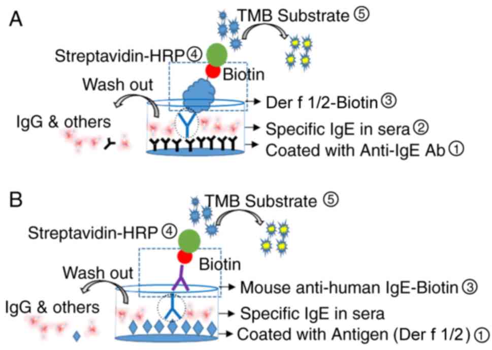 | Figure 4.Schematic diagrams of direct capture
IgE-ELISA and indirect IgE-ELISA. (A) Direct capture and (B)
indirect IgE-ELISAs for the detection of specific IgEs in
HDM-allergic sera. For capture IgE-ELISA, 96-well plates were
coated with anti-IgE Ab (omalizumab), blocked and incubated with
HDM-allergic sera containing HDM-specific IgE, prior to incubations
with rDer f 1/2-Biotin and streptavidin-HRP. For indirect
IgE-ELISA, plates were coated with rDer f 1/2, blocked and
incubated with HDM-allergic sera, prior to incubations with
biotinylated mouse anti-human IgE and streptavidin-HRP. For the two
methods, TMB substrate was subsequently added; the reaction was
terminated following the addition of 2 M H2SO4. Ab, antibody; Der
f, allergen from Dermatophagoides farina; Der f 1/2-biotin,
biotinylated Der f 1/2 protein; HDM, house dust mite; HDM-allergic
sera, sera from patients with HDM allergies; HRP, horseradish
peroxidase; Ig, immunoglobulin; r, recombinant; TMB,
3,3′,5,5′-tetramethylbenzidine. |
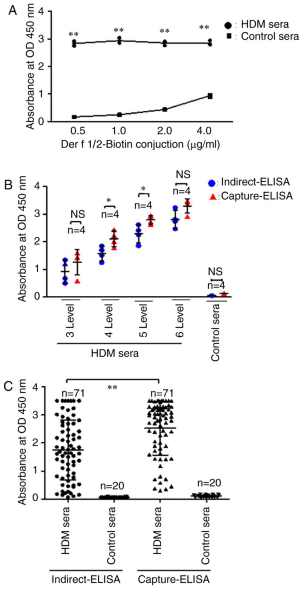 | Figure 5.Capture-ELISA detection of specific
IgEs in HDM sera. (A) Identification of optimal Der f1/2-biotin
conjugate concentration for capture ELISA. (B) Sensitivity of
indirect and capture ELISAs in the detection of allergen-specific
IgE in HDM serum samples with varying levels of IgE as determined
by ImmunoCAP. IgE levels were defined as follows: Level 3, 3.5–17.5
IU/ml; level 4, 17.5–50.0 IU/ml; level 5, 50.0–100 IU/ml and level
6, >100 IU/ml. (C) Sensitivity of indirect and capture ELISAs in
the detection of allergen-specific IgE in a random cohort of HDM
serum samples (n=71). Data are presented as the mean ± standard
error of the mean. *P<0.05, **P<0.01. Control sera, sera from
non-allergic control individuals; Der f, allergen from
Dermatophagoides farina; Der f 1/2-biotin, biotinylated Der f 1/2
protein; HDM, house dust mite; HDM sera, sera from patients with
HDM allergies; Ig, immunoglobulin; OD, optical density. |
Subsequently, the relative sensitivity of rDer f
1/2-based capture IgE-ELISA compared with indirect IgE-ELISA for
the detection of specific IgEs was determined using HDM-allergic
serum samples containing various levels of IgE (as defined by
ImmunoCAP). Significantly increased OD values were reported
following capture IgE-ELISA compared with indirect IgE-ELISA using
level 4 and 5 serum samples (P<0.05; Fig. 5B).
Finally, a random cohort of 71 HDM-allergic serum
samples was used to determine the specific IgE detection
reliability of the two ELISA methods. The mean OD value for rDer f
1/2-specific IgE detected by capture IgE-ELISA was significantly
increased compared with indirect IgE-ELISA; indirect and capture
ELISAs yielded mean OD values of 1.754±0.25 and 2.52±0.23,
respectively (P<0.01; Fig. 5C).
The P/N value was defined as the ratio of the optical density
values of positive samples compared with the negative samples. The
positive rates for specific IgE detection in the random
HDM-allergic serum cohort were 68/71 (95.8%) for indirect IgE-ELISA
and 71/71 (100%) for capture IgE-ELISA when the cutoff was set to
P/N value >2.1 (Table III).
Collectively, the results suggested that capture IgE-ELISA based on
rDer f 1/2 protein exhibits good sensitivity for HDM
allergen-specific IgEs.
| Table III.Positive rate of IgE-ELISA for sera
from patients with HDM-allergic disease as determined using
indirect-ELISA and capture-ELISA. |
Table III.
Positive rate of IgE-ELISA for sera
from patients with HDM-allergic disease as determined using
indirect-ELISA and capture-ELISA.
| Methods | Positive rate
[cutoff (P/N>2.1)] (%) | Positive rate
(cutoff=0.7)a
(%) |
|---|
| Indirect ELISA | 68/71 (95.8) | 56/71 (78.9) |
| Capture ELISA | 71/71 (100) | 66/71 (93.0) |
Discussion
The sensitivity of indirect ELISA for the detection
of allergen-specific IgE is decreased by competition from
high-titer IgG, reducing the accuracy of indirect ELISAs in
determining the levels of allergen-specific IgE in the sera of
patients (30). To overcome this
limitation, capture ELISA was employed, using anti-IgE antibodies
to capture total IgE antibodies in sera, thereby enhancing
sensitivity for the detection of allergen-specific IgEs (31). In a random HDM-allergic serum
cohort, positive rDer f 1/2-specific IgE detection rates of 68/71
(95.8%) and 71/71 (100%) were reported with indirect and capture
ELISA, respectively, demonstrating the increased sensitivity of
capture IgE-ELISA for allergen-specific IgE detection compared with
indirect IgE-ELISA.
The sensitivity of the capture ELISA method reported
in the present study was enhanced by the high efficiency of the
rDer f 1/2 fusion protein in detecting IgE. Antigen coatings
produced from HDM extract mixtures exhibit low sensitivity due to
the inclusion of very small amounts of effective allergen
components. Compared with natural antigen extraction, recombinant
antigens possess the benefits of high antigenic consistency and low
risk of contamination by impurities (32). Recombinant allergens mimic the
properties of natural allergens or modified variants; however, they
can be manipulated to improve safety or efficacy (33).
It was previously reported that Der f 1- and Der f
2-specific IgEs were detected in 86.2 and 89.4% of patients with
HDM-sensitive respiratory allergy, respectively, whereas IgEs that
bound to Der f 1 and Der f 2 were detected in 92.3% of the patient
population (33). Multi-allergen
fusion proteins produced by genetic engineering are a novel
strategy for the diagnosis or treatment of allergies.
Allergen fusion proteins enable various allergen
components to be combined into a single molecule, thereby
simplifying molecular diagnostics. The development of a recombinant
molecule comprising four major dog allergens for the diagnosis and
vaccination of patients with dog allergies has been reported
(34). Additionally, the two major
allergens associated with Japanese cedar pollen were expressed as a
fusion protein and conjugated to polyethylene glycol to improve
solubility and create a safer vaccine (35). These advancements promote the
replacement of cruder allergen extract mixtures with recombinant
allergen proteins for the diagnosis of allergic diseases.
The findings of the present study require further
validation with increased sample sizes. Additionally, the
efficacies of rDer f 1/2 and the capture IgE-ELISA method were only
investigated compared with the fusion proteins Der f 1 and Der f 2,
antigens from two highly dominant HDM allergen groups. The high
sensitivity of the capture IgE-ELISA method reported in the present
study indicated that it may be particularly useful for identifying
minor allergens, such as HDM allergens from groups beyond groups 1
and 2. The sensitivity of this method for the detection of minor
allergens requires further investigation.
In conclusion, rDer f 1/2 was successfully expressed
and purified, and was demonstrated to exhibit high IgE-binding
activity in HDM-allergic sera. The novel capture IgE-ELISA based
upon the biotinylation of Der f 1/2 exhibited increased efficacy
for the detection of HDM allergen-specific IgEs compared with
indirect IgE-ELISA. Thus, it was demonstrated to be a reliable
method for the accurate detection of specific anti-allergen IgEs in
allergic sera. Further studies employing this method may aid to
improve the diagnosis of HDM-induced allergic disease and the
development of anti-allergy vaccines.
Acknowledgements
Not applicable.
Funding
The present study was supported in part by research
funding from the National Natural Science Foundation of China
(grant no. 81571570), Guangdong Province (grant nos.
2014A030313563, 2016A020215176, 2016A030313039 and 2017A010105014),
and Shenzhen City (grant no. JCYJ20150626141652681 and 2016
Biochemistry Discipline Construction).
Availability of data and materials
All data generated or analyzed in the present study
are available from the corresponding author upon reasonable
request.
Authors' contributions
ZZ performed the experiments and drafted the
manuscript. ZC, YiH, JH, and YoH participated in the experiments.
JC and KJ made substantial contributions to the design of the
present study and wrote the manuscript. All the authors reviewed
and approved the final version of the manuscript.
Ethics approval and consent to
participate
Permission to conduct the present study was obtained
from the Ethics Committee of The First Affiliated Hospital of
Guangzhou Medical College (Guangzhou, China). All procedures
involving human participants were in accordance with the ethical
standards of the committee. Informed consent was obtained from all
participants.
Patient consent for publication
All procedures involving human participants were in
accordance with the ethical standards of the committee.
Competing interests
The authors declare that they have no competing
interests.
Glossary
Abbreviations
Abbreviations:
|
Der f 1
|
Group 1 allergen of
Dermatophagoides farina
|
|
Der f 2
|
Group 2 allergen of
Dermatophagoides farina
|
|
rDer f 1/2
|
recombinant Der f 1 and Der f 2 fusion
protein
|
|
DSM
|
Difco skim milk
|
|
HDM
|
house dust mite
|
|
HDM sera/HDM-allergic sera
|
sera from HDM-sensitive
individuals
|
|
control sera
|
sera from non-allergic individuals
|
|
HRP
|
horseradish peroxidase
|
|
IPTG
|
isopropyl-β-D-thiogalactopyranoside
|
|
PBST
|
phosphate-buffered saline containing
0.05% Tween 20
|
|
TBST
|
Tris buffered saline containing 0.05%
Tween 20
|
References
|
1
|
Chan TF, Ji KM, Yim AK, Liu XY, Zhou JW,
Li RQ, Yang KY, Li J, Li M, Law PT, et al: The draft genome,
transcriptome, and microbiome of Dermatophagoides farinae reveal a
broad spectrum of dust mite allergens. J Allergy Clin Immunol.
135:539–548. 2015. View Article : Google Scholar : PubMed/NCBI
|
|
2
|
Elkady A: Allergy to Dermatophagoides
pteronyssinus (Der p1) and Dermatophagoides farina (Der f1) in
patients with atopic asthma. Int J Sci Res. 4:1896–1902. 2015.
|
|
3
|
An S, Chen L, Long C, Liu X, Xu X, Lu X,
Rong M, Liu Z and Lai R: Dermatophagoides farinae allergens
diversity identification by proteomics. Mol Cell Proteomics.
12:1818–1828. 2013. View Article : Google Scholar : PubMed/NCBI
|
|
4
|
Chang ML, Cui C, Liu YH, Pei LC and Shao
B: Analysis of total immunoglobulin E and specific immunoglobulin E
of 3,721 patients with allergic disease. Biomed Rep. 3:573–577.
2015. View Article : Google Scholar : PubMed/NCBI
|
|
5
|
Williams P, Sewell WA, Pumphrey R, Read G
and Jolles S: Clinical immunology review series: An approach to the
use of the immunology laboratory in the diagnosis of clinical
allergy. Clin Exp Immunol. 153:10–18. 2010. View Article : Google Scholar
|
|
6
|
Platts-Mills TA, Snajdr MJ, Ishizaka K and
Frankland AW: Measurement of IgE antibody by an antigen-binding
assay: Correlation with PK activity and IgG and IgA antibodies to
allergens. J Immunol. 120:1201–1210. 1978.PubMed/NCBI
|
|
7
|
De Amici M and Ciprandi G: The age impact
on serum total and allergen-specific IgE. Allergy Asthma Immunol
Res. 5:170–174. 2013. View Article : Google Scholar : PubMed/NCBI
|
|
8
|
Honigberg BM: Trichomonads parasitic in
humans. J Parasitol. 10:1991.
|
|
9
|
Jeong KY, Hongb CS and Yong TS:
Recombinant allergens for diagnosis and immunotherapy of allergic
disorders, with emphasis on cockroach allergy. Curr Protein Pept
Sci. 7:57–71. 2006. View Article : Google Scholar : PubMed/NCBI
|
|
10
|
Howard R, Belgrave D, Papastamoulis P,
Simpson A, Rattray M and Custovic A: Evolution of IgE responses to
multiple allergen components throughout childhood. J Allergy Clin
Immunol. 142:1322–1330. 2018. View Article : Google Scholar : PubMed/NCBI
|
|
11
|
Mas S, Boissy P, Monsalve RI,
Cuesta-Herranz J, Díaz-Perales A, Fernández J, Colás C, Rodríguez
R, Barderas R and Villalba M: A recombinant Sal k 1 isoform as an
alternative to the polymorphic allergen from Salsola kali pollen
for allergy diagnosis. Int Arch Allergy Immunol. 167:83–93. 2015.
View Article : Google Scholar : PubMed/NCBI
|
|
12
|
He J, Xiu B, Wang G, Chen K, Feng X, Song
X, Zhu C, Yang X, Bai G, Ling S and Zhang H: Construction,
expression, purification and biotin labeling of a single
recombinant multi-epitope antigen for double-antigen sandwich ELISA
to detect hepatitis C virus antibody. Protein Pept Lett.
18:839–847. 2011. View Article : Google Scholar : PubMed/NCBI
|
|
13
|
Dai Z, Liu Z, Xiu B, Yang X, Zhao P, Zhang
X, Duan C, Que H, Zhang H and Feng X: A multiple-antigen detection
assay for tuberculosis diagnosis based on broadly reactive
polyclonal antibodies. Iran J Basic Med Sci. 20:360–367.
2017.PubMed/NCBI
|
|
14
|
Babu KS, Polosa R and Morjaria JB:
Anti-IgE-emerging opportunities for Omalizumab. Expert Opin Biol
Ther. 13:765–777. 2013. View Article : Google Scholar : PubMed/NCBI
|
|
15
|
Hayashi N, Tsukamoto Y, Sallas WM and Lowe
PJ: A mechanism-based binding model for the population
pharmacokinetics and pharmacodynamics of omalizumab. Br J Clin
Pharmacol. 63:548–561. 2007. View Article : Google Scholar : PubMed/NCBI
|
|
16
|
Bai Y, Ji K, Liu Z and Cai C: Polymorphic
analysis of gene encoding Der f 1 allergen and identification on
the bioactivity of its prokaryotic expression products. Chin J
Zoonoses. 23:156–160. 2007.(In Chinese).
|
|
17
|
Zhu JQ, Liu ZG, Gao B, Ji KM and Xing M:
Cloning, expression, purification, and identification of Der f II
gene and its immunological characteristics. Immunol J. 24:213–216.
2006.(In Chinese).
|
|
18
|
Froger A and Hall JE: Transformation of
plasmid DNA into E. coli using the heat shock method. J Visual Exp.
6:2532007.
|
|
19
|
Candiano G, Bruschi M, Musante L, Santucci
L, Ghiggeri GM, Carnemolla B, Orecchia P, Zardi L and Righetti PG:
Blue silver: A very sensitive colloidal Coomassie G-250 staining
for proteome analysis. Electrophoresis. 25:1327–1333. 2010.
View Article : Google Scholar
|
|
20
|
Banerjee S, Weber M, Blatt K, Swoboda I,
Focke-Tejkl M, Valent P, Valenta R and Vrtala S: Conversion of Der
p 23, a new major house dust mite allergen, into a hypoallergenic
vaccine. J Immunol. 192:4867–4875. 2014. View Article : Google Scholar : PubMed/NCBI
|
|
21
|
Asturias JA, Ibarrola I, Arilla MC, Vidal
C, Ferrer A, Gamboa PM, Viñuela JE, Sanz ML, Andreu C and Martínez
A: Engineering of major house dust mite allergens Der p 1 and Der p
2 for allergen-specific immunotherapy. Clin Exp Allergy.
39:1088–1098. 2009. View Article : Google Scholar : PubMed/NCBI
|
|
22
|
Yang Z, Zhang L, Zhang Y, Zhang T, Feng Y,
Lu X, Lan W, Wang J, Wu H, Cao C and Wang X: Highly efficient
production of soluble proteins from insoluble inclusion bodies by a
two-step-denaturing and refolding method. PLoS One. 6:e229812011.
View Article : Google Scholar : PubMed/NCBI
|
|
23
|
Tsumoto K, Umetsu M, Kumagai I, Ejima D,
Philo JS and Arakawa T: Role of arginine in protein refolding,
solubilization, and purification. Biotechnol Prog. 20:1301–1308.
2004. View Article : Google Scholar : PubMed/NCBI
|
|
24
|
Gagnon M, Bergeron MJ, Lavertu G,
Castonguay A, Tripathy S, Bonin RP, Perez-Sanchez J, Boudreau D,
Wang B, Dumas L, et al: Chloride extrusion enhancers as novel
therapeutics for neurological diseases. Nat Med. 19:1524–1528.
2013. View
Article : Google Scholar : PubMed/NCBI
|
|
25
|
Gieras A, Linhart B, Roux KH, Dutta M,
Khodoun M, Zafred D, Cabauatan CR, Lupinek C, Weber M, Focke-Tejkl
M, et al: IgE epitope proximity determines immune complex shape and
effector cell activation capacity. J Allergy Clin Immunol.
137:1557–1565. 2016. View Article : Google Scholar : PubMed/NCBI
|
|
26
|
de Halleux S, Stura E, VanderElst L,
Carlier V, Jacquemin M and Saint-Remy JM: Three-dimensional
structure and IgE-binding properties of mature fully active Der p
1, a clinically relevant major allergen. J Allergy Clin Immunol.
117:571–576. 2006. View Article : Google Scholar : PubMed/NCBI
|
|
27
|
Jeannin P, Didierlaurent A, Gras-Masse H,
Elass AA, Delneste Y, Cardot E, Joseph M, Tartar A, Vergoten G and
Pestel J: Specific histamine release capacity of peptides selected
from the modelized Der p I protein, a major allergen of
Dermatophagoides pteronyssinus. Mol Immunol. 29:739–749. 1992.
View Article : Google Scholar : PubMed/NCBI
|
|
28
|
Takai T, Yuuki T, Okumura Y, Mori A and
Okudaira H: Determination of the N- and C-terminal sequences
required to bind human IgE of the major house dust mite allergen
Der f 2 and epitope mapping for monoclonal antibodies. Mol Immunol.
34:255–261. 1997. View Article : Google Scholar : PubMed/NCBI
|
|
29
|
Takeda A, Wu JJ and Maizel AL: Evidence
for monomeric and dimeric forms of CD45 associated with a 30-kDa
phosphorylated protein. J Biol Chem. 267:16651–16659.
1992.PubMed/NCBI
|
|
30
|
Nilsson OB, Neimert-Andersson T, Bronge M,
Grundström J, Sarma R, Uchtenhagen H, Kikhney A, Sandalova T,
Holmgren E, Svergun D, et al: Designing a multimer allergen for
diagnosis and immunotherapy of dog allergic patients. PLoS One.
9:e1110412014. View Article : Google Scholar : PubMed/NCBI
|
|
31
|
Waritani T, Chang J, Mckinney B and Terato
K: An ELISA protocol to improve the accuracy and reliability of
serological antibody assays. MethodsX. 4:153–165. 2017. View Article : Google Scholar : PubMed/NCBI
|
|
32
|
Vidal C, Lojo S, Juangorena M and
Gonzalezquintela A: Association between asthma and sensitization to
allergens of Dermatophagoides pteronyssinus. J Investig Allergol
Clin Immunol. 26:304–309. 2016. View Article : Google Scholar : PubMed/NCBI
|
|
33
|
Jeong KY, Lee JY, Son M, Yi MH, Yong TS,
Shin JU, Lee KH, Kim YJ, Park KH, Park HJ, et al: Profiles of IgE
sensitization to Der f 1, Der f 2, Der f 6, Der f 8, Der f 10, and
Der f 20 in Korean house dust mite allergy patients. Allergy Asthma
Immunol Res. 7:483–488. 2015. View Article : Google Scholar : PubMed/NCBI
|
|
34
|
Woodfolk JA: T-cell responses to
allergens. J Allergy Clin Immunol. 119:280–294. 2007. View Article : Google Scholar : PubMed/NCBI
|
|
35
|
Little SA and Warner JO: Improved
diagnosis of ABPA using gp66 (formerly antigen 7) of Aspergillus
fumigatus for specific IgE detection. J Allergy Clin Immunol.
98:55–63. 1996. View Article : Google Scholar : PubMed/NCBI
|