Introduction
Tight junctions (TJs) form the apical junctional
complex in epithelial and endothelial cellular sheets in
cooperation with adherens junctions and desmosomes (1). TJs are important for the tight
structure of cellular sheets, which enables monitored paracellular
ion flux and the maintenance of tissue homeostasis (2). At present, >40 diverse proteins
have been identified in the TJs of epithelia and endothelia
(3,4). TJs exhibit a cement-like function and
prevent the detachment of epithelial cells (5). An important step during the
initiation of cancer metastasis is contact with and dissemination
of the vascular endothelium by disconnected tumor cells (6); therefore, TJs are the first obstacle
that tumor cells are required to overcome to metastasize (7). TJs comprise three major types of
fundamental membrane protein: Occludin, claudins (CLDNs) and
junctional adhesion molecules (8,9). The
precise characteristics of these proteins remain unclear; however,
improved understanding of the molecular construction of TJs led to
the development of models that identified TJs as present in diverse
tissues and responsive to fluctuating natural, pathological or
experimental surroundings (10,11).
The CLDN family of transmembrane proteins serves
important roles in the formation of TJs and comprises ~27 members,
the majority of which bind with PDZ domain-containing proteins
(12,13). The theory of TJs as a purely
paracellular barrier has been altered to consider its involvement
in signaling cascades that regulate the proliferation and
differentiation of cells (14).
Thus, CLDNs are associated with multimolecular complexes and the
transduction of cell signaling pathways (15–17).
CLDNs have been reported to be associated with the regulation of
proliferation, differentiation and other cellular functions via
interactions with signaling proteins (18,19).
The expression profile of CLDNs is tissue-specific; however, the
majority of tissues express various CLDNs, which can recruit
homotypic and heterotypic CLDNs for the formation of TJs (20). The combination of CLDNs comprising
TJs determine their selectivity and strength (21). CLDNs assemble as polymers inside
cells that cooperate with the CLDNs of adjacent cells to form
adhesive structures (22). The
expression levels of CLDNs have been reported as altered in
numerous types of tumor (23).
Tumor cells commonly exhibit uncharacteristic CLDN expression
profiles, in addition to reduced differentiation and cell polarity
(24,25). CLDN1 has been reported to be
downregulated in breast cancer and colon cancer (26); CLDN2 expression is also reduced in
invasive breast cancer (27,28).
Studies demonstrating decreased TJ protein expression in various
types of cancer are consistent with the hypothesis that
tumorigenesis is associated with the interruption of TJs, which may
contribute to the damaged interconnectivity and suppressed
differentiation of tumor cells.
Conversely, it has been reported that the expression
levels of certain CLDNs are increased in tumor cells, suggesting
that these proteins promote tumorigenesis (29,30).
For example, studies employing serial analyses of gene expression
have identified the expression of CLDN3 and CLDN4 as increased in
ovarian cancer (31,32). The roles of CLDNs in cancer may be
tissue-specific and depend on the precise molecular circuitry of
the cell. In summary, abnormality of the CLDNs has been accepted as
a factor that endows transformed epithelial cells with metastatic
capability (22). However, the
expression profiles of the CLDNs in laryngeal squamous carcinoma
have yet to be determined (33).
Thus, the aims of the present study were to investigate the
expression of CLDNs in adjacent non-neoplastic laryngeal tissues
and laryngeal squamous carcinoma tissues, and to determine
associations between alterations of CLDNs and the
clinicopathological characteristics of patients with laryngeal
squamous carcinoma.
Materials and methods
Patients
Biopsies were obtained from 80 patients with a
pathologically confirmed diagnosis of laryngeal squamous carcinoma
who received treatment at Jilin Cancer Hospital (Changchun, China)
between June 2007 and May 2012. The patients were selected based
upon the following criteria: No history of radiotherapy and
chemotherapy, and no prior malignant disease. The grade and
classification of the laryngeal squamous patients were determined
according to the American Joint Committee on Cancer
tumor-node-metastasis staging system (34). Histologically verified
non-neoplastic laryngeal tissues were collected >3 cm from the
tumors. The patients included in the study comprised 46 males and
34 females and were between 32 and 76 years old, with an average
age of 52 years. The medical records of the patients, including
Ki67 expression, were reviewed to determine the clinical and
pathological characteristics.
Reverse transcription-quantitative
polymerase chain reaction (RT-qPCR)
RT-qPCR was used to investigate the expression of
CLDNs in laryngeal squamous tissues and adjacent non-neoplastic
tissues from 6 patients. Total RNA was extracted using a RNAiso
Plus (Takara Bio, Inc., Otsu, Japan) according to the
manufacturer's protocols. qPCR was conducted as previously
described (35). The cDNA reaction
products of RT were subjected to qPCR using a CTFX 96 Real-time
system (Bio-Rad Laboratories, Inc., Hercules, CA, USA) and
SYBR® Green Supermix (Bio-Rad Laboratories, Inc.)
according to the manufacturer's protocol. The thermocycling
conditions were: 95°C for 30 sec, followed by 30 cycles of 95°C for
10 sec, 60°C for 32 sec, 95°C for 15 sec, 60°C for 60 sec and 95°C
for 15 sec. The following primer pairs were used for qPCR: CLDN1,
forward 5′-GCCACAGCAAGGTATGGTAAC-3′, reverse,
5′-AGTAGGGCACCTCCCAGAAG-3′; CLDN2, forward
5′-TTCATCGGCAACAGCATCG-3′, reverse, 5′-GGTTATAGAAGTCCCGGATGA-3′;
CLDN3, forward 5′-AGTGCAAGGTGTACGACTC-3′, reverse,
5′-AGTCCCGGATAATGGTGTTG-3′; CLDN4, forward
5′-TTGTCACCTCGCAGACCATC-3′, reverse, 5′-GCAGCGAGTCGTACACCTTG-3′;
CLDN5, forward 5′-AACATCGTGACGGCGCAGACCA-3′, reverse,
5′-TCAGAGCCAGCACCGAGTCGTACA-3′; CLDN6, forward
5′-GGCAACAGCATCGTCGTGG-3′, reverse, 5′-GAAGTCCTGGATGATAGAGTGGGC-3′;
CLDN7, forward 5′-TTTTCATCGTGGCAGGTCTT-3′, reverse,
5′-GGCCAAACTCATACTTAATGTTGG-3′; CLDN8, forward
5′-TCTGCAGTAGGACATAGAAACCCCTAA-3′, reverse,
5′-CGTTTAGGGGTTTCTATGTCCTACTGC-3′; CLDN9, forward
5′-CTAGCACTAGTTTCGAAATGGCTTCGACCGGCTTAG-3′, reverse,
5′-TCTCGAGCTAGTCGACTCACACGTAGTCCCTCTTGTC-3′; CLDN10, forward
5′-GGAGGCTCCGATAAAGCCAA-3′, reverse, 5′-GTGGCCCCGTTGTATGTGTA-3′;
CLDN11, forward 5′-TGACCTGCAGCTACACCATC-3′, reverse,
5′-GGGGTTTGCAGTGGTAGAGA-3′; CLDN12, forward
5′-CCGTGATGTCCTTCTTGGCTTTC-3′, reverse,
5′-CTCTGATGATGGCATTGGCAACC-3′; and GAPDH, forward
5′-AACGTGTCAGTCGTGGACCTG-3′ and reverse,
5′-AGTGGGTGTCGCTGTFGAAGT-3′. Relative levels of mRNA expression
were quantified using the 2−ΔΔCq method and were
normalized to GAPDH (30).
Western blotting
Western blotting was performed to detect the
expression of CLDNs in laryngeal squamous tissues and adjacent
non-neoplastic tissues from 6 patients. Total protein was extracted
from laryngeal tissues using radioimmunoprecipitation assay lysis
buffer (Beyotime Institute of Biotechnology, Shanghai, China)
containing phenylmethylsulfonyl fluoride (Beyotime Biotechnology)
and proteinase inhibitor cocktail solution (Roche Diagnostics,
Basel, Switzerland). The total protein was washed with ice-cold PBS
three times, and cell lysates were prepared with a lysis buffer
containing 10 mM Tris-HCl (pH 7.4), 1% SDS and 1 mM
Na3VO4. A Bicinchoninic Acid Protein Assay
Kit (Pierce; Thermo Fisher Scientific, Inc.) was used to determine
protein concentration. Total protein (30 µg) was separated via 10%
SDS-PAGE and then transferred to a nitrocellulose membrane.
Membranes were blocked with 5% fat-free dried milk at room
temperature for 1 h and then incubated with the following primary
antibodies (all 1:1,000 dilution) for 1 h at room temperature:
Rabbit anti-human CLDN1 (sc-81796, Santa Cruz Biotechnology, Inc.,
Dallas, TX, USA); rabbit anti-human CLDN3 (sc-517546, Santa Cruz
Biotechnology, Inc.); rabbit anti-human CLDN7 (ab27487, Abcam,
Cambridge, UK); rabbit anti-human CLDN8 (ab183738, Abcam) and mouse
anti-human β-actin (ab8226, Abcam). The membranes were subsequently
washed three times with PBS and incubated with horseradish
peroxidase-conjugated secondary antibodies (sc-2537; 1:1,000; Santa
Cruz Biotechnology, Inc.) for 1 h at room temperature.
Immunoreactive bands were visualized using enhanced
chemiluminescence western blotting reagents (GE Healthcare,
Chicago, IL, USA) and quantified using Image Lab 6.0.1 software
(Bio-Rad Laboratories, Inc.).
Immunohistochemistry
Immunohistochemistry was performed to investigate
the expression profiles of CLDNs in laryngeal squamous tissues and
adjacent non-neoplastic tissues. Experiments were conducted as
previously described (36),
briefly, the immunohistochemical analysis was performed according
to the manufacturer's protocols of UltraSensitive™ SP
(Mouse/Rabbit) IHC Kit (cat. no. KIT-9710, Maixin Biological
Technology Development Company, Fujian, China). The tissue was
fixed overnight with 10% formaldehyde and embedded in paraffin wax.
Following deparaffining, rehydration in a graded ethanol series and
antigen retrieval with Citrate Buffer pH6.0 (1:300 dilution;
ZLI-9065, OriGene Technologies, Inc., Beijing, China), paraffin
sections (1.5 mm thick) were incubated at 4°C overnight with the
following antibodies: Rabbit anti-human CLDN1 antibody (1:300),
rabbit anti-human CLDN3 antibody (1:400), rabbit anti-human CLDN7
antibody (1:400) and rabbit anti-human CLDN8 antibody (1:300). The
levels of protein expression were determined based upon the
percentage of positively stained tumor cells combined with the
staining intensity as previously described (37). Subsequently, the slides were
incubated with goat anti-rabbit amplification reagent (included in
the IHC kit) for 30 min at room temperature and followed by
incubation with diaminobenzidine (DAB) for 5 min at room
temperature and counterstaining with hematoxylin. For negative
controls, the tissue sections were incubated with isotype
antibodies (diluted at same concentration with primary antibodies)
the at 4°C overnight. All sections were scored by two pathologists
using a light microscope (E100; Nikon Corporation, Tokyo, Japan;
magnification, ×400).
Follow-up
Patients with a pathologically confirmed diagnosis
of laryngeal carcinoma were followed-up from the beginning of
diagnosis to 60 months for the analysis of occurrence, metastasis
and survival. The mortal status of patients was obtained via a
telephone interview or on an outpatient basis prior July 2017.
Statistical analysis
All experiments were repeated three times, and all
data were presented as the mean ± standard deviation of at least
three experimental results. Origin 7.5 laboratory data analysis
software (OriginLab, Northampton, MA, USA) and image processing
software (Image-Pro Plus 6.0, Media Cybernetics, Inc., Rockville,
MD, USA) were used to quantify the data. The results were analyzed
by a paired Student's t-test. P<0.05 was considered to indicate
a statistically significant difference. The
χ2/χ2 goodness-of-fit tests were applied for
the analysis of associations with clinicopathological indicators.
In addition, the cohort was separated into tumors that were
positive for CLDNs and those negative for CLDNs, and associations
between CLDNs expression and clinical survival were analyzed via
the Kaplan-Meier method and compared using log-rank tests.
Results
Expression levels of CLDN family
members in laryngeal squamous tissues and adjacent non-neoplastic
tissues
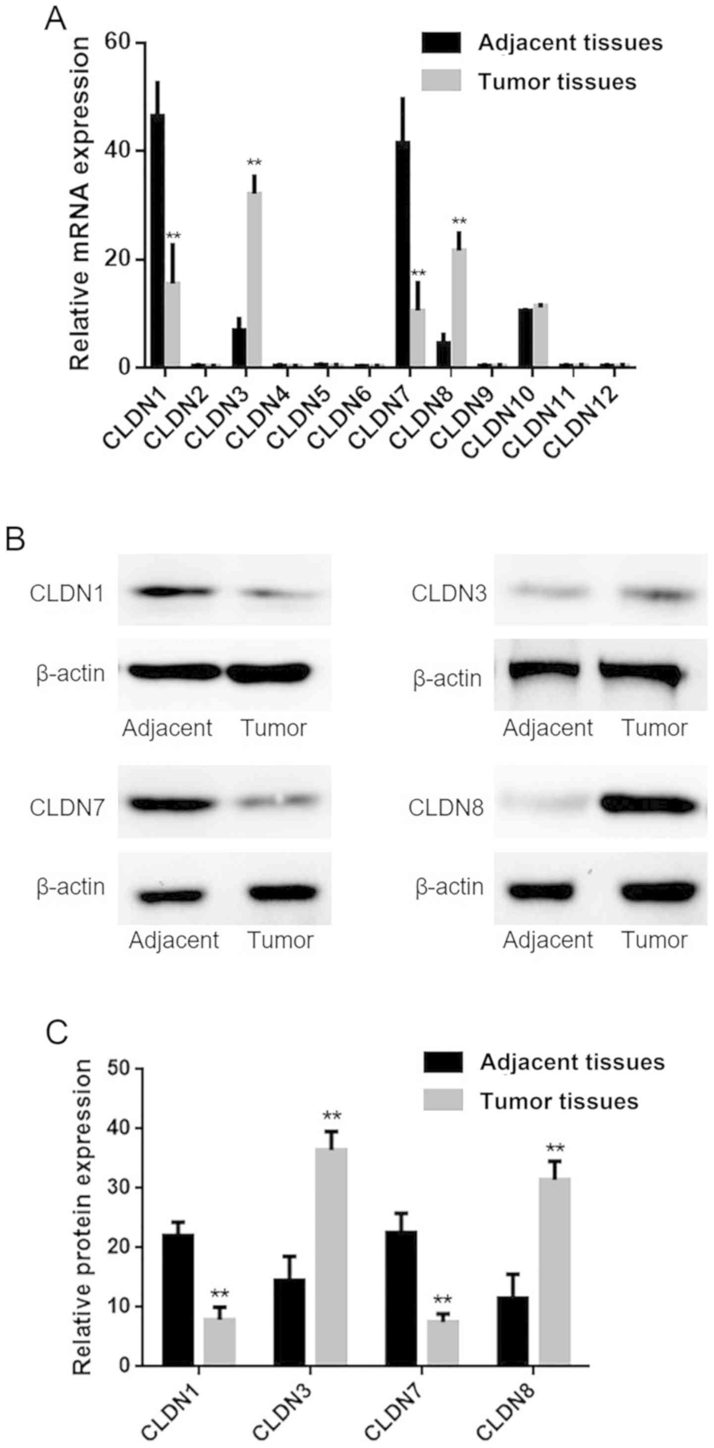
RT-qPCR and western blotting were performed to
analyze the expression of CLDN family members in laryngeal squamous
tissues and adjacent non-neoplastic tissues. As presented in
Fig. 1A, the expression of CLDN2,
CLDN4, CLDN5, CLDN6, CLDN9, CLDN11 or CLDN12 was not detected at
the mRNA in the samples of laryngeal squamous carcinoma or adjacent
non-neoplastic tissues. CLDN10 mRNA was expressed in laryngeal
squamous carcinoma and non-neoplastic tissues; however, there was
no significant difference was observed. Conversely, the levels of
CLDN1 and CLDN7 expression were significantly downregulated at the
mRNA and protein levels in laryngeal squamous carcinoma tissues
compared with adjacent non-neoplastic tissues, whereas those of
CLDN3 and CLDN8 were significantly upregulated (Fig. 1A-C).
CLDN1 and CLDN7 are downregulated,
while CLDN3 and CLDN8 are upregulated in laryngeal squamous
carcinoma
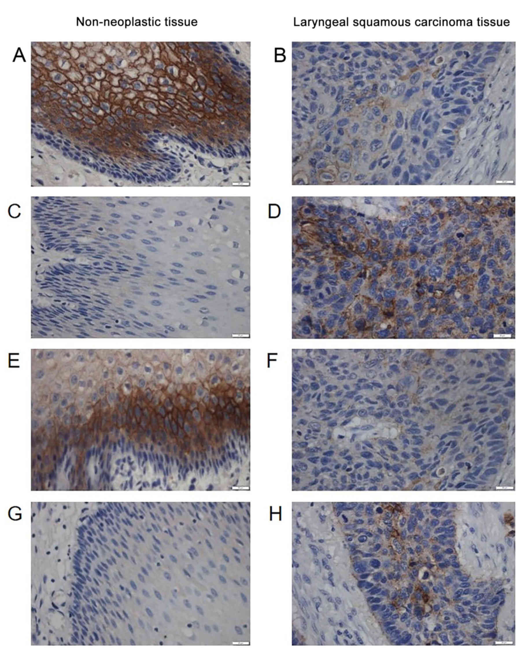
CLDN1 expression was evaluated in 80 pairs of
laryngeal squamous carcinoma tissue and adjacent non-neoplastic
tissue specimens via immunohistochemistry. CLDNs were expressed in
the cell membrane (Fig. 2). CLDN1
expression was observed in 28.8% (23/80) of laryngeal squamous
carcinoma tissues and in 61.3% (49/80) of adjacent non-neoplastic
tissues (P<0.01; Fig. 2A and B;
Table I). As presented in Table I, the expression of CLDN1 was not
associated with age (P>0.999), gender (P>0.999), the
expression of Ki67 (P>0.999), histological grade (P>0.999) or
distant metastasis (P=0.296).
 | Table I.Expression of CLDN1 and CLDN7
clinicopathological characteristics of patients with laryngeal
squamous carcinoma. |
Table I.
Expression of CLDN1 and CLDN7
clinicopathological characteristics of patients with laryngeal
squamous carcinoma.
| Factor | n | CLDN1 (+) | CLDN1 (−) | χ2 | P-value | n | CLDN7 (+) | CLDN7 (−) | χ2 | P-value |
|---|
| Laryngeal squamous
carcinoma tissue | 80 | 23 | 57 | 8.673 | <0.01 | 80 | 18 | 62 | 9.493 | <0.01 |
| Adjacent
non-neoplastic tissue | 80 | 49 | 31 |
|
| 80 | 52 | 28 |
|
|
| Gender |
|
Male | 46 | 13 | 33 | 0.178 |
>0.999a | 46 | 8 | 38 | 0.164 |
>0.999a |
|
Female | 34 | 10 | 24 |
|
| 34 | 10 | 24 |
|
|
| Age (years) |
|
≤60 | 58 | 15 | 43 | 0.116 |
>0.999a | 58 | 14 | 44 | 0.422 | 0.462a |
|
>60 | 22 | 8 | 14 |
|
| 22 | 4 | 18 |
|
|
| Histological
grade |
| Well-
differentiated | 42 | 14 | 28 | 0.243 |
>0.999a | 42 | 10 | 32 | 0.096 |
>0.999a |
|
Moderately and poor
differentiated | 38 | 9 | 29 |
|
| 38 | 8 | 30 |
|
|
| Distant
metastasis |
| + | 47 | 13 | 34 | 1.568 | 0.296a | 47 | 6 | 41 | 9.286 | <0.01 |
| - | 33 | 10 | 23 |
|
| 33 | 12 | 21 |
|
|
| Ki67 |
| + | 27 | 7 | 20 | 0.234 |
>0.999a | 29 | 7 | 20 | 0.448 | 0.496a |
| - | 53 | 16 | 37 |
|
| 43 | 11 | 42 |
|
|
The membrane staining of CLDN3 and CLDN8 was
increased in laryngeal squamous carcinoma tissues compared with
adjacent non-neoplastic tissues (Fig.
2). CLDN3 was expressed in 67.5% (54/80) of laryngeal squamous
carcinoma tissues and in 30.0% (24/80) of adjacent non-neoplastic
tissues (P<0.01; Fig. 2C and D;
Table II). As presented in
Table II, the expression of CLDN3
was not associated with gender (P=0.243), age (P=0.276),
histological grade (P>0.999) or the expression of Ki67
(P=0.175), but was associated with distant metastasis
(P<0.01).
| Table II.Expression of CLDN3 and CLDN8 and
clinicopathological characteristics of patients with laryngeal
squamous carcinoma. |
Table II.
Expression of CLDN3 and CLDN8 and
clinicopathological characteristics of patients with laryngeal
squamous carcinoma.
| Factor | n | CLDN3 (+) | CLDN8 CLDN3
(−) | χ2 | P-value | n | (+) | CLDN8 (−) | χ2 | P-value |
|---|
| Laryngeal squamous
carcinoma tissue | 80 | 54 | 26 | 8.078 | <0.01 | 80 | 39 | 41 | 9.451 | <0.01 |
| Adjacent
non-neoplastic tissue | 80 | 24 | 56 |
|
| 80 | 17 | 63 |
|
|
| Gender |
|
Male | 46 | 28 | 18 | 1.216 | 0.243a | 46 | 23 | 23 | 0.112 |
>0.999a |
|
Female | 34 | 26 | 8 |
|
| 34 | 16 | 18 |
|
|
| Age (years) |
|
≤60 | 58 | 40 | 18 | 1.382 | 0.276a | 58 | 27 | 31 | 2.218 | 0.146a |
|
>60 | 22 | 12 | 10 |
|
| 22 | 12 | 10 |
|
|
| Histological
grade |
| Well-
differentiated | 42 | 28 | 14 | 0.124 |
>0.999a | 42 | 16 | 26 | 4.326 | <0.05 |
|
Moderately and poor
differentiated | 38 | 26 | 12 |
|
| 38 | 23 | 15 |
|
|
| Distant
metastasis |
| + | 47 | 37 | 10 | 9.624 | <0.01 | 47 | 20 | 27 | 0.943 | 0.124a |
| - | 33 | 17 | 16 |
|
| 33 | 19 | 14 |
|
|
| Ki67 |
| + | 27 | 20 | 7 | 1.023 | 0.175a | 27 | 18 | 9 | 8.652 | <0.01 |
| - | 53 | 34 | 19 |
|
| 53 | 21 | 32 |
|
|
Membrane expression of CLDN7 protein was observed in
22.5% (18/80) of laryngeal squamous carcinoma tissues and in 65.0%
(52/80) of adjacent non-neoplastic tissues (P<0.01; Fig. 2E and F; Table I). As presented in Table I, the expression of CLDN7 was not
associated with age (P=0.462), gender (P>0.999), histological
grade (P>0.999) or the expression of Ki67 (P=0.496), but was
associated with distant metastasis (P<0.01). The results
suggested that CLDN1 and CLDN7 are downregulated in laryngeal
squamous carcinoma.
As presented in Fig. 2G
and H, the membrane staining of CLDN8 was increased in
laryngeal squamous carcinoma tissues compared with in
non-neoplastic tissues. CLDN8 was expressed in 48.8% (39/80) of
laryngeal squamous carcinoma tissues and in 21.3% (17/80) of
adjacent non-neoplastic tissues (P<0.01; Table II). As presented in Table II, the expression of CLDN8 was not
associated with age (P=0.146), gender (P>0.999) or distant
metastasis (P=0.124), but was associated with histological grade
(P<0.05) and the expression of Ki67 (P<0.01). The results
suggested that CLDN3 and CLDN8 are upregulated in laryngeal
squamous carcinoma.
CLDN1 and CLDN7 are concurrently
expressed in laryngeal squamous carcinoma tissue
As presented in Table
III, a significant association between CLDN1 and CLDN7
expression was observed in laryngeal squamous carcinoma tissues
(ϕ=0.897, P<0.01).
| Table III.Association between the levels of
CLDN1 and CLDN7 expression in laryngeal squamous carcinoma
tissues. |
Table III.
Association between the levels of
CLDN1 and CLDN7 expression in laryngeal squamous carcinoma
tissues.
| Expression | CLDN7 (+) | CLDN7 (−) | ϕ | P-value |
|---|
| CLDN1 (+) | 14 | 9 | 0.897 | <0.01 |
| CLDN1 (−) | 4 | 53 |
|
|
Associations with survival and
clinical outcomes
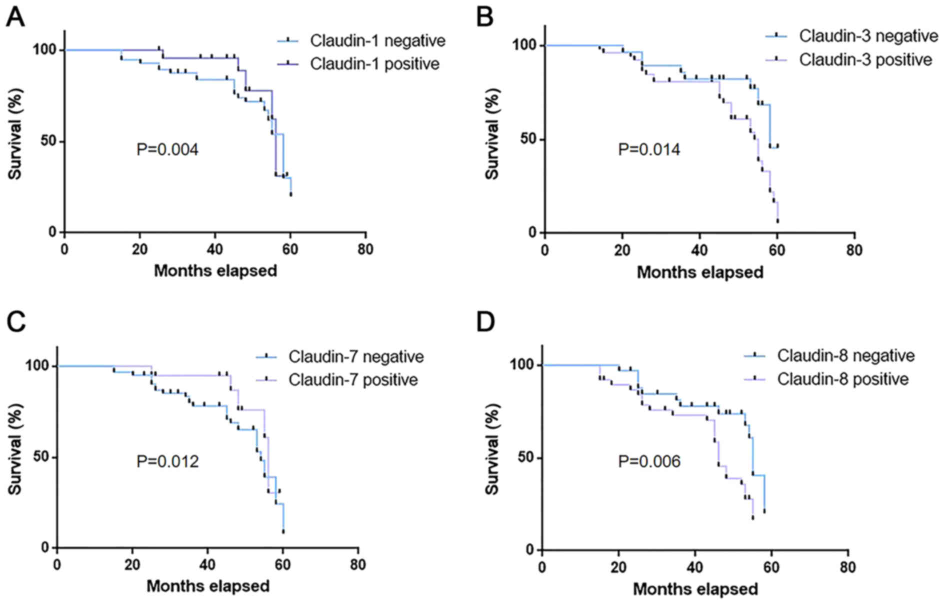
As presented in Fig.
3, patients with tumors that were positive for CLDN1 and CLDN7
(median survival, 51.42 and 52.43 months, respectively) exhibited
significantly increased survival times (P=0.004 and P=0.012,
respectively) compared with those negative for CLDN1 and CLDN7
(median survival, 45.29 and 45.56 months, respectively). Patients
with tumors that were positive for CLDN3 and CLDN8 (median
survival, 44.32 and 44.87 months, respectively) exhibited
significantly reduced survival times (P=0.014 and P=0.006,
respectively) compared with those negative for CLDN3 and CLDN8
(median survival, 52.27 and 52.13 months, respectively). The
results indicated that the expression profiles of CLDNs are
associated with the survival of patients with laryngeal squamous
carcinoma.
Discussion
Alterations in cell-to-cell adhesion are commonly
reported as early events in metastasis, permitting the release of
individual tumor cells from the primary tumor (6). Cell-to-cell adhesion of epithelial
cells is primarily maintained via adherens junctions and TJs
(1,3). Studies have previously investigated
the adherens junction protein E-cadherin (38); deficiencies in E-cadherin function
have been reported to lead to an enhancement in cell motility and
confer invasive abilities in various cell types (39,40).
Thus, E-cadherin is considered a tumor suppressor in a variety of
tissues and has been demonstrated to be a valuable prognostic
marker for numerous human cancers (41), indicating the important role of
cell-to-cell adhesion proteins in tumorigenesis.
Interruption of TJs, which serve crucial roles in
the penetrability and polarity of cells, is hypothesized to lead to
epithelial tumorigenesis (42).
Alterations in the structure and function of TJs have been reported
in numerous types of tumor (22).
Dysregulated expression of CLDN proteins may lead to the disruption
of TJs, and subsequently affect the polarity and interconnectivity
of cells (21). Reduced or
aberrant expression of CLDNs has been hypothesized to be associated
with pathophysiological consequences (43). CLDN1-deficient mice exhibit
lethality 1 day following birth due to the loss of epidermal
barrier function (44).
Abnormalities in TJ integrity induced by dysregulated CLDN
expression may serve major roles in permitting the dispersion of
nutrients and other factors essential for the maintenance and
progression of tumor cells (45).
Disruption of CLDNs in tumors has been suggested to be a mechanism
underlying reduced cell adhesion and an important event in the
progression of tumor cells toward metastasis (46–48).
Consistent with this theory, a previous study demonstrated that the
expression of CLDN4 in pancreatic carcinoma cells decreased the
metastatic phenotype of these cells (49). Additionally, CLDN1 overexpression
in tumor cells promotes the apoptosis of cells in three-dimensional
cultures (50). The physiological
ratio of CLDNs serves an important role in maintaining the
structure and function of TJs in epithelial cells (19); however, the mechanisms by which
altered CLDN expression and damage to TJs enhance tumor formation,
and the effects of these alterations on the progression of tumors
remain unclear.
The primary cause of cancer-associated mortality is
malignancy due to the metastasis of tumor cells from primary tumor
locations to distant organs (21).
An important step in the metastasis of tumor cells is hypothesized
to be the epithelial-mesenchymal transition (EMT) (51,52).
Downregulation of certain CLDNs in tumors is associated with the
interruption of TJs during tumorigenesis and EMT (53,54).
Furthermore, enforced initiation of EMT in epithelial cells leads
to loss of function of TJs and abnormal expression of CLDNs
(55). Additionally, CLDNs have
been revealed to interact with the TJ protein zonula occludens-1
(ZO-1) via their C-termini (15).
Notably, ZO-1 binds to numerous proteins that are involved in cell
signaling and transcriptional regulation (56,57),
suggesting that CLDNs may serve roles in these processes. Providing
that CLDN expression profiles are tumor-specific, it has been
proposed that CLDNs may be valuable biomarkers for various types of
tumor. For example, a set of four indicators that included CLDN3
was reported as an effective biomarker to precisely classify 158
cases of ovarian cancer (58).
Furthermore, CLDNs may serve as prognostic markers. CLDN1
expression is associated with the poor prognosis of stage II colon
cancer (59). In addition, CLDN10
expression has been reported to be an autonomous prognostic
indicator of hepatocellular carcinoma recurrence (60); however, at present, the association
between laryngeal squamous carcinogenesis and the expression of
CLDNs remains unclear. In the present study, the expression of
CLDNs was determined in laryngeal squamous carcinoma samples and
adjacent non-neoplastic tissues from 80 patients. It was revealed
that the levels of CLDN1 and CLDN7 expression were decreased in
laryngeal squamous carcinoma compared with adjacent non-neoplastic
tissues, whereas those of CLDN3 and CLDN8 were increased. CLDN3 was
the most commonly expressed of these proteins in laryngeal squamous
carcinoma tissue; 67.5% of laryngeal squamous carcinoma cases
exhibited CLDN3 reactivity, whereas the expression of CLDN1, CLDN7
and CLDN8 was observed in 28.8, 22.5 and 48.8% of cases,
respectively. The functional roles of CLDN1, −3, −7 and −8 in
laryngeal squamous carcinoma are yet to be determined; however,
based upon their roles in cell-to-cell adhesion, abnormalities in
these proteins may contribute to metastasis.
In conclusion, the present study revealed that the
expression of CLDN1, 3, −7 and −8 varied between human laryngeal
squamous carcinomas and adjacent non-neoplastic tissues, and
expression of these proteins was associated with the survival of
patients. Additionally, CLDN3 and CLDN7 expression was associated
with distant metastasis. Furthermore, CLDN1 and CLDN7 were reported
to exhibit significant co-expression in laryngeal squamous
carcinoma tissues; however, the mechanisms underlying these
observations require further investigation.
Acknowledgements
Not applicable.
Funding
No funding was received.
Availability of data and materials
The datasets used and/or analyzed during the present
study are available from the corresponding author upon reasonable
request.
Authors' contributions
SZ and XP performed the experiments. CW and RW
contributed to the conception and design of the study. ZS revised
the manuscript critically and analyzed the data. All authors read
and approved the final manuscript.
Ethics approval and consent to
participate
All procedures performed in studies involving human
participants were in accordance with the ethical standards of the
institutional and/or national research committee and with the 1964
Declaration of Helsinki and its later amendments or comparable
ethical standards. The present study was approved by the Ethics
Committee of Jilin Cancer Hospital (approval no. JLCH01224).
Informed consent for participation was obtained from all patients
and/or their parents.
Patient consent for publication
Not applicable.
Competing interests
The authors declare that they have no competing
interests.
References
|
1
|
Brandner JM, Haftek M and Niessen CM:
Adherens junctions, desmosomes and tight junctions in epidermal
barrier function. Open Dermatol J. 4:14–20. 2010. View Article : Google Scholar
|
|
2
|
Green KJ, Getsios S, Troyanovsky S and
Godsel LM: Intercellular junction assembly, dynamics, and
homeostasis. Cold Spring Harb Perspect Biol. 2:a0001252010.
View Article : Google Scholar : PubMed/NCBI
|
|
3
|
Niessen CM: Tight junctions/adherens
junctions: Basic structure and function. J Invest Dermatol.
127:2525–2532. 2007. View Article : Google Scholar : PubMed/NCBI
|
|
4
|
Schneeberger EE and Lynch RD: The tight
junction: A multifunctional complex. Am J Physiol Cell Physiol.
286:C1213–C1228. 2004. View Article : Google Scholar : PubMed/NCBI
|
|
5
|
Shin K, Fogg VC and Margolis B: Tight
junctions and cell polarity. Annu. Rev. Cell Dev. Biol. 22:207–235.
2006.
|
|
6
|
Oliveira SS and Morgado-Díaz JA: Claudins:
Multifunctional players in epithelial tight junctions and their
role in cancer. Cell Mol Life Sci. 64:17–28. 2007. View Article : Google Scholar : PubMed/NCBI
|
|
7
|
Martin TA and Jiang WG: Loss of tight
junction barrier function and its role in cancer metastasis.
Biochim Biophys Acta. 1788:872–891. 2009. View Article : Google Scholar : PubMed/NCBI
|
|
8
|
Van Itallie CM and Anderson JM: The
molecular physiology of tight junction pores. Physiology
(Bethesda). 19:331–338. 2004.PubMed/NCBI
|
|
9
|
Saitou M, Furuse M, Sasaki H, Schulzke JD,
Fromm M, Takano H, Noda T and Tsukita S: Complex phenotype of mice
lacking occludin, a component of tight junction strands. Mol Biol
Cell. 11:4131–4142. 2000. View Article : Google Scholar : PubMed/NCBI
|
|
10
|
Günzel D and Yu AS: Claudins and the
modulation of tight junction permeability. Physiol Rev. 93:525–569.
2013. View Article : Google Scholar : PubMed/NCBI
|
|
11
|
Hadj-Rabia S, Baala L, Vabres P,
Hamel-Teillac D, Jacquemin E, Fabre M, Lyonnet S, De Prost Y,
Munnich A, Hadchouel M and Smahi A: Claudin-1 gene mutations in
neonatal sclerosing cholangitis associated with ichthyosis: A tight
junction disease. Gastroenterology. 127:1386–1390. 2004. View Article : Google Scholar : PubMed/NCBI
|
|
12
|
Morin PJ: Claudin proteins in human
cancer: Promising new targets for diagnosis and therapy. Cancer
Res. 65:9603–9606. 2005. View Article : Google Scholar : PubMed/NCBI
|
|
13
|
Morita K, Furuse M, Fujimoto K and Tsukita
S: Claudin multigene family encoding four-transmembrane domain
protein components of tight junction strands. Proc Natl Acad Sci
USA. 96:511–516. 1999. View Article : Google Scholar : PubMed/NCBI
|
|
14
|
González-Mariscal L, Tapia R and Chamorro
D: Crosstalk of tight junction components with signaling pathways.
Biochim Biophys Acta. 1778:729–756. 2008. View Article : Google Scholar : PubMed/NCBI
|
|
15
|
Itoh M, Furuse M, Morita K, Kubota K,
Saitou M and Tsukita S: Direct binding of three tight
junction-associated MAGUKs, ZO-1, ZO-2, and ZO-3, with the COOH
termini of claudins. J Cell Biol. 147:1351–1363. 1999. View Article : Google Scholar : PubMed/NCBI
|
|
16
|
Swisshelm K, Macek R and Kubbies M: Role
of claudins in tumorigenesis. Adv Drug Deliv Rev. 57:919–928. 2005.
View Article : Google Scholar : PubMed/NCBI
|
|
17
|
Miwa N, Furuse M, Tsukita S, Niikawa N,
Nakamura Y and Furukawa Y: Involvement of claudin-1 in the
beta-catenin/Tcf signaling pathway and its frequent upregulation in
human colorectal cancers. Oncol Res. 12:469–476. 2001. View Article : Google Scholar : PubMed/NCBI
|
|
18
|
Angelow S, Ahlstrom R and Yu AS: Biology
of claudins. Am J Physiol Renal Physiol. 295:F867–F876. 2008.
View Article : Google Scholar : PubMed/NCBI
|
|
19
|
Krause G, Winkler L, Mueller SL, Haseloff
RF, Piontek J and Blasig IE: Structure and function of claudins.
Biochim Biophys Acta. 1778:631–645. 2008. View Article : Google Scholar : PubMed/NCBI
|
|
20
|
Günzel D and Fromm M: Claudins and other
tight junction proteins. Compr Physiol. 2:1819–1852.
2012.PubMed/NCBI
|
|
21
|
Osanai M, Takasawa A, Murata M and Sawada
N: Claudins in cancer: Bench to bedside. Pflugers Arch. 469:55–67.
2017. View Article : Google Scholar : PubMed/NCBI
|
|
22
|
Tabariès S and Siegel PM: The role of
claudins in cancer metastasis. Oncogene. 36:1176–1190. 2017.
View Article : Google Scholar : PubMed/NCBI
|
|
23
|
Escudero-Esparza A, Jiang WG and Martin
TA: The Claudin family and its role in cancer and metastasis. Front
Biosci (Landmark Ed). 16:1069–1083. 2010. View Article : Google Scholar
|
|
24
|
Lal-Nag M and Morin PJ: The claudins.
Genome Biol. 10:2352009. View Article : Google Scholar : PubMed/NCBI
|
|
25
|
Ouban A and Ahmed AA: Claudins in human
cancer: A review. Histol Histopathol. 25:83–90. 2010.PubMed/NCBI
|
|
26
|
Morohashi S, Kusumi T, Sato F, Odagiri H,
Chiba H, Yoshihara S, Hakamada K, Sasaki M and Kijima H: Decreased
expression of claudin-1 correlates with recurrence status in breast
cancer. Int J Mol Med. 20:139–143. 2007.PubMed/NCBI
|
|
27
|
Kinugasa T, Huo Q, Higashi D, Shibaguchi
H, Kuroki M, Tanaka T, Futami K, Yamashita Y, Hachimine K, Maekawa
S, et al: Selective up-regulation of claudin-1 and claudin-2 in
colorectal cancer. Anticancer Res. 27:3729–3734. 2007.PubMed/NCBI
|
|
28
|
Tabariès S, Dong Z, Annis MG, Omeroglu A,
Pepin F, Ouellet V, Russo C, Hassanain M, Metrakos P, Diaz Z, et
al: Claudin-2 is selectively enriched in and promotes the formation
of breast cancer liver metastases through engagement of integrin
complexes. Oncogene. 30:1318–1328. 2011. View Article : Google Scholar : PubMed/NCBI
|
|
29
|
Jääskeläinen A, Soini Y, Jukkola-Vuorinen
A, Auvinen P, Haapasaari KM and Karihtala P: High-level cytoplasmic
claudin 3 expression is an independent predictor of poor survival
in triple-negative breast cancer. BMC Cancer. 18:2232018.
View Article : Google Scholar : PubMed/NCBI
|
|
30
|
Zhang L, Wang Y, Zhang B, Zhang H, Zhou M,
Wei M, Dong Q, Xu Y, Wang Z, Gao L, et al: Claudin-3 expression
increases the malignant potential of lung adenocarcinoma cells:
Role of epidermal growth factor receptor activation. Oncotarget.
8:23033–23047. 2017.PubMed/NCBI
|
|
31
|
Agarwal R, D'Souza T and Morin PJ:
Claudin-3 and claudin-4 expression in ovarian epithelial cells
enhances invasion and is associated with increased matrix
metalloproteinase-2 activity. Cancer Res. 65:7378–7385. 2005.
View Article : Google Scholar : PubMed/NCBI
|
|
32
|
Rangel LB, Agarwal R, D'Souza T, Pizer ES,
Alò PL, Lancaster WD, Gregoire L, Schwartz DR, Cho KR and Morin PJ:
Tight junction proteins claudin-3 and claudin-4 are frequently
overexpressed in ovarian cancer but not in ovarian cystadenomas.
Clin Cancer Res. 9:2567–2575. 2003.PubMed/NCBI
|
|
33
|
Blackwell KE, Calcaterra TC and Fu YS:
Laryngeal dysplasia: Epidemiology and treatment outcome. Ann Otol
Rhinol Laryngol. 104:596–602. 1995. View Article : Google Scholar : PubMed/NCBI
|
|
34
|
Edge SB and Compton CC: The American Joint
Committee on Cancer: The 7th edition of the AJCC cancer staging
manual and the future of TNM. Ann Surg Oncol. 17:1471–1474. 2010.
View Article : Google Scholar : PubMed/NCBI
|
|
35
|
Livak KJ and Schmittgen TD: Analysis of
relative gene expression data using real-time quantitative PCR and
the 2(-Delta Delta C(T)) method. Methods. 25:402–408. 2001.
View Article : Google Scholar : PubMed/NCBI
|
|
36
|
Jiang L, Yang YD, Fu L, Xu W, Liu D, Liang
Q, Zhang X, Xu L, Guan XY, Wu B, et al: CLDN3 inhibits cancer
aggressiveness via Wnt-EMT signaling and is a potential prognostic
biomarker for hepatocellular carcinoma. Oncotarget. 5:7663–7676.
2014. View Article : Google Scholar : PubMed/NCBI
|
|
37
|
Gao M, Li W, Wang H and Wang G: The
distinct expression patterns of claudin-10, −14, −17 and E-cadherin
between adjacent non-neoplastic tissues and gastric cancer tissues.
Diagn Pathol. 8:2052013. View Article : Google Scholar : PubMed/NCBI
|
|
38
|
Berx G, Becker KF, Höfler H and van Roy F:
Mutations of the human E-cadherin (CDH1) gene. Hum Mutat.
12:226–237. 1998. View Article : Google Scholar : PubMed/NCBI
|
|
39
|
Christofori G and Semb H: The role of the
cell-adhesion molecule E-cadherin as a tumour-suppressor gene.
Trends Biochem Sci. 24:73–76. 1999. View Article : Google Scholar : PubMed/NCBI
|
|
40
|
Berx G and Van Roy F: The
E-cadherin/catenin complex: An important gatekeeper in breast
cancer tumorigenesis and malignant progression. Breast Cancer Res.
3:289–293. 2001. View
Article : Google Scholar : PubMed/NCBI
|
|
41
|
Hirohashi S: Inactivation of the
E-cadherin-mediated cell adhesion system in human cancers. Am J
Pathol. 153:333–339. 1998. View Article : Google Scholar : PubMed/NCBI
|
|
42
|
Kwon MJ: Emerging roles of claudins in
human cancer. Int J Mol Sci. 14:18148–18180. 2013. View Article : Google Scholar : PubMed/NCBI
|
|
43
|
Turksen K and Troy TC: Junctions gone bad:
Claudins and loss of the barrier in cancer. Biochim Biophys Acta.
1816:73–79. 2011.PubMed/NCBI
|
|
44
|
Furuse M, Hata M, Furuse K, Yoshida Y,
Haratake A, Sugitani Y, Noda T, Kubo A and Tsukita S: Claudin-based
tight junctions are crucial for the mammalian epidermal barrier a
lesson from claudin-1-deficient mice. J Cell Biol. 156:1099–1111.
2002. View Article : Google Scholar : PubMed/NCBI
|
|
45
|
Singh AB, Sharma A and Dhawan P: Claudin
family of proteins and cancer: An overview. J Oncol.
2010:5419572010. View Article : Google Scholar : PubMed/NCBI
|
|
46
|
Findley MK and Koval M: Regulation and
roles for claudin-family tight junction proteins. IUBMB Life.
61:431–437. 2009. View Article : Google Scholar : PubMed/NCBI
|
|
47
|
Hewitt KJ, Agarwal R and Morin PJ: The
claudin gene family: Expression in normal and neoplastic tissues.
BMC Cancer. 6:1862006. View Article : Google Scholar : PubMed/NCBI
|
|
48
|
Kominsky SL: Claudins: Emerging targets
for cancer therapy. Expert Rev Mol Med. 8:1–11. 2006. View Article : Google Scholar : PubMed/NCBI
|
|
49
|
Michl P, Barth C, Buchholz M, Lerch MM,
Rolke M, Holzmann KH, Menke A, Fensterer H, Giehl K, Löhr M, et al:
Claudin-4 expression decreases invasiveness and metastatic
potential of pancreatic cancer. Cancer Res. 63:6265–6271.
2003.PubMed/NCBI
|
|
50
|
Chang TL, Ito K, Ko TK, Liu Q,
Salto-Tellez M, Yeoh KG, Fukamachi H and Ito Y: Claudin-1 has tumor
suppressive activity and is a direct target of RUNX3 in gastric
epithelial cells. Gastroenterology. 138:255–265. e1-3. 2010.
View Article : Google Scholar : PubMed/NCBI
|
|
51
|
De Craene B and Berx G: Regulatory
networks defining EMT during cancer initiation and progression. Nat
Rev Cancer. 13:97–110. 2013. View Article : Google Scholar : PubMed/NCBI
|
|
52
|
Kaufhold S and Bonavida B: Central role of
Snail1 in the regulation of EMT and resistance in cancer: A target
for therapeutic intervention. J Exp Clin Cancer Res. 33:622014.
View Article : Google Scholar : PubMed/NCBI
|
|
53
|
Bhat AA, Pope JL, Smith JJ, Ahmad R, Chen
X, Washington MK, Beauchamp RD, Singh AB and Dhawan P: Claudin-7
expression induces mesenchymal to epithelial transformation (MET)
to inhibit colon tumorigenesis. Oncogene. 34:4570–4580. 2015.
View Article : Google Scholar : PubMed/NCBI
|
|
54
|
Lu Z, Ding L, Hong H, Hoggard J, Lu Q and
Chen YH: Claudin-7 inhibits human lung cancer cell migration and
invasion through ERK/MAPK signaling pathway. Exp Cell Res.
317:1935–1946. 2011. View Article : Google Scholar : PubMed/NCBI
|
|
55
|
Yilmaz M and Christofori G: EMT, the
cytoskeleton, and cancer cell invasion. Cancer Metastasis Rev.
28:15–33. 2009. View Article : Google Scholar : PubMed/NCBI
|
|
56
|
Balda MS, Garrett MD and Matter K: The
ZO-1-associated Y-box factor ZONAB regulates epithelial cell
proliferation and cell density. J Cell Biol. 160:423–432. 2003.
View Article : Google Scholar : PubMed/NCBI
|
|
57
|
Hamazaki Y, Itoh M, Sasaki H, Furuse M and
Tsukita S: Multi-PDZ domain protein 1 (MUPP1) is concentrated at
tight junctions through its possible interaction with claudin-1 and
junctional adhesion molecule. J Biol Chem. 277:455–461. 2002.
View Article : Google Scholar : PubMed/NCBI
|
|
58
|
Bose CK and Mukhopadhyay A: Claudin and
ovarian cancer. J Turk Ger Gynecol Assoc. 11:48–54. 2010.PubMed/NCBI
|
|
59
|
Ouban A: Claudin-1 role in colon cancer:
An update and a review. Histol Histopathol. 33:1013–1019.
2018.PubMed/NCBI
|
|
60
|
Cheung ST, Leung KL, Ip YC, Chen X, Fong
DY, Ng IO, Fan ST and So S: Claudin-10 expression level is
associated with recurrence of primary hepatocellular carcinoma.
Clin Cancer Res. 11:551–556. 2005.PubMed/NCBI
|