Introduction
Anaplastic thyroid cancer (ATC) accounts for 2% of
all thyroid cancers in the worldwide (1). ATC is a very aggressive malignancy in
humans; it is resistant to numerous chemotherapy treatments and is
associated with a high prevalence of mortality (2). The prognosis of ATC is poor. The
majority of patients succumb within 5 months following the
diagnosis; <20% survive for >1 year (3). Most patients with ATC are diagnosed
at an advanced stage and can develop distant metastasis as ATC
progresses (4). There is a lack of
standard therapy for ATC (5). A
multimodal approach based on extensive resection followed by
adjuvant chemotherapy and radiotherapy may improve the prognosis
(6). However, numerous patients
with late-stage ATC cannot undergo surgery and receive therefore
chemotherapy drugs only (7,8).
Adriamycin (ADM) is the main chemotherapy drug used
to treat ATC. The guidelines set by the American Thyroid
Association and National Comprehensive Cancer Network state that
ADM should be considered a first-line anticancer drug in ATC
treatment (3,9). However, ADM causes several serious
adverse drug reactions, including cardiotoxicity, myelosuppression
and digestive tract injury, which depend on the dose (10). Increasing attention has therefore
been applied to the development of novel synergistic formulations
in order to increase the specific anti-tumor efficacy and to reduce
ADM toxicity.
The role of Reactive oxygen species (ROS) in
apoptosis induction has been described for a diverse collection of
xenobiotics. ROS plays an important role in apoptosis induced by
chemo drug, 1,25(OH)2D3 can modulate ROS production. ROS production
is believed to be the major mechanism underlying its cytotoxicity
to cancer cells in most major organs, including thyroid (11). Increased levels of ROS may activate
the mitochondrial cascade of apoptosis. The role of oxidative
stress is ambivalent in cancer. The proper amount of reactive
oxygen species in normal cells can protect cells from damage.
However, the excessive generation of ROS in the drug treatment may
result in cell damage via apoptosis, necrosis and autophagy. It has
been demonstrated that 1,25-dihydroxyvitamin D3
(1,25(OH)2D3) increases the anticancer
effects of numerous chemotherapy drugs in synergistic or additive
manners, including ADM, cisplatin, paclitaxel and docetaxel, in
various types of malignant somatic cell in vitro and in
vivo (12,13). In addition,
1,25(OH)2D3 can intensify the apoptotic
effect of chemotherapy drugs, including ADM, the dose of which can
be reduced when combined with 1,25(OH)2D3
(14,15). However, whether
1,25(OH)2D3 can promote the ADM-induced
apoptosis of ATC cells remains known.
The present study aimed to investigate the apoptotic
effects of 1,25(OH)2D3 pretreatment upon ADM
treatment in ATC cells in vitro. In addition, the molecular
mechanism of action of this drug combination was examined as a
novel therapeutic strategy.
Materials and methods
Cell lines and cultures
The ATC cell lines 8305c and 8505c were obtained
from the Department of Endocrinology and Metabolism, Institute of
Endocrinology, Liaoning Provincial Key Laboratory of Endocrine
Diseases from the First Affiliated Hospital of China Medical
University. Cells were cultured in minimum essential medium (MEM;
HyClone; GE Healthcare Life Sciences) supplemented with 10% fetal
bovine serum (PAN-Biotech GmbH) and placed at 37°C in a humidified
incubator containing 5% CO2 (Thermo Fisher Scientific,
Inc.).
Cell viability assessment
Cell viability was assessed using the Cell Counting
Kit-8 (CCK-8; Dojindo Molecular Technologies, Inc.). ATC cells were
transferred to 96-well plates (2×10³ cells/well) and
incubated with 1,25(OH)2D3 (Selleck
Chemicals; dissolved in DMSO) at final concentrations of 0, 1, 10
and 100 nM at 37°C for 48 h.
Subsequently, the cells were washed with PBS for
twice and incubated in fresh MEM containing 100 ng/ml ADM (Selleck
Chemicals; dissolved in sterile distilled water) for 24 h. In order
to assess cell viability,10 µl CCK-8 solution was added into the
medium and the ATC cells were incubated for another 2.5 h at 37°C
before the absorbance at 450 nm was measured with a microplate
reader.
Apoptosis assessment
Cells were seeded in 6-well plates (1×10⁵
cells/well). Following overnight incubation, cells were treated
with 1,25(OH)2D3 at final concentrations of 0
or 10 nM for 48 h at 37°C. Treatment was then removed and replaced
by a solution of ADM (100 ng/ml) dissolved in MEM for 24 h at 37°C.
Subsequently, cells were suspended in PBS and stained by 5 µl
Annexin V-FITC and 5 µl Propidium Iodide (PI; BD Biosciences) in
the dark for 15 min at 25°C. Positively stained cells were detected
by flow cytometry (BD Biosciences) within 1 h. Compensation and
data analysis were conducted using FlowJo 7.6.1 (FlowJo LLC).
Nuclear morphology was evaluated by staining the
cells with Hoechst 33342 (Beyotime Institute of Biotechnology).
Briefly, cells were seeded in 6-well plates (1×10⁵
cells/well), treated with the appropriate drugs as aforementioned,
were stained with Hoechst 33342 at room temperature for 5 min and
immediately imaged with a fluorescence microscope (magnification,
×1,000; Olympus Corporation). Cells with changes in nuclear
morphology, including condensation and fragmentation of chromatin,
were considered to be apoptotic. Data were presented as the
percentage of apoptotic cells.
Caspase-3 activity assessment by
ELISA
Caspase-3 activity was determined using a Caspase-3
ELISA kit (c1115; Beyotime Institute of Biotechnology) according to
the manufacturer's instructions. To evaluate caspase-3 activity,
cells were treated with the aforementioned drugs, and cell lysates
were prepared and homogenized in 100 ml fluorescent substrate
Ac-DEVD-pNA (Beyotime Institute of Biotechnology) containing 10 µl
caspase-3 substrate (2 mM) and incubated at 37°C overnight.
Caspase-3 activity was detected with an enzyme-linked immunosorbent
assay at 405 nm using a microplate reader. Caspase-3 activity was
expressed as the percentage of enzyme activity compared with
control.
Measurement of intracellular reactive
oxygen species (ROS)
ATC cells were seeded into 96-well flat-bottom
plates, cultured with MEM containing 10% fetal bovine serum and
1,25(OH)2D3 (10 nM) and placed for 48 h at
37°C in a humidified incubator containing 5% CO2. ADM
(100 ng/ml) was added to the wells, and the ROS level in each well
was measured by the cell-permeant probe H2-DCFDA (Beyotime
Institute of Biotechnology) at 2 h after adding ADM. After
incubation with 10 µM H2-DCFDA at 37°C for 30 min, fluorescence
intensity was measured using a fluorescence microplate reader
(excitation wavelength, 488 nm; emission wavelength, 525 nm). Data
were presented as the mean of three independent experiments.
Western blotting
ATC cells were treated with
1,25(OH)2D3 (10 nM) for 48 h and with ADM
(100 ng/ml) for 24 h as aforementioned. Cells were then washed
twice in PBS. To determine protein expression by western blotting,
cells were harvested and lysed in RIPA buffer (Beyotime Institute
of Biotechnology) containing 1 mM phenylmethylsulfonyl fluoride
(Nanjing KeyGen Biotech Co., Ltd.) on ice for 1 h. Cell lysate was
mixed with 5X loading buffer (1:5; CW Biotech) and immediately
boiled for 10 min. Protein concentrations were quantified by a BCA
assay kit (Beyotime Institute of Biotechnology). Proteins (10 µg)
were separated on 12% SDS-PAGE and transferred onto nitrocellulose
membranes (Bio-Rad Laboratories, Inc.) and blocked in 5% skimmed
milk for 2 h at room temperature. Membranes were incubated with
antibodies against caspase-3 (1:1,000 dilution; 9962, Cell
Signaling Technology, Inc.), cleaved caspase-3 (1:1,000 dilution;
9964, Cell Signaling Technology, Inc.) and β-actin (1:1,000; 4790,
Cell Signaling Technologies, Inc.) at 4°C overnight. Then, the
membranes were incubated with horseradish peroxidase (HRP) labelled
secondary antibody (1:10,000 dilution; ZB-2301, OriGene
Technologies, Inc.) for another 45 min at 37°C. Proteins were
visualized using an enhanced chemiluminescence system (Thermo
Fisher Scientific, Inc.). Bands intensity was normalized to
expression of the internal control β-actin.
N-acetyl-L-cysteine (NAC)
treatment
The antioxidant NAC (Beyotime Institute of
Biotechnology) was dissolved in PBS to produce a 1 mM stock
solution. This solution was used to decrease ROS production. Cell
viability, caspase 3 activity and expression, intracellular ROS
level measurement and apoptosis were measured in cell cultures that
were treated in the presence or absence of 1 mM NAC for 24 h.
Statistical analyses
Statistical analyses were conducted using SPSS v21.0
(IBM Corp.) and Prism v5.0 (GraphPad Software, Inc.). Data are the
means ± standard error of the mean. Each experiment was repeated
independently three times. Differences between the groups were
analyzed using the Student's t-test. The data from three or more
groups were analyzed by one-way analysis of variance followed by
Bonferroni's multiple comparison test. P<0.05 was considered to
indicate a statistically significant difference.
Results
1,25(OH)2D3
enhances ADM-induced cell toxicity in ATC cell lines
1,25(OH)2D3 treatment alone
did not reduce ATC cell viability compared with control; however,
ADM significantly inhibited ATC cell viability (Fig. 1A and B). In addition, ATC cell
viability was significantly reduced by various concentrations of
1,25(OH)2D3 followed by ADM treatment
(Fig. 1A and B) in a
dose-dependent manner. Notably, pretreatment with 10 nM
1,25(OH)2D3 followed by treatment with 100 ng/ml ADM
significantly reduced ATC cell viability compared with cells
treated with ADM alone (P<0.05). A greater effect was achieved
with 100 ng/ml ADM combined with 100 nM
1,25(OH)2D3 (P<0.05). In addition, 8505c
was more sensitive than 8305c to the treatments. These results
suggested that 1,25(OH)2D3 increased the
potent toxic effect of ADM on ATC cells.
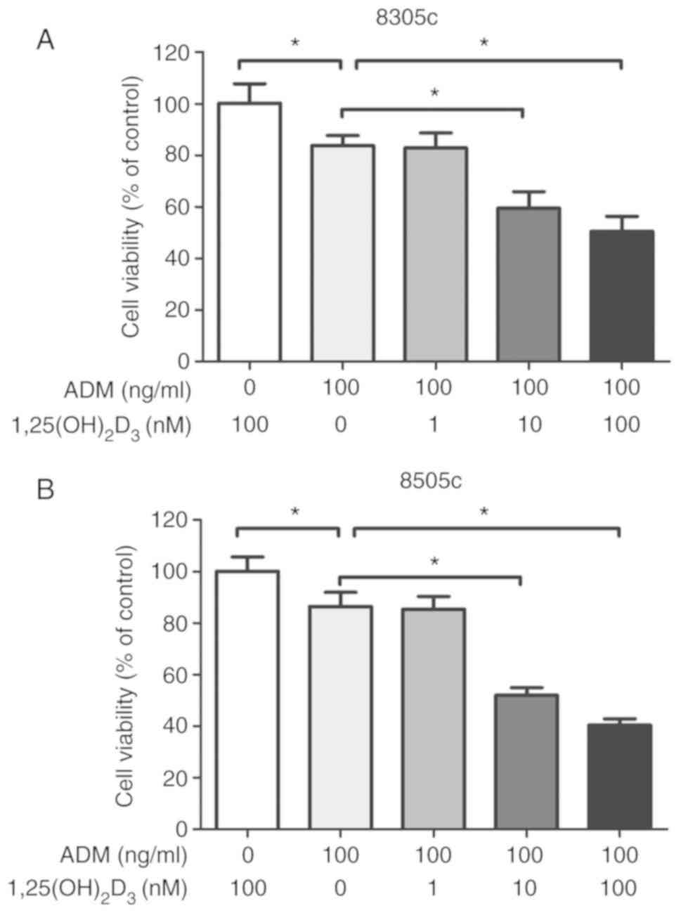 | Figure 1.Effects of ADM of ATC cell viability
are increased following 1,25(OH)2D3
pretreatment. 1,25(OH)2D3 enhanced
ADM-induced viability inhibition of (A) 8305c and (B) 8505c ATC
cell lines. Cells were incubated with ADM (100 ng/ml) for 24 h with
or without various concentrations of
1,25(OH)2D3 for 48 h prior to assessing cell
viability using a Cell Counting Kit-8 assay. Results are the means
± standard error of the mean of three independent experiments.
*P<0.05. 1,25(OH)2D3,
1,25-dihydroxyvitamin D3; ADM, Adriamycin; ATC,
anaplastic thyroid cancer. Cells were treated with 100 ng/ml ADM or
10 nM of 1,25(OH)2D3 followed by ADM. (A)
Flow cytometric analyses of cell apoptosis in anaplastic thyroid
cancer cell lines. Annexin V-FITC/PI double-staining was used to
label cells. (B) Morphologic assessment of apoptosis detected by
Hoechst 33342 under fluorescence microscopy. Following staining,
cells presenting nuclear fragmentation were counted as apoptotic
cells (white arrow). Magnification, ×1,000. Scale bar, 10 µm.
Results are the means ± standard error of the mean of three
independent experiments. *P<0.05.
1,25(OH)2D3, 1,25-dihydroxyvitamin
D3; ADM, adriamycin; PI, propidium iodide. |
1,25(OH)2D3
increases ADM-induced apoptosis. Apoptosis was evaluated to
determine the mechanism underlying ATC cell viability
inhibition
The results demonstrated that
1,25(OH)2D3 did not induce ATC cell apoptosis
(Fig. S1). However, ADM
significantly increased ATC cell apoptosis (Fig. 2A). No increase in apoptosis was
observed in cells incubated with 1 nM
1,25(OH)2D3 followed by ADM compared with ADM
alone (data not shown); however, apoptosis was significantly
increased in cells pretreated with 10 nM
1,25(OH)2D3 followed by ADM. Morphologic
changes were detected by staining cells with Hoechst 33342. Cells
incubated with ADM displayed apoptotic nuclei with DNA
fragmentation, chromatin condensation and apoptotic body formation.
Cells incubated with 1,25(OH)2D3 followed by ADM (P<0.05;
Fig. 2B) presented a higher number
of apoptotic nuclei.
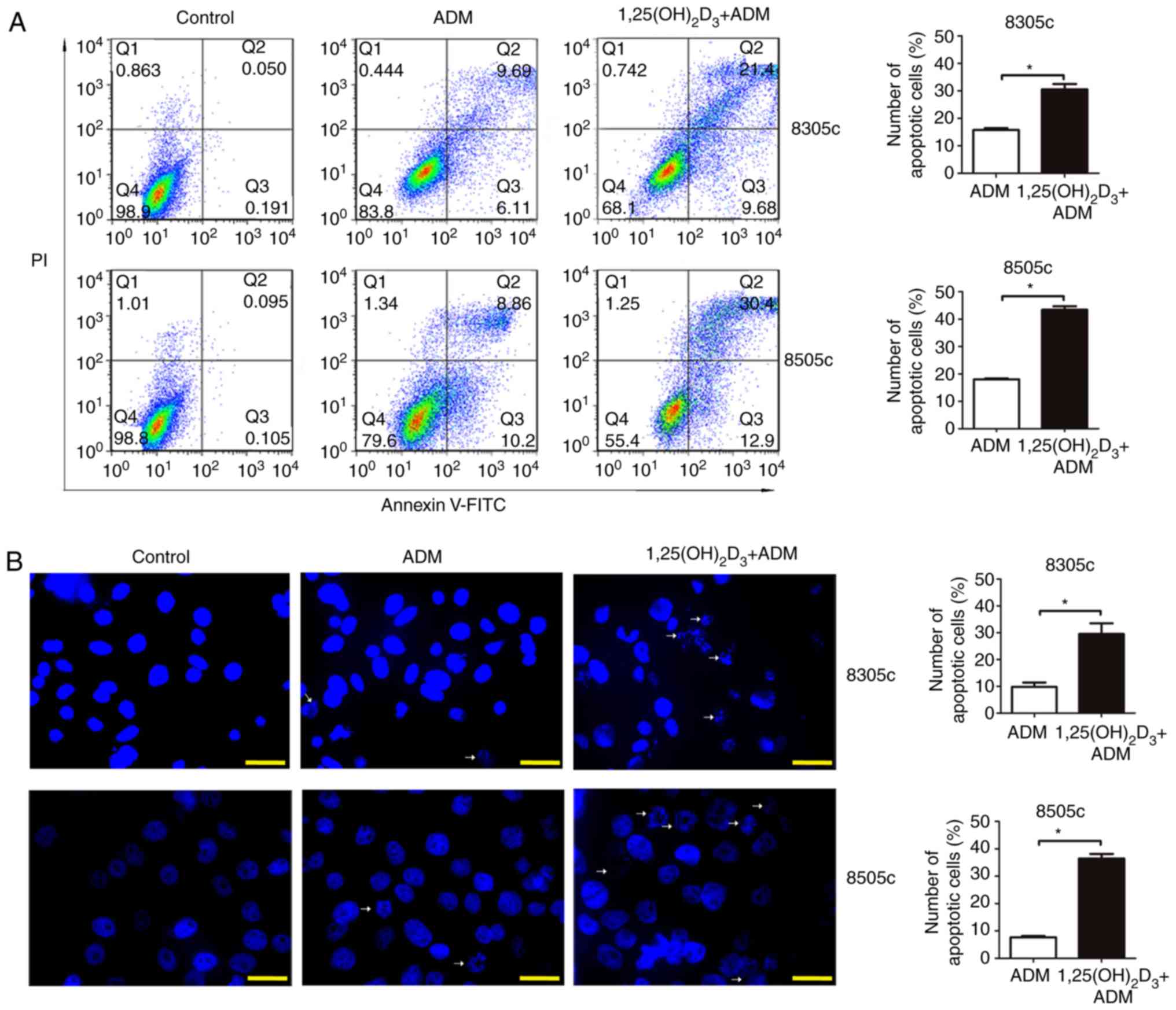 | Figure 2.1,25(OH)2D3
pretreatment promotes ADM-induced apoptosis. Cells were treated
with 100 ng/ml ADM or 10 nM of 1,25(OH)2D3 followed by ADM. (A)
Flow cytometric analyses of cell apoptosis in anaplastic thyroid
cancer cell lines. Annexin V-FITC/PI double-staining was used to
label cells. (B) Morphologic assessment of apoptosis detected by
Hoechst 33342 under fluorescence microscopy. Following staining,
cells presenting nuclear fragmentation were counted as apoptotic
cells (white arrow). Magnification, ×1,000. Scale bar, 10 µm.
Results are the means ± standard error of the mean of three
independent experiments. *P<0.05. 1,25(OH)2D3,
1,25-dihydroxyvitamin D3; ADM, adriamycin; PI, propidium
iodide. |
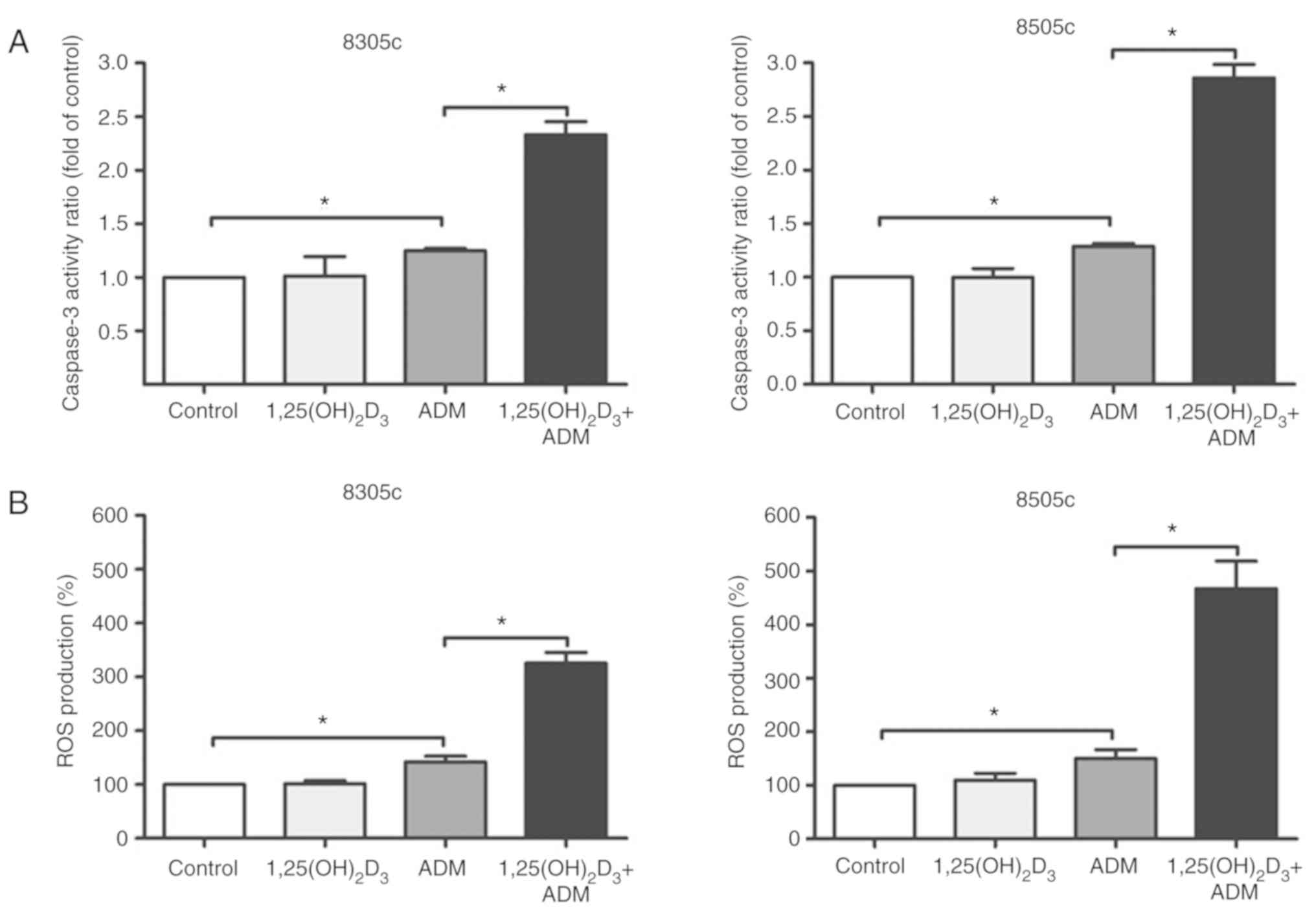
1,25(OH)2D3
increases ADM-induced caspase-3 activation
1,25(OH)2D3 treatment did not
increase caspase-3 activation in ATC cells; however, ADM
significantly increased caspase-3 activation compared with the
control. In addition, caspase-3 activity was significantly
increased following 1,25(OH)2D3 treatment
combined with ADM (P<0.05; Fig.
3A). These results indicated that ADM induced ATC cell
apoptosis through caspase-3 activation. In addition,
1,25(OH)2D3 pretreatment enhanced ADM-induced
caspase-3 activation.
1,25(OH)2D3
increases ADM-induced ROS production
To determine whether the apoptotic effect of
1,25(OH)2D3 combined ADM was caused by an
increase in ROS production, the impact of ADM alone or
1,25(OH)2D3 plus ADM on intracellular ROS
generation was measured. The results demonstrated that
1,25(OH)2D3 did not increase intracellular
ROS production in ATC cells. However, ADM significantly increased
ROS generation compared with the control. In addition,
1,25(OH)2D3 (10 nM) followed by ADM
significantly increased intracellular ROS generation (P<0.05;
Fig. 3B).
NAC attenuates the increased ROS
generation and apoptosis induced by
1,25(OH)2D3 plus ADM
To establish the association between the increased
oxidative stress and the apoptosis induced by the combined drugs,
the antioxidant NAC was used as an intracellular ROS scavenger.
Cell viability was examined via a CCK-8 assay (Fig. 4A) and apoptosis was detected by
flow cytometry and western blotting. The results demonstrated that
NAC abolished the apoptotic effects induced by treatment with
1,25(OH)2D3 followed by ADM (Fig. 4B). In addition, NAC attenuated the
ROS production and upregulation of cleaved caspase-3 mediated by
the combined drug treatment (Fig. 5A
and B). These results indicated that
1,25(OH)2D3 increased ADM-induced apoptosis
in ATC cells, which may be mediated by an increase in intracellular
ROS generation.
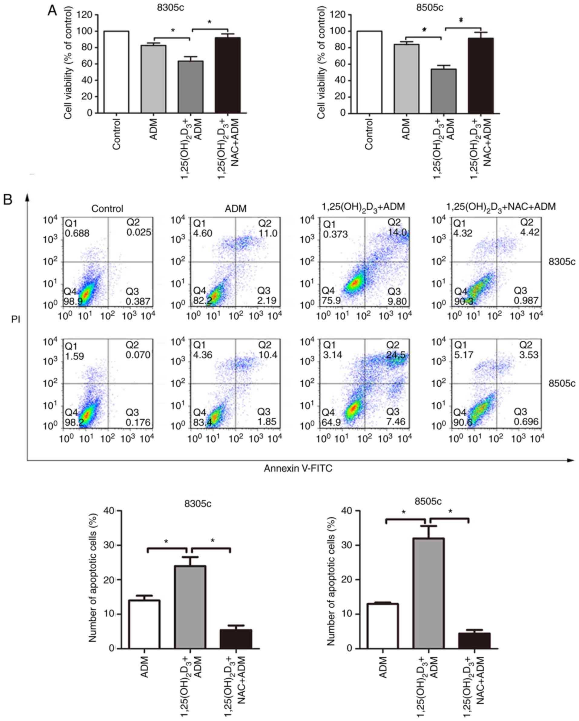 | Figure 4.NAC attenuates
1,25(OH)2D3 pretreatment-induced enhancement
of ADM cytotoxicity. Cells were treated with 100 ng/ml ADM, 10 nM
1,25(OH)2D3 followed by ADM, or
1,25(OH)2D3 plus ADM and 1 mM NAC. (A) Cell
viability was measured using Cell Counting Kit-8 assay. (B)
Apoptosis was detected by flow cytometry. Results are the means ±
standard error of the mean of three independent experiments.
*P<0.05. 1,25(OH)2D3,
1,25-dihydroxyvitamin D3; ADM, adriamycin; NAC,
N-acetyl-L-cysteine. |
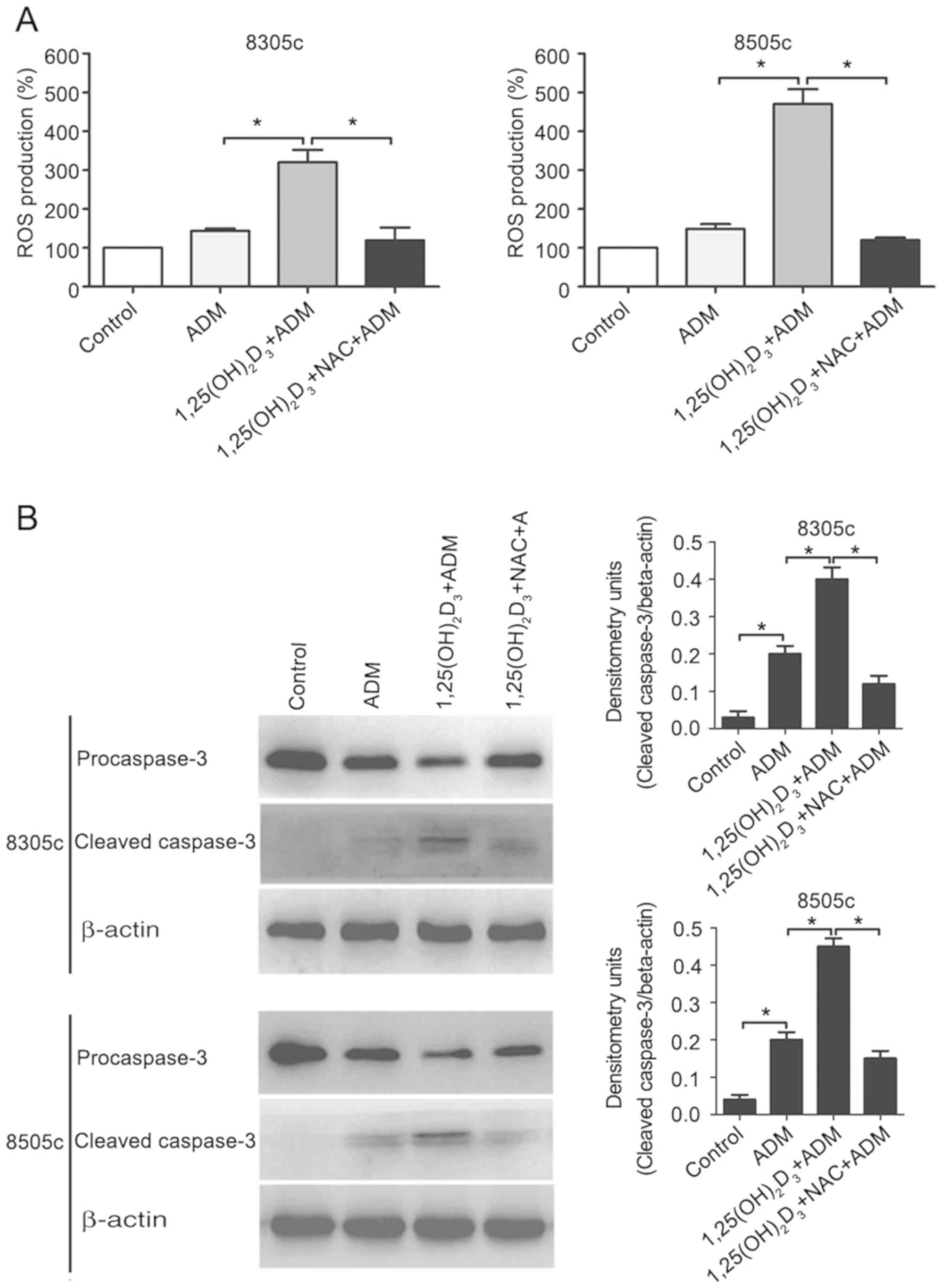 | Figure 5.NAC attenuates
1,25(OH)2D3 pretreatment-induced enhancement
of ADM-induced ROS production and cleaved caspase-3. Cells were
treated with 100 ng/ml ADM, 10 nM 1,25(OH)2D3
followed by ADM, or 1,25(OH)2D3 plus ADM and
1 mM NAC. (A) Intracellular ROS production in anaplastic thyroid
cancer cell lines was determined by H2-DCFDA. (B)
Expression of cleaved caspase-3 was measured using western blotting
and β-actin protein was used for normalization. Results are the
means ± standard error of the mean of three independent
experiments. *P<0.05. 1,25(OH)2D3,
1,25-dihydroxyvitamin D3; ADM, adriamycin; NAC,
N-acetyl-L-cysteine; ROS, reactive oxygen species. |
Discussion
ADM belongs to the anthracycline family of antitumor
antibiotics (16). Most patients
with late-stage ATC cannot undergo surgery and receive ADM
treatment. However, patients treated with high doses of ADM usually
suffer from serious adverse drug reactions, including
cardiotoxicity, inhibition of bone-marrow function, nausea,
vomiting, alopecia and increased transaminase levels (17). As these side effects are partly
dose-dependent, a reduction in the ADM dose without reducing its
therapeutic effect would therefore be of high clinical value.
Therapeutic results could be improved if cancer cells were killed
by ADM combined with a ‘chemotherapy sensitizer’ (18).
Previous studies reported that vitamin D receptors
are highly expressed in ATC cell lines, which suggests that
1,25(OH)2D3 could have a role in the
treatment of malignant tumors (19–21).
1,25(OH)2D3 could therefore be a chemotherapy
sensitizer candidate for ADM and exert anticancer effects in the
clinical treatment of ATC. The present study combined
1,25(OH)2D3 with ADM to develop a therapeutic
strategy that could allow ADM dose reduction, toxicity alleviation
and potential inhibition of ATC cell proliferation. The results
demonstrated that 1,25(OH)2D3 amplified the
apoptotic effect of ADM and reduced ATC cell viability in a
synergistic manner.
The mechanisms underlying ADM-induced cytotoxicity
are multiple such as cardiac injury and myelosuppression. However,
ROS generation has been of high interest for a long time. Previous
studies reported that ADM can stimulate intracellular ROS
production and apoptosis in a dose-dependent manner (22,23).
ROS are oxygen-containing chemically reactive species (24). The role of oxidative stress is
ambivalent in cancer. A moderate increase in ROS production can
promote cell proliferation and differentiation (25). However, an acute and excessive
increase in ROS production can induce cell apoptosis by activating
DNA damage response and the p53 pathway (26). The role of ROS in cancer is not
completely understood; however, certain studies have suggested that
ROS serve vital roles in the proliferation, differentiation and
apoptosis of cancer cells through the regulation of protein
activity (27,28).
To the best of our knowledge, the present study
reported for the first time that 1,25(OH)2D3
enhanced ADM susceptibility in ATC cells. Subsequently, whether the
drugs worked in synergy was investigated. The results demonstrated
that ATC cells pretreatment with 1,25(OH)2D3
significantly enhanced ADM-induced cytotoxicity in a dose-dependent
manner. Furthermore, by using Annexin V-FITC/PI double-staining and
Hoechst 33342, this study demonstrated that
1,25(OH)2D3 enhanced ADM-induced apoptosis.
In addition, ADM induced ATC cell apoptosis, potentially by
generating increased levels of ROS. The results demonstrated that
10 nM 1,25(OH)2D3 did not increase ROS
production compared with control; however, when combined with ADM,
ROS generation was increased in ATC cells. This finding has been
previously demonstrated in breast cancer cell lines (29). In addition, the apoptotic cell rate
in ATC cells was reduced following NAC addition in the two-drug
combination group. Furthermore, cleaved caspase-3 expression was
significantly reduced following NAC addition in the two-drug
combination group. Taken together, these results suggested that ROS
may induce ATC cell apoptosis through cleaved caspase-3 activation.
In addition, the enhancement of ADM-induced apoptosis by
1,25(OH)2D3 in ATC cells may be closely
associated with ROS. It has been demonstrated that ROS is
implicated in the crosstalk between
1,25(OH)2D3 and ADM (30). The specific mechanism underlying
ADM-induced ROS generation by 1,25(OH)2D3
remains unknown; however, it has been hypothesized that
1,25(OH)2D3 could inhibit superoxide
dismutase expression and therefore increase ROS generation
(31). The excessive generation of
ROS in the drug treatment can also be involved in promoting the
activation of multiple signaling mechanisms. ROS generation
precedes apoptosis and is required for the progression to apoptosis
(32). ROS could subsequently
affect the function of apoptosis-associated proteins, including
proteins from the caspase family (33,34).
The present study demonstrated that combination of
ADM chemotherapy drug with 1,25(OH)2D3 increased ADM-induced
apoptosis. Pretreatment with 1,25(OH)2D3
followed by ADM may achieve the same therapeutic effect against ATC
and reduce the ADM-associated adverse reactions. These data
suggested that 1,25(OH)2D3 may be considered
as a sensitizer for ADM chemotherapy and allow a reduction in ADM
dose, which may be a novel therapeutic strategy for ATC. Whether
the combination of 1,25(OH)2D3 and ADM in the
same cell culture, or 1,25(OH)2D3
pretreatment followed by ADM administration can affect ATC cells
and induce autophagy will be further investigated, alongside with
the downstream molecular mechanisms involved.
In conclusion, ADM had high antineoplastic activity
and induced apoptosis through activation of cleaved caspase-3 in
ATC cells and increased ROS generation. The present study
illustrated that 1,25(OH)2D3 increased
ADM-induced apoptosis in ATC cells, potentially by increasing ROS
production.
Supplementary Material
Supporting Data
Acknowledgements
Not applicable.
Funding
This study was supported by the Natural Science
Foundation of Liaoning Province, China (grant no. 20180530090).
Availability of data and materials
The datasets used or analyzed during the current
study are available from the corresponding author on reasonable
request.
Authors' contributions
HZ conceived the study and edited the final
manuscript. TZ contributed significantly to the study design and
preparation of the manuscript. LH and WS conducted data collection
and literature research. YQ and PZ analyzed data. All authors
approved the final version of the manuscript.
Ethics approval and consent to
participate
Not applicable.
Patient consent for publication
Not applicable.
Competing interests
The authors declare that they have no competing
interests.
References
|
1
|
Kasmann L, Bolm L, Janssen S and Rades D:
Prognostic factors for survival in patients treated with multimodal
therapy for anaplastic thyroid cancer. Anticancer Res.
36:4697–4700. 2016. View Article : Google Scholar : PubMed/NCBI
|
|
2
|
Nagaiah G, Hossain A, Mooney CJ,
Parmentier J and Remick SC: Anaplastic thyroid cancer: A review of
epidemiology, pathogenesis, and treatment. J Oncol.
2011:5423582011. View Article : Google Scholar : PubMed/NCBI
|
|
3
|
Smallridge RC, Ain KB, Asa SL, Bible KC,
Brierley JD, Burman KD, Kebebew E, Lee NY, Nikiforov YE, Rosenthal
MS, et al: American thyroid association guidelines for management
of patients with anaplastic thyroid cancer. Thyroid. 22:1104–1139.
2012. View Article : Google Scholar : PubMed/NCBI
|
|
4
|
Akaishi J, Sugino K, Kitagawa W, Nagahama
M, Kameyama K, Shimizu K and Ito K and Ito K: Prognostic factors
and treatment outcomes of 100 cases of anaplastic thyroid
carcinoma. Thyroid. 21:1183–1189. 2011. View Article : Google Scholar : PubMed/NCBI
|
|
5
|
Jiménez-Fonseca P, Gómez Saez JM,
Santamaria Sandi J, Capdevila J, Navarro Gonzalez E, Zafon Llopis
C, Ramón Y, Cajal Asensio T, Riesco-Eizaguirre G, Grande E and
Galofré JC: Spanish consensus for the management of patients with
anaplastic cell thyroid carcinoma. Clin Transl Oncol. 19:12–20.
2017. View Article : Google Scholar : PubMed/NCBI
|
|
6
|
Kebebew E, Greenspan FS, Clark OH, Woeber
KA and McMillan A: Anaplastic thyroid carcinoma. Treatment outcome
and prognostic factors. Cancer. 103:1330–1335. 2005. View Article : Google Scholar : PubMed/NCBI
|
|
7
|
Mitchell AL, Gandhi A, Scott-Coombes D and
Perros P: Management of thyroid cancer: United Kingdom National
Multidisciplinary Guidelines. J Laryngol Otol. 130:S150–S160. 2016.
View Article : Google Scholar : PubMed/NCBI
|
|
8
|
Smallridge RC: Approach to the patient
with anaplastic thyroid carcinoma. J Clin Endocrinol Metab.
97:2566–2572. 2012. View Article : Google Scholar : PubMed/NCBI
|
|
9
|
Haddad RI, Lydiatt WM, Ball DW, Busaidy
NL, Byrd D, Callender G, Dickson P, Duh QY, Ehya H, Haymart M, et
al: Anaplastic thyroid carcinoma, version 2.2015. J Natl Compr Canc
Netw. 13:1140–1150. 2015. View Article : Google Scholar : PubMed/NCBI
|
|
10
|
Aiken MJ, Suhag V, Garcia CA, Acio E,
Moreau S, Priebat DA, Chennupati SP and Van Nostrand D:
Doxorubicin-induced cardiac toxicity and cardiac rest gated blood
pool imaging. Clin Nucl Med. 34:762–767. 2009. View Article : Google Scholar : PubMed/NCBI
|
|
11
|
Yang L, Wu L, Du S, Hu Y, Fan Y and Ma J:
1,25(OH)2D3 inhibits high glucose-induced apoptosis and ROS
production in human peritoneal mesothelial cells via the MAPK/P38
pathway. Mol Med Rep. 14:839–844. 2016. View Article : Google Scholar : PubMed/NCBI
|
|
12
|
Ma Y, Trump DL and Johnson CS: Vitamin D
in combination cancer treatment. J Cancer. 1:101–107. 2010.
View Article : Google Scholar : PubMed/NCBI
|
|
13
|
Xian M, Cao H, Cao J, Shao X, Zhu D, Zhang
N, Huang P, Li W, Yang B, Ying M and He Q: Bortezomib sensitizes
human osteosarcoma cells to adriamycin-induced apoptosis through
ROS-dependent activation of p-eIF2α/ATF4/CHOP axis. Int J Cancer.
141:1029–1041. 2017. View Article : Google Scholar : PubMed/NCBI
|
|
14
|
Chaudhry M, Sundaram S, Gennings C, Carter
H and Gewirtz DA: The vitamin D3 analog, ILX-23-7553, enhances the
response to adriamycin and irradiation in MCF-7 breast tumor cells.
Cancer Chemother Pharmacol. 47:429–436. 2001. View Article : Google Scholar : PubMed/NCBI
|
|
15
|
Hanušová V, Caltová K, Svobodová H, Ambrož
M, Skarka A, Murínová N, Králová V, Tomšík P and Skálová L: The
effects of β-caryophyllene oxide and trans-nerolidol on the
efficacy of doxorubicin in breast cancer cells and breast
tumor-bearing mice. Biomed Pharmacother. 95:828–836. 2017.
View Article : Google Scholar : PubMed/NCBI
|
|
16
|
Aung LHH, Li R, Prabhakar BS and Li P:
Knockdown of Mtfp1 can minimize doxorubicin cardiotoxicity by
inhibiting Dnm1l-mediated mitochondrial fission. J Cell Mol Med.
21:3394–3404. 2017. View Article : Google Scholar : PubMed/NCBI
|
|
17
|
Sherman EJ, Lim SH, Ho AL, Ghossein RA,
Fury MG, Shaha AR, Rivera M, Lin O, Wolden S, Lee NY and Pfister
DG: Concurrent doxorubicin and radiotherapy for anaplastic thyroid
cancer: A critical re-evaluation including uniform pathologic
review. Radiother Oncol. 101:425–430. 2011. View Article : Google Scholar : PubMed/NCBI
|
|
18
|
Garg M, Kanojia D, Mayakonda A, Ganesan
TS, Sadhanandhan B, Suresh S, S S, Nagare RP, Said JW, Doan NB, et
al: Selinexor (KPT-330) has antitumor activity against anaplastic
thyroid carcinoma in vitro and in vivo and enhances sensitivity to
doxorubicin. Sci Rep. 7:97492017. View Article : Google Scholar : PubMed/NCBI
|
|
19
|
Sharma V, Fretwell D, Crees Z, Kerege A
and Klopper JP: Thyroid cancer resistance to vitamin D receptor
activation is associated with 24-hydroxylase levels but not the ff
FokI polymorphism. Thyroid. 20:1103–1111. 2010. View Article : Google Scholar : PubMed/NCBI
|
|
20
|
Clinckspoor I, Verlinden L, Overbergh L,
Korch C, Bouillon R, Mathieu C, Verstuyf A and Decallonne B:
1,25-dihydroxyvitamin D3 and a superagonistic analog in combination
with paclitaxel or suberoylanilide hydroxamic acid have potent
antiproliferative effects on anaplastic thyroid cancer. J Steroid
Biochem Mol Biol. 124:1–9. 2011. View Article : Google Scholar : PubMed/NCBI
|
|
21
|
Chiang KC, Kuo SF, Chen CH, Ng S, Lin SF,
Yeh CN, Chen LW, Takano M, Chen TC, Juang HH, et al: MART-10, the
vitamin D analog, is a potent drug to inhibit anaplastic thyroid
cancer cell metastatic potential. Cancer Lett. 369:76–85. 2015.
View Article : Google Scholar : PubMed/NCBI
|
|
22
|
Wang Z, Wang J, Xie R, Liu R and Lu Y:
Mitochondria-derived reactive oxygen species play an important role
in Doxorubicin-induced platelet apoptosis. Int J Mol Sci.
16:11087–11100. 2015. View Article : Google Scholar : PubMed/NCBI
|
|
23
|
Rogalska A, Gajek A, Szwed M, Jozwiak Z
and Marczak A: The role of reactive oxygen species in WP
631-induced death of human ovarian cancer cells: A comparison with
the effect of doxorubicin. Toxicol In Vitro. 25:1712–1720. 2011.
View Article : Google Scholar : PubMed/NCBI
|
|
24
|
Atashi F, Modarressi A and Pepper MS: The
role of reactive oxygen species in mesenchymal stem cell adipogenic
and osteogenic differentiation: A review. Cells Dev. 24:1150–1163.
2015. View Article : Google Scholar
|
|
25
|
Zhang L, Wang G, Chen X, Xue X, Guo Q, Liu
M and Zhao J: Formyl peptide receptors promotes neural
differentiation in mouse neural stem cells by ROS generation and
regulation of PI3K-AKT signaling. Sci Rep. 7:2062017. View Article : Google Scholar : PubMed/NCBI
|
|
26
|
Blanco J, Tomás-Hernández S, García T,
Mulero M, Gómez M, Domingo JL and Sánchez DJ: Oral exposure to
silver nanoparticles increases oxidative stress markers in the
liver of male rats and deregulates the insulin signalling pathway
and p53 and cleaved caspase 3 protein expression. Food Chem
Toxicol. 115:398–404. 2018. View Article : Google Scholar : PubMed/NCBI
|
|
27
|
Zhou D, Shao L and Spitz DR: Reactive
oxygen species in normal and tumor stem cells. Adv Cancer Res.
122:1–67. 2014. View Article : Google Scholar : PubMed/NCBI
|
|
28
|
Redza-Dutordoir M and Averill-Bates DA:
Activation of apoptosis signalling pathways by reactive oxygen
species. Biochim Biophys Acta. 1863:2977–2992. 2016. View Article : Google Scholar : PubMed/NCBI
|
|
29
|
Pan WY, Lin KJ, Huang CC, Chiang WL, Lin
YJ, Lin WC, Chuang EY, Chang Y and Sung HW: Localized
sequence-specific release of a chemopreventive agent and an
anticancer drug in a time-controllable manner to enhance
therapeutic efficacy. Biomaterials. 101:241–250. 2016. View Article : Google Scholar : PubMed/NCBI
|
|
30
|
Bondza-Kibangou P, Millot C, El Khoury V
and Millot JM: Antioxidants and doxorubicin supplementation to
modulate CD14 expression and oxidative stress induced by vitamin D3
and seocalcitol in HL60 cells. Oncol Rep. 18:1513–1519.
2007.PubMed/NCBI
|
|
31
|
Teixeira TM, da Costa DC, Resende AC,
Soulage CO, Bezerra FF and Daleprane JB: Activation of
Nrf2-antioxidant signaling by 1,25-Dihydroxycholecalciferol
prevents leptin-induced oxidative stress and inflammation in human
endothelial cells. J Nutr. 147:506–513. 2017. View Article : Google Scholar : PubMed/NCBI
|
|
32
|
Rogalska A, Koceva-Chyła A and Jóźwiak Z:
Aclarubicin-induced ROS generation and collapse of mitochondrial
membrane potential in human cancer cell lines. Chem Biol Interact.
176:58–70. 2008. View Article : Google Scholar : PubMed/NCBI
|
|
33
|
Gillissen B, Richter A, Richter A,
Preissner R, Schulze-Osthoff K, Essmann F and Daniel PT:
Bax/Bak-independent mitochondrial depolarization and reactive
oxygen species induction by sorafenib overcome resistance to
apoptosis in renal cell carcinoma. J Biol Chem. 292:6478–6492.
2017. View Article : Google Scholar : PubMed/NCBI
|
|
34
|
Guo N and Peng Z: MG132, a proteasome
inhibitor, induces apoptosis in tumor cells. Asia Pac J Clin Oncol.
9:6–11. 2013. View Article : Google Scholar : PubMed/NCBI
|