Introduction
Osteoarthritis (OA) has a high clinical incidence
rate and is a common chronic degenerative joint disease (1). Musculoskeletal disorders, including
OA, are the leading cause of disability worldwide and significantly
contribute to increased health care costs (2). There are a number of risk factors for
the development of OA, including previous joint injury, obesity,
genetics, sex and anatomical factors related to joint shape and
alignment; however, the most prominent risk factor is an increasing
age (3). Advanced-stage OA often
requires joint replacement to relieve pain and disability, and the
number of knee replacement surgeries has markedly increased over
the past 20 years. The aging of the population will increase the
number of older adults who are disabled by OA and require joint
replacements (4). OA that occurs
in young individuals is usually caused by a previous joint injury,
such as an anterior cruciate ligament or meniscal tear, which is
common in athletes and induces the early onset of OA in patients
(5). During OA, chondrocytes
become highly active, with increased matrix synthesis and
inflammatory cytokine-induced catabolic pathways (6). Early intervention strategies
targeting pathological changes may attenuate or halt disease
progression.
Glutathione peroxidase (GPX) is a family of
antioxidant enzymes with eight types, GPX1-8. Together with
superoxide dismutase and catalase, GPX forms an enzymatic
antioxidant system that reduces reactive oxygen species (ROS)
(7). GPX7 is mainly located in the
endoplasmic reticulum (ER) and has two unique ER-secreted proteins,
namely the N-terminal ER signal peptide and the C-terminal atypical
KDEL sequence (8). Unlike other
family members, GPX7 does not apply GSH as a substrate to
participate in redox reactions, but promotes signal transduction
and release by targeting proteins, such as GRP78 (9). In osteoporosis, GPX7 promotes
osteoblastogenesis in bone marrow-derived mesenchymal stem cells by
inhibiting ER stress, and can also regulate bone mass and bone
turnover by blocking signals through the gp130 transducer protein
(10). It has been demonstrated
that GPX7 overexpression in hepatocytes inhibits ROS production and
reduces the expression of pro-fibrotic and pro-inflammatory genes,
improving non-alcoholic steatohepatitis (11). Furthermore, GPX7 protects cells
from oxidative stress during epidermal keratinogenesis (12). Nevertheless, the role of GPX7 in OA
remains unclear.
Previous studies have indicated that D-mannose
alleviates the progression of OA by inhibiting the HIF-2α-mediated
sensitivity of chondrocytes to ferroptosis (13), while GPX7 knockdown enhances
ferroptosis in glioma (14).
Therefore, it was hypothesized that in OA, increasing GPX7
expression could alleviate the progression of the disease by
inhibiting ferroptosis. The present study established a research
model by inducing chondrocytes with interleukin (IL)-1β, and
explored the effects of GPX7 on intracellular inflammation,
extracellular matrix (ECM) breakdown and ferroptosis by increasing
intracellular GPX7 expression. The findings of the present study
may lay a theoretical foundation for subsequent OA-related
research.
Materials and methods
Cells, cell culture and treatment
The immortalized human chondrocytes, C28/I2 [cat.
no. BFN60803901; BLUEFBIO Life Sciences; Qingqi (Shanghai)
Biotechnology Development Co., Ltd.], were cultured in Dulbecco's
modified Eagle's medium (DMEM) supplemented with 10% fetal bovine
serum and 1% penicillin/streptomycin under 5% CO2 at
37°C. The C28/I2 cells were stimulated with IL-1β (10 ng/ml,
MilliporeSigma) for 24 h to mimic OA in vitro (15). Additionally, to explore the
mediating effects of ferroptosis, the C28/I2 cells were pre-treated
with erastin (5 µM, Selleck Chemicals), a type of ferroptosis
inducer (16), for 24 h.
Cell transfection
The C28/I2 cells were transfected with plasmids
[cat. no. GM-1013P001, Genomeditech (Shanghai) Co., Ltd.] carrying
GPX7 to achieve gene overexpression. The cells transfected with
empty vectors were regarded as the negative control (named oe-NC).
The C28/I2 cells were seeded in a six-well plate the day prior to
transfection, 5 µg plasmids were mixed with Lipofectamine
3000® transfection reagent (Thermo Fisher Scientific,
Inc.) and were then were added to the wells. The cells were
harvested following 48 h of transfection at 37°C to assess the
transfection efficiency.
Western blot analysis
The concentration of protein extracted from the
C28/I2 cells with RIPA (Shanghai Maokang BioTec Co., Ltd.) was
determined using a Nanodrop 2000 spectrophotometer (Thermo Fisher
Scientific, Inc.). These proteins (20 µg/lane) were subsequently
resolved on 15% sodium dodecyl sulfate-polyacrylamide gels and
transferred onto PVDF membranes (MilliporeSigma). The membranes
were blocked at room temperature with 5% skimmed milk for 1 h,
incubated at 4°C overnight with primary antibodies [anti-GPX7 (cat.
no. 13501-1-AP; Proteintech Group, Inc.), aggrecan (cat. no.
MA3-16888; Invitrogen), collagen II (cat. no. PA5-99159;
Invitrogen; Thermo Fisher Scientific, Inc.), MMP13 (cat. no.
18165-1-AP; Proteintech Group, Inc.), A disintegrin and
metalloproteinase with thrombospondin motifs 5 (ADAMTS5; cat. no.
PA5-32142; 3,000 dilution; Invitrogen; Thermo Fisher Scientific,
Inc.), Bcl2 (cat. no. 66799-1-Ig; Proteintech Group, Inc.), Bax
(cat. no. 50599-2-Ig; 8,000 dilution; Proteintech Group, Inc.),
cleaved caspase-3 (cat. no. 25128-1-AP; Proteintech Group, Inc.),
caspase-3 (cat. no. 19677-1-AP; Proteintech Group, Inc.), cleaved
caspase-9 (cat. no. PA5-77889; Invitrogen; Thermo Fisher
Scientific, Inc.), caspase-9 (cat. no. 10380-1-AP; Proteintech
Group, Inc.), ferritin heavy chain 1 (FTH1; cat. no. 11682-1-AP;
Proteintech Group, Inc.), GPX4 (cat. no. 67763-1-Ig; Proteintech
Group, Inc.), transferrin (Tf; cat. no. 17435-1-AP; Proteintech
Group, Inc.) and GAPDH (cat. no. 60004-1-Ig; 200,000 dilution;
Proteintech Group, Inc.)]; and then incubated with HRP-linked goat
secondary antibody (cat. no. SA00001-1 and SA00001-2; 10,000
dilution; Proteintech Group, Inc.) for 1.5 h at 37°C. Unless
otherwise stated, antibodies were diluted a 1,000-fold. The bands
were visualized using enhanced chemifluorescence substrate (GE
Healthcare; Cytiva) and semi-quantified using ImageJ software
(version 1.53; National Institutes of Health).
Reverse transcription-quantitative PCR
(RT-qPCR)
Complementary DNA was acquired by converting total
RNA from the C28/I2 cells (extracted with TRIzol; Invitrogen;)
using the Evo M-MLV reverse transcription assay kit [Accurate
Biology (Changsha) Co., Ltd] according to the manufacturer's
instructions. qPCR was performed using the QuantiTect SYBR-Green
PCR kit (Qiagen GmbH). qPCR was performed in a total volume of
20-µl reaction system, containing 10 µl Master Mix, 10 ng DNA
template and 500 nM specific forward and reverse primers for GPX7.
The thermocycling conditions were as follows: 95°C for 15 min, and
40 cycles of 94°C for 15 sec, 55°C for 30 sec and 72°C for 30 sec.
The expression levels of mRNA were quantified using the
2−∆∆Cq method (17) and
normalized to the levels of actin. Primer sequences were as
follows: GPX7 forward, 5′-TACGACTTCAAGGCGGTCAA-3′ and reverse,
5′-CCACCAGGGACACCGATCC-3′; and actin forward,
5′-CTTCGCGGGCGACGAT-3′ and reverse,
5′-CCACATAGGAATCCTTCTGACC-3′.
ELISA
The levels of TNF-α and IL-6 were detected using
ELISA kits (cat. nos. PT518 and PI330; Beyotime Institute of
Biotechnology) to assess the degree of inflammation. The
supernatant of C28/I2 cells was obtained by centrifugation at 2,000
× g, 4°C for 10 min. The collected supernatant was incubated at
room temperature with biotin-labeled antibodies for 1 h,
avidin-peroxidase complex for 20 min, TMB chromogenic solution for
20 min, and stop solution provided with the kit in sequence.
Absorbance at 450 nm was measured using a microplate reader
(Molecular Devices, LLC).
TUNEL assay
TUNEL assay was performed to determine apoptosis
using a TUNEL kit (cat. no. C1086; Beyotime Institute of
Biotechnology). The C28/I2 cells were fixed with 4%
paraformaldehyde for 30 min and incubated with PBS containing 0.3%
Triton X-100 for 5 min at room temperature. The C28/I2 cells were
then incubated at 37°C with TUNEL working fluid for 1 h in the
dark. Microcopies were captured under a fluorescence microscope
(Olympus Corporation).
C11-BODIPY
The C11 BODIPY probe (cat. no. ajci64572; Amgicam;
Wuhan Anjiekai Biomedical Science and Technology Co., Ltd.) was
used to indicate lipid peroxidation. The C28/I2 cells were washed
with PBS thrice and incubated with C11 BODIPY for 30 min. Following
washing with PBS twice, the cells were observed under a
fluorescence microscope (Olympus Corporation).
FerroOrange
The ferrous iron level in the C28/I2 cells was
determined using FerroOrange probes (cat. no. F374; Dojindo
Laboratories, Inc.). The cells were washed with PBS thrice and
incubated at 37°C with FerroOrange for 30 min. Following washing
with PBS twice, the cells were observed under a fluorescence
microscope (Olympus Corporation).
Statistical analysis
Data are presented as the mean ± standard deviation
and were analyzed using GraphPad 8.0 software (Dotmatics).
Significant differences between two groups were determined using a
two-tailed unpaired Student's t test and those between multiple
groups were determined using one-way ANOVA followed by Tukey's post
hoc test. A value of P<0.05 was considered to indicate a
statistically significant difference.
Results
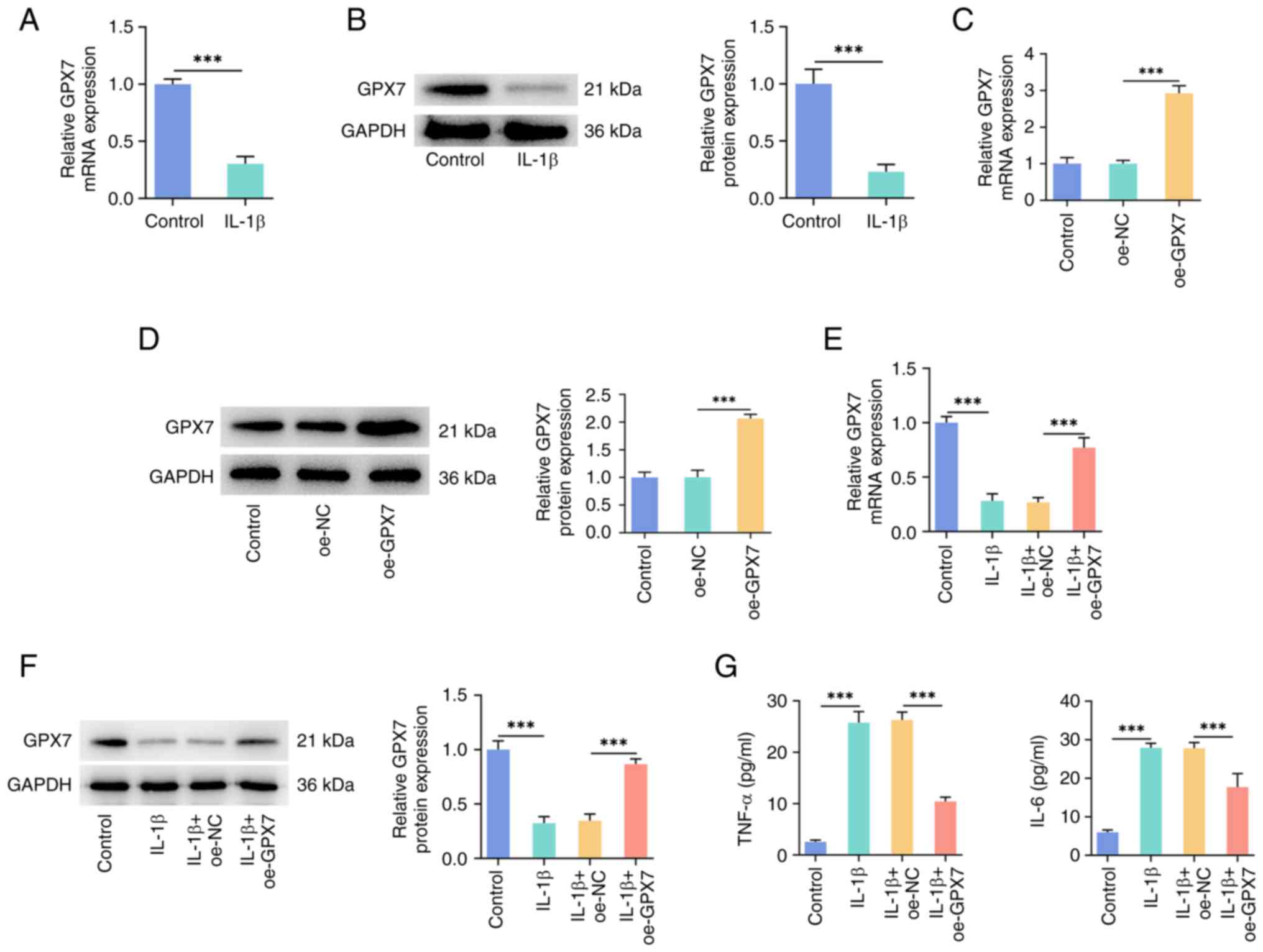
GPX7 overexpression inhibits
IL-1β-induced chondrocyte inflammation
The results of RT-qPCR and western blot analysis
demonstrated that the GPX7 level was decreased in chondrocytes in
response to IL-1β treatment, compared with the control group
(Fig. 1A and B). In order to
explore the specific role of GPX7 in chondrocytes, the C28/I2 cells
were transfected with plasmids overexpressing GPX7. The results of
RT-qPCR and western blot analysis confirmed that the GPX7 level in
the oe-GPX7 group was significantly higher than that in the oe-NC
group, indicating that the transfection was successful (Fig. 1C and D). Similarly, following
treatment with IL-1β, the expression of GPX7 in the oe-GPX7 group
was higher than that in the oe-NC group (Fig. 1E and F). In addition, ELISA was
used to measure the levels of inflammatory factors to evaluate the
degree of the inflammatory response in cells. The levels of TNF-α
and IL-6 in the cell supernatant were significantly increased upon
IL-1β treatment, and a similar increase was observed in the IL-1β +
oe-NC group. Compared with the IL-1β + oe-NC group, the levels of
TNF-α and IL-6 decreased in the IL-1β + oe-GPX7 group (Fig. 1G).
GPX7 overexpression inhibits
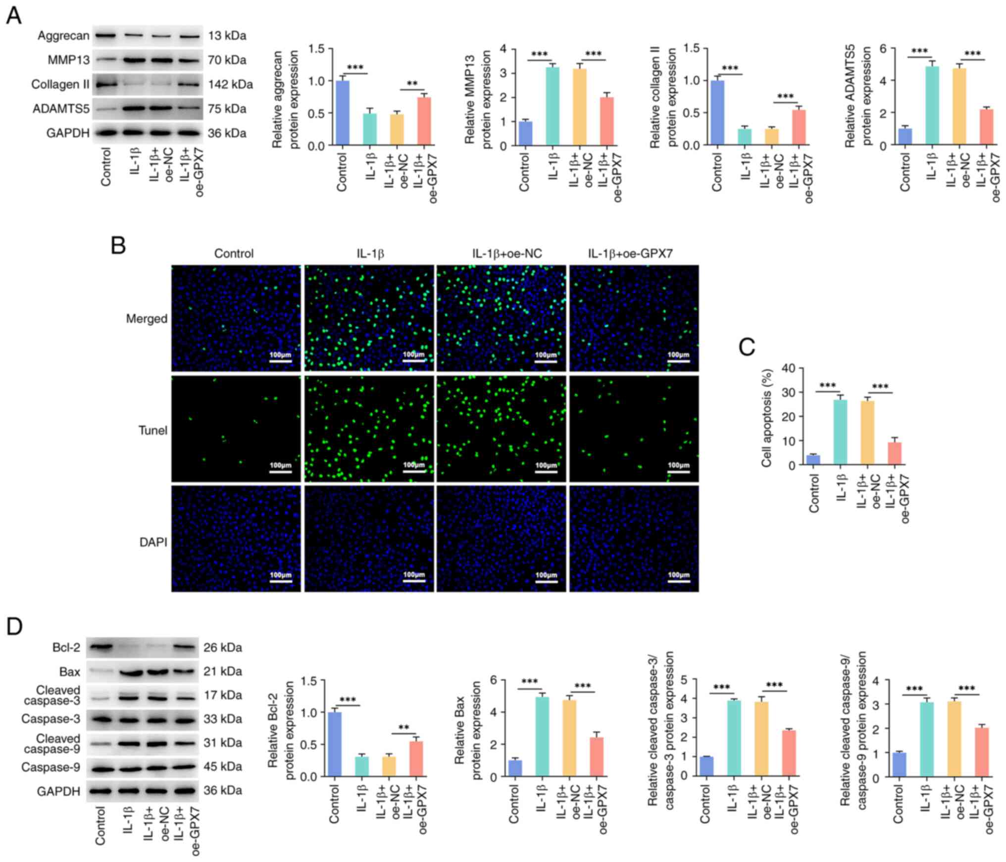
IL-1β-induced cartilage ECM degradation and apoptosis
Western blot analysis was used to determine the
levels of ECM-related proteins in the C28/I2 cells. In response to
IL-1β treatment, the protein levels of aggrecan and collagen II in
the cells decreased, while those of MMP13 and ADAMTS5 increased.
Compared with the IL-1β + oe-NC group, GPX7 overexpression
attenuated the decline in aggrecan and collagen II protein levels,
and reduced MMP13 and ADAMTS5 protein accumulation (Fig. 2A). TUNEL staining indicated the
trend of cell apoptosis, and the fluorescence increased upon IL-1β
treatment, indicating an increased cell apoptosis. Notably,
compared with the IL-1β + oe-NC group, the apoptosis of
GPX7-overexpressing cells was significantly reduced following
treatment with IL-1β (Fig. 2B and
C). In addition, the levels of the pro-apoptotic proteins, Bax,
cleaved caspase-3/caspase-3 and cleaved caspase-9/caspase-9, were
significantly increased in the cells under the influence of IL-1β,
while the level of the anti-apoptotic protein, Bcl-2, decreased.
The alterations in the levels of apoptosis-related proteins in the
IL-1β + oe-GPX7 group were attenuated following the overexpression
of GPX7, indicating the function of GPX7 in inhibiting apoptosis
(Fig. 2D).
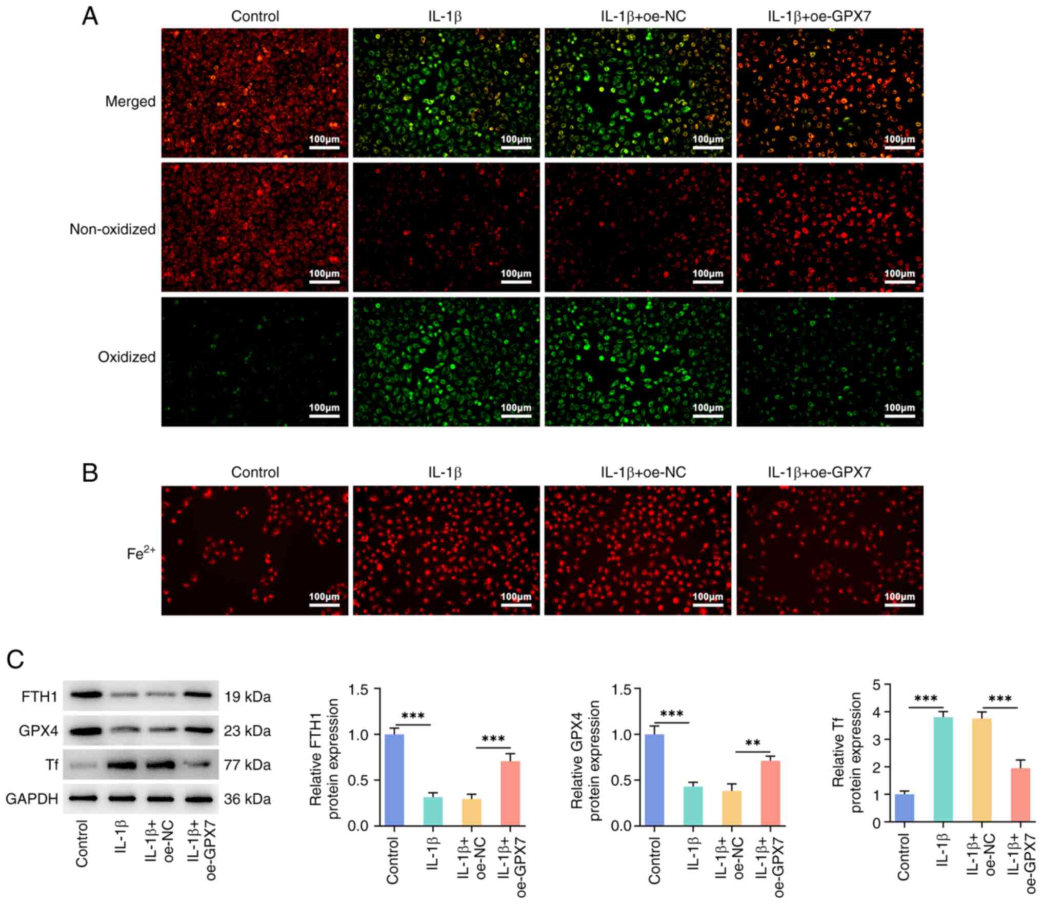
GPX7 overexpression inhibits
IL-1β-induced lipid peroxidation and ferroptosis in
chondrocytes
In the C11-BODIPY assay, the fluorescence in the
IL-1β group increased, indicating that the level of lipid
peroxidation increased, and GPX7 overexpression reduced the degree
of this oxidation (Fig. 3A). IL-1β
treatment also increased the ferrous ion levels in the C28/I2
cells, with the increase being less pronounced in the cells
overexpressing GPX7 (Fig. 3B). By
determining the levels of ferroptosis-related proteins, GPX7
overexpression was found to increase the FTH1 and GPX4 levels, and
reduce the Tf protein levels, suggesting that it reversed
IL-1β-induced ferroptosis (Fig.
3C).
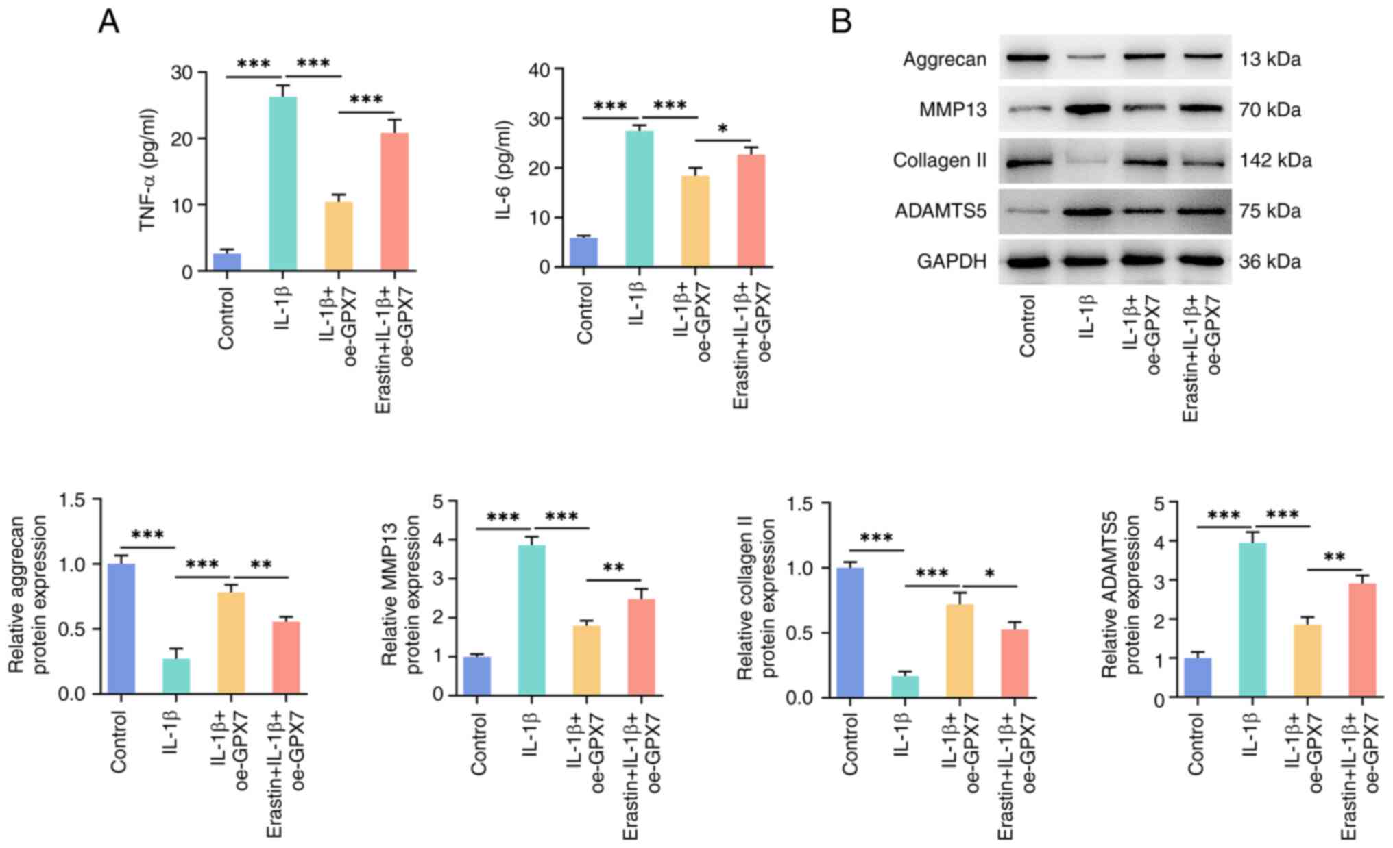
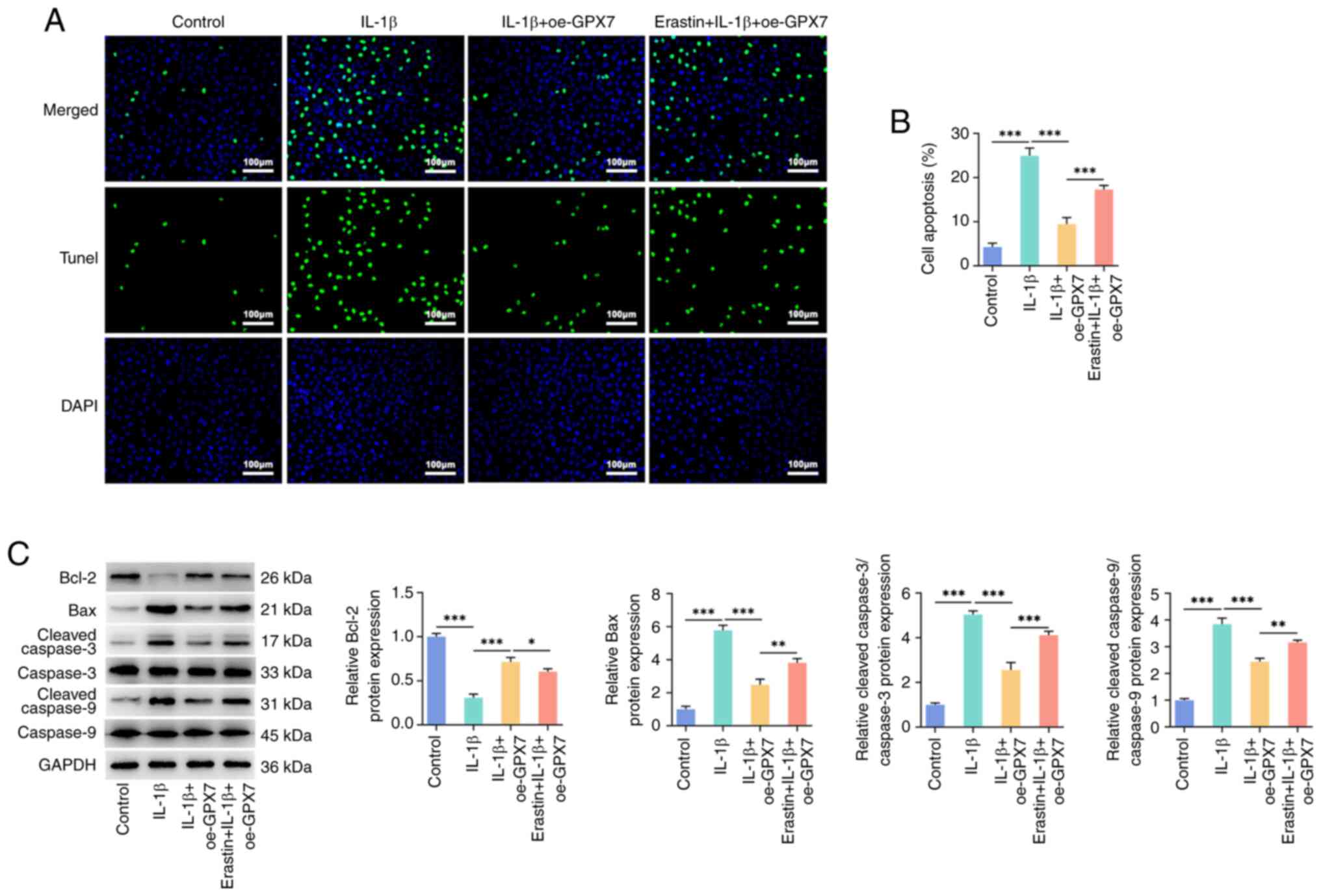
Effects of a ferroptosis inducer on
the regulatory effects of GPX7 overexpression
To verify the role of ferroptosis in the regulation
of multiple phenotypes aforementioned in chondrocytes, the
transfected cells were pre-treated with erastin followed by the
treatment with IL-1β. Compared with the IL-1β + oe-GPX7 group, the
addition of erastin increased the levels of the inflammatory
factors, TNF-α and IL-6 (Fig. 4A).
Furthermore, the addition of erastin inhibited the increase in the
aggrecan and collagen II protein levels induced by the
overexpression of GPX7, and elevated the levels of MMP13 and
ADAMTS5 (Fig. 4B). These findings
suggested that erastin promoted ECM degradation. The enhancement of
TUNEL fluorescence in the erastin-treated group indicated that
erastin reversed the inhibitory effects of GPX7 overexpression on
cell apoptosis (Fig. 5A and B).
This was supported by the increase in the levels of the
apoptosis-related proteins, Bax, cleaved caspase-3/caspase-3 and
cleaved caspase-9/caspase-9, and the decrease in the level of
Bcl-2, as a consequence of erastin treatment (Fig. 5C).
Discussion
The crux to the pathogenesis of OA is the
destruction of cartilage cells, the degradation of cartilage ECM
and the imbalance of the synthesis of subchondral bone components,
which in turn cause cartilage tissue defects and ultimately lead to
the occurrence of OA (18). In
terms of genetic factors, the heritability of OA is enhanced within
families, and the genetic probability of offspring developing OA is
as high as 40–65% (19). At the
molecular biological level, the pathogenesis of OA is closely
related to various inflammatory factors, MMPs, signaling pathways
and growth factors (20,21). Focusing on inflammatory factors,
the present study found that GPX7 overexpression suppressed the
TNF-α and IL-6 levels. The synthesis of these two factors is
related to the development of arthritis and is considered a key
drug target (22). Additionally,
IL-6 is associated with aging (23), with the levels of IL-6 in the
systemic circulation increasing with age, and being closely
associated with the risk of OA progression. Subsequently, the
levels of ECM-related proteins were determined. GPX7 overexpression
promoted the accumulation of the ECM components, aggrecan and
collagen II, and reduced the levels of MMP13 and ADAMTS5, which are
key enzymes involved in cartilage degradation (24). The experimental results indicated
that GPX7 has the ability to regulate and inhibit the
aforementioned pathogenic factors.
Increased chondrocyte apoptosis is a key
manifestation of OA. In advanced-stage OA, chondrocytes become
sparse and form cavities, and the ECM is also reduced due to
chondrocyte apoptosis (25). It
has been confirmed that in OA cartilage tissue, the apoptotic rate
of chondrocytes is ~20% (26).
Therefore, mechanisms through which to inhibit chondrocyte
apoptosis has become a practical strategy in the treatment of OA.
The present study demonstrated that the overexpression of GPX7
inhibited apoptosis, as shown using TUNEL staining and by the
detection of apoptosis-related proteins. In addition, previous
research has indicated that ferroptosis and apoptosis are closely
linked, and ferroptosis promotes cell sensitivity to apoptosis
(27). A previous study on
stigmasterol demonstrated that inhibiting ferroptosis can alleviate
IL-1β-induced chondrocyte damage and may have potential as a novel
treatment for knee OA (28).
Autophagic degradation of ferritin can cause chondrocyte
ferroptosis and ECM degradation (29). High concentrations of iron can
promote joint degeneration and promote the development of OA
(30). Hence, the present study
considered whether GPX7 can also affect ferroptosis. The detection
of lipid peroxidation, ferrous ions and transporters revealed that
GPX7 overexpression inhibited ferroptosis. An increasing number of
studies have shown that ROS-induced lipid peroxidation is related
to the pathogenesis of OA (31,32).
Under pathological conditions, the intracellular cysteine
metabolism pathway is blocked, indirectly inhibiting the activity
of GPX4, thereby leading to the accumulation of lipid peroxides
(33). The iron storage protein
FTH1, which maintains iron homeostasis to prevent oxidative damage,
decreases and transferrin receptors further enhance ferroptosis
through additional uptake of iron-loaded Tf (34). In addition, inflammation, ECM
degradation and apoptosis were promoted in the cells following
treatment with the ferroptosis-inducing agent, erastin. This
suggests that the regulatory role of GPX7 in these cell phenotypes
may be partially mediated by a pathway involving ferroptosis.
Erastin can also affect the biosynthesis of iron-sulfur clusters
and thus mitochondrial function, interfering with cellular energy
metabolism pathways and antioxidant systems (35). These combined mechanisms lead to
changes in the response to erastin treatment and ultimately affect
cell survival. Future detailed studies of these mechanisms will
help to understand the role of GPX7 and erastin in ferroptosis and
other cellular processes. In short, GPX4 has been widely recognized
to be responsible for clearing peroxidized lipids, reducing cell
damage, and delaying or inhibiting the occurrence of ferroptosis
(36). Based on the results of the
present study, GPX7 appears to be involved in inhibiting
ferroptosis as well. However, the mechanisms through which GPX7
regulates ferroptosis specifically remain unclear. Additionally,
the chondrocyte phenotypes cannot adequately represent the
pathological environment of OA. Further research is required in the
future to shed further light on this matter.
In conclusion, the present study reveals that GPX7
reduces IL-1β-induced chondrocyte inflammation, apoptosis and ECM
degradation partially through a pathway involving ferroptosis.
Exploring the association between OA and cell death may provide a
theoretical basis and may enable the development of translational
strategies for the treatment of OA. Future medicine development can
target GPX7 (or the GPX family) and design compounds or biological
agents to enhance or inhibit its activity. Screening and
optimization of their modulators could lead to the development of
medicine for modulating ferroptosis.
Acknowledgements
Not applicable.
Funding
The present study was supported by the Research Program of
Teacher Education Curriculum Reform in Henan (grant no.
2024-JSJYYB-094), the Philosophy and Social Science Planning
Program in Henan (grant no. 2021BTY006), and the Students'
Innovation and Entrepreneurship Training Program in Henan
University of Science and Technology (grant no. 2023461).
Availability of data and materials
The data generated in the present study may be
requested from the corresponding author.
Authors' contributions
BC and WF designed and performed the experiments.
CJ, GZ, ZL, YL and SZ participated in experiments and analyzed the
data. BC wrote the manuscript. All authors read and approved the
final manuscript. BC and WF confirm the authenticity of all the raw
data.
Ethics approval and consent to
participate
Not applicable.
Patient consent for publication
Not applicable.
Competing interests
The authors declare that they have no competing
interests.
References
|
1
|
Yunus MHM, Nordin A and Kamal H:
Pathophysiological perspective of osteoarthritis. Medicina
(Kaunas). 56:6142020. View Article : Google Scholar : PubMed/NCBI
|
|
2
|
Yao Q, Wu X, Tao C, Gong W, Chen M, Qu M,
Zhong Y, He T, Chen S and Xiao G: Osteoarthritis: Pathogenic
signaling pathways and therapeutic targets. Signal Transduct Target
Ther. 8:562023. View Article : Google Scholar : PubMed/NCBI
|
|
3
|
Hawker GA and King LK: The burden of
osteoarthritis in older adults. Clin Geriatr Med. 38:181–192. 2022.
View Article : Google Scholar : PubMed/NCBI
|
|
4
|
Katz JN, Arant KR and Loeser RF: Diagnosis
and treatment of hip and knee osteoarthritis: A review. JAMA.
325:568–578. 2021. View Article : Google Scholar : PubMed/NCBI
|
|
5
|
Krause M, Freudenthaler F, Frosch KH,
Achtnich A, Petersen W and Akoto R: Operative versus conservative
treatment of anterior cruciate ligament rupture. Dtsch Arztebl Int.
115:855–862. 2018.PubMed/NCBI
|
|
6
|
Molnar V, Matišić V, Kodvanj I, Bjelica R,
Jeleč Ž, Hudetz D, Rod E, Čukelj F, Vrdoljak T, Vidović D, et al:
Cytokines and chemokines involved in osteoarthritis pathogenesis.
Int J Mol Sci. 22:92082021. View Article : Google Scholar : PubMed/NCBI
|
|
7
|
Xie Y, Kang R, Klionsky DJ and Tang D:
GPX4 in cell death, autophagy, and disease. Autophagy.
19:2621–2638. 2023. View Article : Google Scholar : PubMed/NCBI
|
|
8
|
Kanemura S, Sofia EF, Hirai N, Okumura M,
Kadokura H and Inaba K: Characterization of the endoplasmic
reticulum-resident peroxidases GPx7 and GPx8 shows the higher
oxidative activity of GPx7 and its linkage to oxidative protein
folding. J Biol Chem. 295:12772–12785. 2020. View Article : Google Scholar : PubMed/NCBI
|
|
9
|
Wei PC, Hsieh YH, Su MI, Jiang X, Hsu PH,
Lo WT, Weng JY, Jeng YM, Wang JM, Chen PL, et al: Loss of the
oxidative stress sensor NPGPx compromises GRP78 chaperone activity
and induces systemic disease. Mol Cell. 48:747–759. 2012.
View Article : Google Scholar : PubMed/NCBI
|
|
10
|
Hu X, Li B, Wu F, Liu X, Liu M, Wang C,
Shi Y and Ye L: GPX7 Facilitates BMSCs Osteoblastogenesis via ER
Stress and mTOR Pathway. J Cell Mol Med. 25:10454–10465. 2021.
View Article : Google Scholar : PubMed/NCBI
|
|
11
|
Kim HJ, Lee Y, Fang S, Kim W, Kim HJ and
Kim JW: GPx7 ameliorates non-alcoholic steatohepatitis by
regulating oxidative stress. BMB Rep. 53:317–322. 2020. View Article : Google Scholar : PubMed/NCBI
|
|
12
|
Hwang HS and Shim JH: Brazilin and
Caesalpinia sappan L. extract protect epidermal keratinocytes from
oxidative stress by inducing the expression of GPX7. Chin J Nat
Med. 16:203–209. 2018.PubMed/NCBI
|
|
13
|
Zhou X, Zheng Y, Sun W, Zhang Z and Liu J,
Yang W, Yuan W, Yi Y, Wang J and Liu J: D-mannose alleviates
osteoarthritis progression by inhibiting chondrocyte ferroptosis in
a HIF-2α-dependent manner. Cell Prolif. 54:e131342021. View Article : Google Scholar : PubMed/NCBI
|
|
14
|
Zhou Y, Wu H, Wang F, Xu L, Yan Y, Tong X
and Yan H: GPX7 Is Targeted by miR-29b and GPX7 knockdown enhances
ferroptosis induced by erastin in glioma. Front Oncol.
11:8021242022. View Article : Google Scholar : PubMed/NCBI
|
|
15
|
Wang BW, Jiang Y, Yao ZL, Chen PS, Yu B
and Wang SN: Aucubin protects chondrocytes against IL-1β-Induced
apoptosis in vitro and inhibits osteoarthritis in mice model. Drug
Des Devel Ther. 13:3529–3538. 2019. View Article : Google Scholar : PubMed/NCBI
|
|
16
|
Dixon SJ, Lemberg KM, Lamprecht MR, Skouta
R, Zaitsev EM, Gleason CE, Patel DN, Bauer AJ, Cantley AM, Yang WS,
et al: Ferroptosis: An iron-dependent form of nonapoptotic cell
death. Cell. 149:1060–1072. 2012. View Article : Google Scholar : PubMed/NCBI
|
|
17
|
Livak KJ and Schmittgen TD: Analysis of
relative gene expression data using real-time quantitative PCR and
the 2(−Delta Delta C(T)) Method. Methods. 25:402–408. 2001.
View Article : Google Scholar : PubMed/NCBI
|
|
18
|
Guilak F, Nims RJ, Dicks A, Wu CL and
Meulenbelt I: Osteoarthritis as a disease of the cartilage
pericellular matrix. Matrix Biol. 71–72. 40–50. 2018.
|
|
19
|
Wilkinson JM and Zeggini E: The genetic
epidemiology of joint shape and the development of osteoarthritis.
Calcif Tissue Int. 109:257–276. 2021. View Article : Google Scholar : PubMed/NCBI
|
|
20
|
Cui N, Hu M and Khalil RA: Biochemical and
biological attributes of matrix metalloproteinases. Prog Mol Biol
Transl Sci. 147:1–73. 2017. View Article : Google Scholar : PubMed/NCBI
|
|
21
|
Huang J, Zhao L and Chen D: Growth factor
signalling in osteoarthritis. Growth Factors. 36:187–195. 2018.
View Article : Google Scholar : PubMed/NCBI
|
|
22
|
Wang T and He C: Pro-inflammatory
cytokines: The link between obesity and osteoarthritis. Cytokine
Growth Factor Rev. 44:38–50. 2018. View Article : Google Scholar : PubMed/NCBI
|
|
23
|
Wang J, Chen J, Zhang B and Jia X: IL-6
regulates the bone metabolism and inflammatory microenvironment in
aging mice by inhibiting Setd7. Acta Histochem. 123:1517182021.
View Article : Google Scholar : PubMed/NCBI
|
|
24
|
Ashruf OS and Ansari MY: Natural
compounds: Potential therapeutics for the inhibition of cartilage
matrix degradation in osteoarthritis. Life (Basel).
13:1022022.PubMed/NCBI
|
|
25
|
Zhu R, Wang Y, Ouyang Z, Hao W, Zhou F,
Lin Y, Cheng Y, Zhou R and Hu W: Targeting regulated chondrocyte
death in osteoarthritis therapy. Biochem Pharmacol. 215:1157072023.
View Article : Google Scholar : PubMed/NCBI
|
|
26
|
Zhang T and Li J: Experimental study on
the ratios of chondrocytes apoptosis in OA. Chin Modern Doctor.
46:35–36. 2008.(In Chinese).
|
|
27
|
Mou Y, Wang J, Wu J, He D, Zhang C, Duan C
and Li B: Ferroptosis, a new form of cell death: Opportunities and
challenges in cancer. J Hematol Oncol. 12:342019. View Article : Google Scholar : PubMed/NCBI
|
|
28
|
Mo Z, Xu P and Li H: Stigmasterol
alleviates interleukin-1beta-induced chondrocyte injury by
down-regulatingsterol regulatory element binding transcription
factor 2 to regulateferroptosis. Bioengineered. 12:9332–9340. 2021.
View Article : Google Scholar : PubMed/NCBI
|
|
29
|
Sun K, Hou L, Guo Z, Wang G, Guo J, Xu J,
Zhang X and Guo F: JNK-JUN-NCOA4 axis contributes to chondrocyte
ferroptosis and aggravates osteoarthritis via ferritinophagy. Free
Radic Biol Med. 200:87–101. 2023. View Article : Google Scholar : PubMed/NCBI
|
|
30
|
Yang J, Hu S, Bian Y, Yao J, Wang D, Liu
X, Guo Z, Zhang S and Peng L: Targeting cell death: Pyroptosis,
ferroptosis, apoptosis and necroptosis in osteoarthritis. Front
Cell Dev Biol. 9:7899482022. View Article : Google Scholar : PubMed/NCBI
|
|
31
|
Zhang X, Hou L, Guo Z, Wang G, Xu J, Zheng
Z, Sun K and Guo F: Lipid peroxidation in osteoarthritis: Focusing
on 4-hydroxynonenal, malondialdehyde, and ferroptosis. Cell Death
Discov. 9:3202023. View Article : Google Scholar : PubMed/NCBI
|
|
32
|
An F, Zhang J, Gao P, Xiao Z, Chang W,
Song J, Wang Y, Ma H, Zhang R, Chen Z and Yan C: New insight of the
pathogenesis in osteoarthritis: The intricate interplay of
ferroptosis and autophagy mediated by mitophagy/chaperone-mediated
autophagy. Front Cell Dev Biol. 11:12970242023. View Article : Google Scholar : PubMed/NCBI
|
|
33
|
Rochette L, Dogon G, Rigal E, Zeller M,
Cottin Y and Vergely C: Lipid peroxidation and iron metabolism: Two
corner stones in the homeostasis control of ferroptosis. Int J Mol
Sci. 24:4492022. View Article : Google Scholar : PubMed/NCBI
|
|
34
|
Hou Y, Wang S, Jiang L, Sun X, Li J, Wang
N, Liu X, Yao X, Zhang C, Deng H and Yang G: Patulin induces acute
kidney injury in mice through autophagy-ferroptosis pathway. J
Agric Food Chem. 70:6213–6223. 2022. View Article : Google Scholar : PubMed/NCBI
|
|
35
|
Cotticelli MG, Xia S, Lin D, Lee T, Terrab
L, Wipf P, Huryn DM and Wilson RB: Ferroptosis as a novel
therapeutic target for friedreich's Ataxia. J Pharmacol Exp Ther.
369:47–54. 2019. View Article : Google Scholar : PubMed/NCBI
|
|
36
|
Weaver K and Skouta R: The selenoprotein
glutathione peroxidase 4: From molecular mechanisms to novel
therapeutic opportunities. Biomedicines. 10:8912022. View Article : Google Scholar : PubMed/NCBI
|