Spandidos Publications Logo
  • About
    • About Spandidos
    • Aims and Scopes
    • Abstracting and Indexing
    • Editorial Policies
    • Reprints and Permissions
    • Job Opportunities
    • Terms and Conditions
    • Contact
  • Journals
    • All Journals
    • Oncology Letters
      • Oncology Letters
      • Information for Authors
      • Editorial Policies
      • Editorial Board
      • Aims and Scope
      • Abstracting and Indexing
      • Bibliographic Information
      • Archive
    • International Journal of Oncology
      • International Journal of Oncology
      • Information for Authors
      • Editorial Policies
      • Editorial Board
      • Aims and Scope
      • Abstracting and Indexing
      • Bibliographic Information
      • Archive
    • Molecular and Clinical Oncology
      • Molecular and Clinical Oncology
      • Information for Authors
      • Editorial Policies
      • Editorial Board
      • Aims and Scope
      • Abstracting and Indexing
      • Bibliographic Information
      • Archive
    • Experimental and Therapeutic Medicine
      • Experimental and Therapeutic Medicine
      • Information for Authors
      • Editorial Policies
      • Editorial Board
      • Aims and Scope
      • Abstracting and Indexing
      • Bibliographic Information
      • Archive
    • International Journal of Molecular Medicine
      • International Journal of Molecular Medicine
      • Information for Authors
      • Editorial Policies
      • Editorial Board
      • Aims and Scope
      • Abstracting and Indexing
      • Bibliographic Information
      • Archive
    • Biomedical Reports
      • Biomedical Reports
      • Information for Authors
      • Editorial Policies
      • Editorial Board
      • Aims and Scope
      • Abstracting and Indexing
      • Bibliographic Information
      • Archive
    • Oncology Reports
      • Oncology Reports
      • Information for Authors
      • Editorial Policies
      • Editorial Board
      • Aims and Scope
      • Abstracting and Indexing
      • Bibliographic Information
      • Archive
    • Molecular Medicine Reports
      • Molecular Medicine Reports
      • Information for Authors
      • Editorial Policies
      • Editorial Board
      • Aims and Scope
      • Abstracting and Indexing
      • Bibliographic Information
      • Archive
    • World Academy of Sciences Journal
      • World Academy of Sciences Journal
      • Information for Authors
      • Editorial Policies
      • Editorial Board
      • Aims and Scope
      • Abstracting and Indexing
      • Bibliographic Information
      • Archive
    • International Journal of Functional Nutrition
      • International Journal of Functional Nutrition
      • Information for Authors
      • Editorial Policies
      • Editorial Board
      • Aims and Scope
      • Abstracting and Indexing
      • Bibliographic Information
      • Archive
    • International Journal of Epigenetics
      • International Journal of Epigenetics
      • Information for Authors
      • Editorial Policies
      • Editorial Board
      • Aims and Scope
      • Abstracting and Indexing
      • Bibliographic Information
      • Archive
    • Medicine International
      • Medicine International
      • Information for Authors
      • Editorial Policies
      • Editorial Board
      • Aims and Scope
      • Abstracting and Indexing
      • Bibliographic Information
      • Archive
  • Articles
  • Information
    • Information for Authors
    • Information for Reviewers
    • Information for Librarians
    • Information for Advertisers
    • Conferences
  • Language Editing
Spandidos Publications Logo
  • About
    • About Spandidos
    • Aims and Scopes
    • Abstracting and Indexing
    • Editorial Policies
    • Reprints and Permissions
    • Job Opportunities
    • Terms and Conditions
    • Contact
  • Journals
    • All Journals
    • Biomedical Reports
      • Information for Authors
      • Editorial Policies
      • Editorial Board
      • Aims and Scope
      • Abstracting and Indexing
      • Bibliographic Information
      • Archive
    • Experimental and Therapeutic Medicine
      • Information for Authors
      • Editorial Policies
      • Editorial Board
      • Aims and Scope
      • Abstracting and Indexing
      • Bibliographic Information
      • Archive
    • International Journal of Epigenetics
      • Information for Authors
      • Editorial Policies
      • Editorial Board
      • Aims and Scope
      • Abstracting and Indexing
      • Bibliographic Information
      • Archive
    • International Journal of Functional Nutrition
      • Information for Authors
      • Editorial Policies
      • Editorial Board
      • Aims and Scope
      • Abstracting and Indexing
      • Bibliographic Information
      • Archive
    • International Journal of Molecular Medicine
      • Information for Authors
      • Editorial Policies
      • Editorial Board
      • Aims and Scope
      • Abstracting and Indexing
      • Bibliographic Information
      • Archive
    • International Journal of Oncology
      • Information for Authors
      • Editorial Policies
      • Editorial Board
      • Aims and Scope
      • Abstracting and Indexing
      • Bibliographic Information
      • Archive
    • Medicine International
      • Information for Authors
      • Editorial Policies
      • Editorial Board
      • Aims and Scope
      • Abstracting and Indexing
      • Bibliographic Information
      • Archive
    • Molecular and Clinical Oncology
      • Information for Authors
      • Editorial Policies
      • Editorial Board
      • Aims and Scope
      • Abstracting and Indexing
      • Bibliographic Information
      • Archive
    • Molecular Medicine Reports
      • Information for Authors
      • Editorial Policies
      • Editorial Board
      • Aims and Scope
      • Abstracting and Indexing
      • Bibliographic Information
      • Archive
    • Oncology Letters
      • Information for Authors
      • Editorial Policies
      • Editorial Board
      • Aims and Scope
      • Abstracting and Indexing
      • Bibliographic Information
      • Archive
    • Oncology Reports
      • Information for Authors
      • Editorial Policies
      • Editorial Board
      • Aims and Scope
      • Abstracting and Indexing
      • Bibliographic Information
      • Archive
    • World Academy of Sciences Journal
      • Information for Authors
      • Editorial Policies
      • Editorial Board
      • Aims and Scope
      • Abstracting and Indexing
      • Bibliographic Information
      • Archive
  • Articles
  • Information
    • For Authors
    • For Reviewers
    • For Librarians
    • For Advertisers
    • Conferences
  • Language Editing
Login Register Submit
  • This site uses cookies
  • You can change your cookie settings at any time by following the instructions in our Cookie Policy. To find out more, you may read our Privacy Policy.

    I agree
Search articles by DOI, keyword, author or affiliation
Search
Advanced Search
presentation
Oncology Letters
Join Editorial Board Propose a Special Issue
Print ISSN: 1792-1074 Online ISSN: 1792-1082
Journal Cover
June-2016 Volume 11 Issue 6

Full Size Image

Sign up for eToc alerts
Recommend to Library

Journals

International Journal of Molecular Medicine

International Journal of Molecular Medicine

International Journal of Molecular Medicine is an international journal devoted to molecular mechanisms of human disease.

International Journal of Oncology

International Journal of Oncology

International Journal of Oncology is an international journal devoted to oncology research and cancer treatment.

Molecular Medicine Reports

Molecular Medicine Reports

Covers molecular medicine topics such as pharmacology, pathology, genetics, neuroscience, infectious diseases, molecular cardiology, and molecular surgery.

Oncology Reports

Oncology Reports

Oncology Reports is an international journal devoted to fundamental and applied research in Oncology.

Experimental and Therapeutic Medicine

Experimental and Therapeutic Medicine

Experimental and Therapeutic Medicine is an international journal devoted to laboratory and clinical medicine.

Oncology Letters

Oncology Letters

Oncology Letters is an international journal devoted to Experimental and Clinical Oncology.

Biomedical Reports

Biomedical Reports

Explores a wide range of biological and medical fields, including pharmacology, genetics, microbiology, neuroscience, and molecular cardiology.

Molecular and Clinical Oncology

Molecular and Clinical Oncology

International journal addressing all aspects of oncology research, from tumorigenesis and oncogenes to chemotherapy and metastasis.

World Academy of Sciences Journal

World Academy of Sciences Journal

Multidisciplinary open-access journal spanning biochemistry, genetics, neuroscience, environmental health, and synthetic biology.

International Journal of Functional Nutrition

International Journal of Functional Nutrition

Open-access journal combining biochemistry, pharmacology, immunology, and genetics to advance health through functional nutrition.

International Journal of Epigenetics

International Journal of Epigenetics

Publishes open-access research on using epigenetics to advance understanding and treatment of human disease.

Medicine International

Medicine International

An International Open Access Journal Devoted to General Medicine.

Journal Cover
June-2016 Volume 11 Issue 6

Full Size Image

Sign up for eToc alerts
Recommend to Library

  • Article
  • Citations
    • Cite This Article
    • Download Citation
    • Create Citation Alert
    • Remove Citation Alert
    • Cited By
  • Similar Articles
    • Related Articles (in Spandidos Publications)
    • Similar Articles (Google Scholar)
    • Similar Articles (PubMed)
  • Download PDF
  • Download XML
  • View XML
Article

CT findings of sclerosing stromal tumor of the ovary: A report of two cases and review of the literature

  • Authors:
    • Tongtong Tian
    • Qingqiang Zhu
    • Wenxin Chen
    • Shouan Wang
    • Weifan Sui
    • Jingtao Wu
  • View Affiliations / Copyright

    Affiliations: Department of Radiology, Subei People's Hospital of Jiangsu Province, Medical School of Yangzhou University, Yangzhou, Jiangsu 225001, P.R. China
  • Pages: 3817-3820
    |
    Published online on: April 15, 2016
       https://doi.org/10.3892/ol.2016.4441
  • Expand metrics +
Metrics: Total Views: 0 (Spandidos Publications: | PMC Statistics: )
Metrics: Total PDF Downloads: 0 (Spandidos Publications: | PMC Statistics: )
Cited By (CrossRef): 0 citations Loading Articles...

This article is mentioned in:


Abstract

Sclerosing stromal tumor (SST) of the ovary, which was first described by Chalvardjian and Scully in 1973, is a rare ovarian neoplasm, occurring predominantly in young women. The most common clinical symptom in patients with SST is menstrual irregularities. Microscopically, the tumor is characterized by the presence of pseudo-lobulated cellular areas, with a prominent tendency to sclerosis, marked vascularity and pronounced variation in cellular size and shape. In the current study, 2 cases of SST of the ovary are presented. These cases were confirmed by imaging, surgical and histological examination. No adjuvant therapy was administered to the patients and the two patients were disease‑free with no imaging findings of recurrence or metastasis 24 months following surgery.

Introduction

Sclerosing stromal tumor (SST) of the ovary is a rare benign tumor that has been classified as a sex cord-stromal tumor and is predominantly observed in young women (1,2). SSTs account for 1.5% of all ovarian tumors (1). The tumor usually occurs in the second and third decades of life (mean age, 21 years). The most common presenting symptoms are menstrual irregularity, abdominal pain and a lower abdominal mass (1,2). Microscopically, the tumor is characterized by pseudo-lobulation of cellular areas, marked vascularity and significant variation in cellular size and shape, and sclerosis is common (1,3). Surgical removal is curative and no local or distant recurrences have been reported following surgery (2,3). To the best of our knowledge, there have been numerous reports regarding the microscopic, ultrastructural and immunohistochemical, but not imaging, findings associated with this tumor (3). The present study describes the computed tomography (CT) findings of 2 patients with ovarian SST, and confirms the correlation between imaging and pathological findings, as well as the reliability of imaging in preoperative characterization. The study was approved by the Ethics Committee of Subei People's Hospital of Jiangsu Province (Yangzhou, China) and the patients provided written informed consent for the publication of the present study.

Case report

Case 1

An 18-year-old Chinese female patient was admitted to Subei People's Hospital of Jiangsu Province in July 2012 with menstrual irregularities for 6 months. The blood test results for blood cell count, biochemistry and tumor markers, including cancer antigen (CA)19-9 (3.57 KU/l; normal range, <35.00 KU/l), carcinoembryonic antigen CA125 (5.15 KU/l; normal range, <35.00 KU/l) and α-fetoprotein (3.57 ng/ml; normal range, <20.00 ng/ml) were all normal. The levels of serum hormones, including estradiol (60 ng/ml; normal range, 22–144 ng/ml), progesterone (8.13 ng/ml; normal range, 5.16–18.56 ng/ml) and testosterone (12.3 ng/ml; normal range, 10.0–75.0 ng/ml), were also normal.

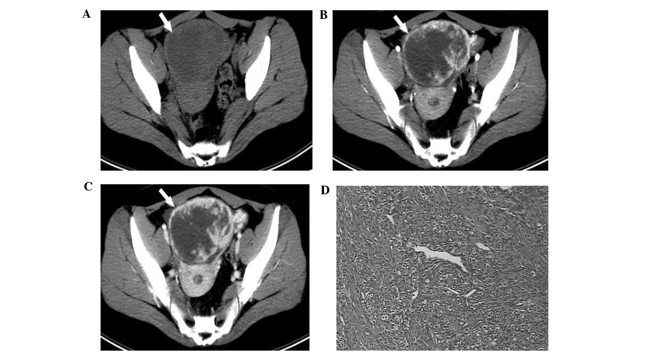
Non-enhanced CT (Lightspeed VCT 64; GE Healthcare, Milwaukee, WI, USA) scan revealed a round soft tissue mass with clear boundaries in the middle of the pelvis. The mass measured 76×77 mm and displayed non-homogeneous density, with solid, non-calcified tissue at the periphery and a fluid, patch-shaped area in the inner zone (Fig. 1A). Plain CT values were 31–45 HU, with a mean value of 37.6 HU. Intravenous bolus injection of iodinated contrast medium (iopromide; Ultravist® 320; Bayer HealthCare Pharmaceuticals, Berlin, Germany) yielded early nodular and ring enhancement of the peripheral portion of the mass, which was as high as that of the vessels in the arterial phase (Fig. 1B). In the venous phase, the degree of enhancement was decreased, but the area of enhancement increased with centripetal progression (Fig. 1C). The CT values in the arterial and venous phases were 145–150 HU, with a mean value of 147 HU, and 128–135 HU, with a mean value of 130 HU, respectively. The cystic components of the tumor were not enhanced in these phases, and no pelvic or lumbo-aortic enlarged lymph nodes were observed.

Figure 1.

Sclerosing stromal tumor of the left ovary in a 18-year-old woman presenting with menstrual irregularities for 6 months. (A) Axial non-enhanced CT scan showing a round, well-defined heterogeneous mass measuring 76×77 mm in size, which was located in the middle of the pelvis (white arrow). The plain CT values were 31–45 HU. (B) Dynamic contrast-enhanced CT scan showing early peripheral ring enhancement associated with patch enhancement in the center of the lesion (white arrow). On the left side of the tumor, multiple circuitous feeding vessels were clearly visible in the arterial phase. The maximum CT value was 145–150 HU, with a mean value of 147 HU. (C) In the venous phase, the tumor demonstrated progressive, centripetal and prolonged enhancement (white arrow). The CT values were 128–135 HU, with a mean value of 130 HU. The central portion of the tumor was not enhanced. (D) Sclerosing and hypocellular areas separated by edematous fibrous bands were observed with hematoxylin and eosin staining. Blood vessels were mildly ectatic. Magnification, ×200. CT, computed tomography.

It was confirmed that the mass originated from the left ovarian tissue. Macroscopic examination revealed a 100×80-mm surgical specimen, which was yellow-orange in color and contained solid, cystic components. Resected tissue specimens were fixed in 10% formalin, paraffin-embedded and cut into 5-mm sections. Sections were stained with hematoxylin and eosin (Shanghai Haiyi Scientific & Trading Co., Ltd., Shanghai, China) for histological examination and staining was visualized using a microscope (JVC1481; Olympus Corporation, Tokyo, Japan). Histological examination of the mass revealed cellular areas separated by hypocellular areas of densely collagenous, edematous or myxoid tissue, alongside prominent vasculature, thus resulting in a pseudo-lobular pattern. The cellular areas were composed of oval to spindle-shaped cells, with a single oval to round-shaped nucleus and a single prominent nucleolus. The cytoplasm was moderately abundant, eosinophilic and occasionally vacuolated (Fig. 1D). Immunohistochemical staining was performed using the avidin biotin peroxidase method, and demonstrated focal positivity for actin, pan-cytokeratin (CK), inhibin and calretinin, and negativity for S100, desmin and epithelial membrane antigen. Based on these findings, a diagnosis of SST of the left ovary was established. The patient exhibited regular menstrual cycles and no signs of local or distal recurrence 2 years post-surgery.

Case 2

A 59-year-old woman was admitted to Subei People's Hospital of Jiangsu Province in May 2013 with continuous pelvic pain that had been ongoing for a year. Clinical examination identified a large mass that was palpable in the right-side of the lower pelvis. Ultrasonography revealed a well-defined solid-cystic echogenic mass in the right ovary measuring 64×55 mm. Laboratory tests and hormonal assays, including CA19-9 (13.11 KU/l; normal range, <35.00 KU/l), carcinoembryonic antigen CA125 (16.15 KU/l; normal range, <35.00 KU/l), α-fetoprotein (1.57 ng/ml; normal range, <20.00 ng/ml), estradiol (31 pg/ml; normal range, 22–144 pg/ml), progesterone (11.13 ng/ml; normal range, 5.16–18.56 ng/ml) and testosterone (21.6 ng/ml; normal range, 10–75 ng/ml) were normal.

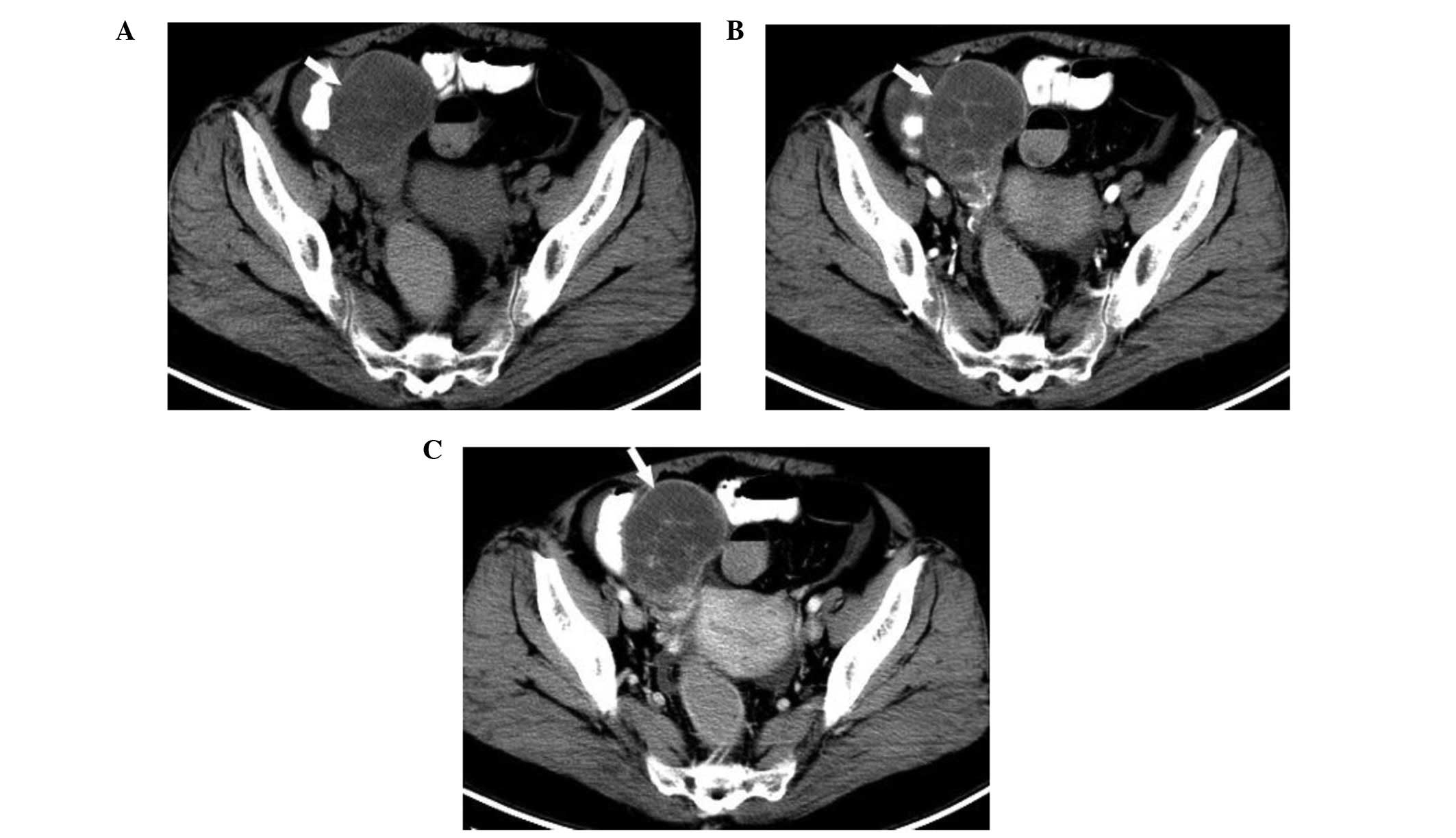
Plain CT scan revealed an ovoid soft tissue mass located in the right pelvis. The tumor, which measured 64×55 mm in size, had clear boundaries and showed non-homogeneous density with solid tissue at the periphery (Fig. 2A). Plain CT scan values were 25–40 HU, with a mean value of 32.6 HU. Intravenous bolus injection of iodinated contrast medium yielded early ring enhancement of the peripheral portion of the mass (Fig. 2B). In the venous phase, the degree of enhancement was decreased, but the area of enhancement increased with centripetal progression (Fig. 2C). The CT values at the arterial and venous phases were 125–140 HU, with a mean value of 132 HU, and 118–130 HU, with a mean value of 128 HU, respectively. The cystic components of the inner region of the lesion were not enhanced in these phases.

Figure 2.

Sclerosing stromal tumor arising from the right ovary in a 59-year-old woman with a history of right-sided pelvic pain. (A) An ovoid solid-cystic mass was observed in the right adnexal area (white arrow). Plain CT values were 25–40 HU, with a mean value of 32.6 HU. (B) Dynamic contrast-enhanced CT scan revealed early septal enhancement in the center, and ring enhancement in the peripheral portion of the mass in the arterial phase (white arrow). The CT value of the solid component was ~132 HU. The CT value of the solid component was 125–140 HU, with a mean value of 132 HU (C) In the venous phase, the tumor demonstrated centripetal, progressive enhancement (white arrow). The CT values were 118–130 HU, with a mean value of 128 HU. The cystic areas of the tumor were observed. CT, computed tomography.

The patient underwent surgical removal of the 70×60-mm mass, which was well-circumscribed and presented a round to oval-shape with a smooth outer surface. The resected section was of pale yellow color with solid cystic areas. Histological examination revealed that the mass was characterized by cellular areas separated by hypocellular areas of densely collagenous, edematous or myxoid tissue and prominent vasculature, thus creating a pseudo-lobular pattern. The cellular areas were composed of oval to spindle-shaped cells, with a single oval to round-shaped nucleus and a single prominent nucleolus. Tumor cells stained positive for smooth muscle actin and vimentin, and focally positive for inhibin. The patient was disease-free with no imaging findings of recurrence or metastasis 24 months following surgery.

Discussion

Ovarian SST is a considerably rare and distinctive sex cord-stromal neoplasm, which was first described by Chalvardjian and Scully in 1973 (1). The histogenesis of this tumor remains unknown, and it predominantly affects young women in the second and third decades of life (mean age, 21 years) (2). To the best of our knowledge, <100 cases of ovarian SST have been reported in the English language literature thus far (2–4). Of these, >80% of SSTs have been observed in young adults, in the second and third decades of life (2,3). The most common clinical symptoms of patients with SST of the ovary are menstrual irregularities, pelvic pain and non-specific symptoms associated with the existence of a mass in the pelvic area (2,4). Due to the rarity of this neoplasm, it is not always possible to predict the presence of this tumor preoperatively based solely on clinical and radiological findings (5).

Microscopically, SST is characterized by cellular areas separated by hypocellular areas of densely collagenous, edematous or myxoid tissue and prominent vasculature, which creates a pseudo-lobular pattern (6,7). In a previous study, the cellular areas were observed to be composed of oval to spindle-shaped cells, with a single oval to round-shaped nucleus and a single prominent nucleolus (8). In addition, the cytoplasm was moderately abundant, eosinophilic and occasionally vacuolated (8). A previous study revealed that the blood vessels within the cellular area were mildly ectatic, and no evidence of malignancy was observed (6). Following surgery, the patient's menstrual condition returned to normal (6).

In previous studies, CT imaging demonstrated that the density and enhancement patterns of SST are associated with the cellularity, vascularity, collagen distribution and necrosis or cystic degeneration observed during histopathological examination of the tumor (6,7). On non-enhanced CT, SST manifests solid densities corresponding to cellularity, vascularity and distribution of collagenous or fibrous stroma, while the areas of necrosis or cystic degeneration exhibit low densities (7). The SSTs of the present 2 cases presented patchy areas of low attenuation.

The appearance of SSTs on imaging scans, particularly the enhancement pattern observed in dynamic contrast-enhanced CT images, may vary widely, depending on the distribution of cellularity, vascularity and collagenous or fibrous stroma (9). Previous studies demonstrated that the heterogeneously enhancing masses observed in contrast-enhanced CT images are associated with the tumor vascularity, cellularity and collagen distribution (7,9). In the present cases, following the administration of intravenous contrast material, the early enhancement of the outer region of the tumor was possibly due to the cellular area with numerous vascular spaces. The maximum enhancement value was 150 HU. In the venous phase, the area of prolonged enhancement observed in the inner portion of the lesion was considered to be associated with the collagenized hypocellular area. The CT values were 110–123 HU, with a mean value of 116 HU. The part of the tumor that did not exhibit any apparent enhancement possibly corresponded to the markedly edematous area. The present CT findings were almost identical to those of previous studies (6,10), and were particularly in agreement with the results of Torricelli et al (11), who reported that the appearance of SST on dynamic contrast-enhanced CT images is considerably similar to that of cavernous hemangioma of the liver, and can be pathologically defined as ‘hemangioma-like’.

In conclusion, differential diagnosis of SSTs should include other thecoma-fibroma, metastatic and malignant epithelial ovarian tumors (12), since, due to the rarity of SST, prospective imaging diagnosis is not possible.

The present case report described certain imaging features of ovarian SST based on a small-population sample. Therefore, further studies are required to confirm the diagnostic accuracy of imaging in cases of SST, and to determine whether the patterns observed in imaging scans may aid the differentiation between SST and other tumors.

Acknowledgements

The present study was supported by the General Program of Yangzhou Natural Science Foundation (Yangzhou, China; grant no., YZ2015100) and National Natural Science Foundation (Beijing, China; grant no., 81571652).

References

1 

Chalvardjian A and Scully RE: Sclerosing stromal tumors of the ovary. Cancer. 31:664–670. 1973. View Article : Google Scholar : PubMed/NCBI

2 

Chang W, Oiseth SJ, Orentlicher R, Agarwal G, Yahr LJ and Cayten CG: Bilateral sclerosing stromal tumor of the ovaries in a premenarchal girl. Gynecol Oncol. 101:342–345. 2006. View Article : Google Scholar : PubMed/NCBI

3 

Kaygusuz EI, Cesur S, Cetiner H, Yavuz H and Koc N: Sclerosing stromal tumour in young women: Clinicopathologic and immunohistochemical spectrum. J Clin Diagn Res. 7:1932–1935. 2013.PubMed/NCBI

4 

Ihara N, Togashi K, Todo G, Nakai A, Kojima N, Ishigaki T, Suginami N, Kinoshita M and Shintaku M: Sclerosing stromal tumor of the ovary: MRI. J Comput Assist Tomogr. 23:555–557. 1999. View Article : Google Scholar : PubMed/NCBI

5 

Outwater EK, Wagner BJ, Mannion C, McLarney JK and Kim B: Sex cord-stromal and steroid cell tumors of the ovary. Radiographics. 18:1523–1546. 1998. View Article : Google Scholar : PubMed/NCBI

6 

Kawauchi S, Tsuji T, Kaku T, Kamura T, Nakano H and Tsuneyoshi M: Sclerosing stromal tumor of the ovary: A clinicopathologic, immunohistochemical, ultrastructural, and cytogenetic analysis with special reference to its vasculature. Am J Surg Pathol. 22:83–92. 1998. View Article : Google Scholar : PubMed/NCBI

7 

Tian TT, Wu JT, Hu XH, Yang GM, Sun J, Chen WX and Tian XC: Imaging findings of solitary fibrous tumor in the abdomen and pelvis. Abdom Imaging. 39:1323–1329. 2014. View Article : Google Scholar : PubMed/NCBI

8 

Matsubayashi R, Matsuo Y, Doi J, Kudo S, Matsuguchi K and Sugimori H: Sclerosing stromal tumor of the ovary: Radiologic findings. Eur Radiol. 9:1335–1338. 1999. View Article : Google Scholar : PubMed/NCBI

9 

Kim JY, Jung KJ, Chung DS, Kim OD, Lee JH and Youn SK: Sclerosing stromal tumor of the ovary: MR-pathologic correlation in three cases. Korean J Radiol. 4:194–199. 2003. View Article : Google Scholar : PubMed/NCBI

10 

Kawamura N, Kamoi I and Shigyo R: Sclerosing stromal tumour of the ovary. Br J Radiol. 60:1031–1033. 1987. View Article : Google Scholar : PubMed/NCBI

11 

Torricelli P, Caruso Lombardi A, Boselli F and Rossi G: Sclerosing stromal tumor of the ovary: US, CT, and MRI findings. Abdom Imaging. 27:588–591. 2002. View Article : Google Scholar : PubMed/NCBI

12 

Jung SE, Lee JM, Rha SE, Byun JY, Jung JI and Hahn ST: CT and MR imaging of ovarian tumors with emphasis on differential diagnosis. Radiographics. 22:1305–1325. 2002. View Article : Google Scholar : PubMed/NCBI

Related Articles

  • Abstract
  • View
  • Download
  • Twitter
Copy and paste a formatted citation
Spandidos Publications style
Tian T, Zhu Q, Chen W, Wang S, Sui W and Wu J: CT findings of sclerosing stromal tumor of the ovary: A report of two cases and review of the literature. Oncol Lett 11: 3817-3820, 2016.
APA
Tian, T., Zhu, Q., Chen, W., Wang, S., Sui, W., & Wu, J. (2016). CT findings of sclerosing stromal tumor of the ovary: A report of two cases and review of the literature. Oncology Letters, 11, 3817-3820. https://doi.org/10.3892/ol.2016.4441
MLA
Tian, T., Zhu, Q., Chen, W., Wang, S., Sui, W., Wu, J."CT findings of sclerosing stromal tumor of the ovary: A report of two cases and review of the literature". Oncology Letters 11.6 (2016): 3817-3820.
Chicago
Tian, T., Zhu, Q., Chen, W., Wang, S., Sui, W., Wu, J."CT findings of sclerosing stromal tumor of the ovary: A report of two cases and review of the literature". Oncology Letters 11, no. 6 (2016): 3817-3820. https://doi.org/10.3892/ol.2016.4441
Copy and paste a formatted citation
x
Spandidos Publications style
Tian T, Zhu Q, Chen W, Wang S, Sui W and Wu J: CT findings of sclerosing stromal tumor of the ovary: A report of two cases and review of the literature. Oncol Lett 11: 3817-3820, 2016.
APA
Tian, T., Zhu, Q., Chen, W., Wang, S., Sui, W., & Wu, J. (2016). CT findings of sclerosing stromal tumor of the ovary: A report of two cases and review of the literature. Oncology Letters, 11, 3817-3820. https://doi.org/10.3892/ol.2016.4441
MLA
Tian, T., Zhu, Q., Chen, W., Wang, S., Sui, W., Wu, J."CT findings of sclerosing stromal tumor of the ovary: A report of two cases and review of the literature". Oncology Letters 11.6 (2016): 3817-3820.
Chicago
Tian, T., Zhu, Q., Chen, W., Wang, S., Sui, W., Wu, J."CT findings of sclerosing stromal tumor of the ovary: A report of two cases and review of the literature". Oncology Letters 11, no. 6 (2016): 3817-3820. https://doi.org/10.3892/ol.2016.4441
Follow us
  • Twitter
  • LinkedIn
  • Facebook
About
  • Spandidos Publications
  • Careers
  • Cookie Policy
  • Privacy Policy
How can we help?
  • Help
  • Live Chat
  • Contact
  • Email to our Support Team