Introduction
Cervical carcinoma is a commonly observed malignant
tumor of the female reproductive system, which occurs in the vagina
and cervical canals. It is the second most common gynecological
tumor globally, following breast cancer (1). In the developing world, cervical
carcinoma belongs to the common multiple gynecological tumor
(2). Approximately 500,000 new
cervical cancer cases are diagnosed annually, accounting for 5% of
all diagnosed cases of cancer worldwide, and 80% of these diagnoses
occur in developing countries (3).
The cause of cervical carcinoma remains to be
elucidated. A large body of epidemiological data and associated
studies has concluded that a major risk factor is infection with
the sexually transmitted human papilloma virus (HPV), particularly
HPV16 and HPV18 (4). However, not all
HPV infections progress to cause cervical carcinoma (5). Alternative risk factors should also be
considered, including sexual disorders, smoking and malnutrition,
which may partly explain the high incidence of cervical carcinoma
in the lower economic levels of society (6). It has been reported that cervical
carcinoma may be associated with dietary factors, and certain
studies have suggested that micronutrients, including folic acid,
vitamin A, vitamin C, β-carotene and vitamin E, may have a role in
the prevention of human malignant tumors, including cervical
carcinoma (7). These above-mentioned
elements are antioxidants (8). During
the process of cervical carcinoma occurrence, reactive oxygen
species (ROS) may cause dysregulation of the chemical properties of
the body, leading to the damaging of DNA and proteins, eventually
causing cancer. In contrast to ROS, the above-mentioned
micronutrients, which demonstrate intracellular antioxidant
ability, cause active oxygen inactivation, preventing oxidative
damage and thus reducing the occurrence of cervical carcinoma
(9).
Inula helenium L. belongs to the Compositae
family and the genus Inula. It is a perennial herbaceous
plant that is widely distributed across Xinjiang and other regions
in China, and in certain areas may be cultivated (10). The dry root of Inula helenium
L. may have medicinal properties, including as an expectorant,
insect repellent and antidiuretic agent (11). Alantolactone is isolated from the
elecampane root, and is one of the main terpene lactone compounds.
It has been confirmed to exert antitumor, anti-inflammatory and
antibacterial effects (12–14). Previous findings have shown that
alantolactone was able to induce U87 glioma cell apoptosis by
mitochondrial injury, via an increase in ROS production (15). Alantolactone has been previously
identified to be able to induce apoptosis of liver cancer cells.
The primary mechanism that allows achievement of this is via
adjustment of Bcl-2 protein expression and activation of the
caspase family. However, the underlying molecular mechanism through
which alantolactone is able to induce anticancer effects and
apoptosis in human cervical cancer cells remains to be elucidated.
The present study aimed to identify the underlying molecular
mechanism and signaling pathway that leads to anticancer effects
and apoptosis in human cervical cancer cells. The present study
identified ROS and glutathione (GSH) as key effectors, which may
lead to activation of the Bcl-2/Bax signaling pathway.
Materials and methods
Reagents
Dulbecco's modified Eagle's medium (DMEM), fetal
bovine serum (FBS) and 3-(4,5-dimethylthiazol-
2-yl)-2,5-diphenyltetrazolium bromide (MTT) were obtained from
Sigma-Aldrich (St. Louis, MO, USA). The apoptosis assay kit,
ROS-sensitive dichlorodihydrofluorescin diacetate (H2DCFDA) kit,
GSH and oxidized GSH (GSSG) kits were purchased from Nanjing KeyGen
Biotech Co., Ltd. (Nanjing, China). BCA protein assay was obtained
from Beyotime Institute of Biotechnology (Haimen, China).
Cell culture
The HeLa human cervical cancer cell line was
obtained from the Cell Bank of the Chinese Academy of Sciences
(Shanghai, China). HeLa cells were cultured in DMEM supplemented
with 10% FBS, 0.03% L-glutamine (Thermo Fisher Scientific, Inc.,
Waltham, MA, USA), penicillin (100 U/ml; Sangon Biotech Co., Ltd.,
Shanghai, China) and streptomycin (100 µg/ml; Sangon Biotech Co.,
Ltd.), and maintained at 37°C with 5% CO2 in a
humidified atmosphere.
MTT assay
Cell growth inhibition was measured using MTT assay
as described previously (16).
Briefly, HeLa cells were dispensed into 96-well flat bottom
microtiter plates (Thermo Fisher Scientific, Inc.) and treated with
alantolactone (Sigma-Alrich; 0, 10, 20, 30, 40, 50 and 60 µM) for
12 h. Subsequently, MTT solution was added to each well for 4 h.
Following incubation with MTT, 150 µl dimethylsulfoxide (GE
Healthcare, Logan, UT, USA) was added to dissolve the formazan
crystals. Optical density was measured with a microplate reader at
570 nm (Multiskan MK3; Thermo Fisher Scientific, Inc.). Cell
viability was calculated based on the following equation: (%) =
(A570sample - A570blank) /
(A570control - A570blank) × 100.
Observation of apoptosis
Cell apoptosis was measured by flow cytometry.
Briefly, HeLa cells were dispensed into 6-well plates (Thermo
Fisher Scientific, Inc.) at a density of 1×10⁶ per
flask. Following 24 h of incubation, HeLa cells were treated with
30 µM of alantolactone for 0, 3, 6 and 12 h. The cells were
harvested using trypsin (Wuhan Amyjet Scientific Co., Ltd., Wuhan,
China) and washed with phosphate-buffered saline (Sangon Biotech
Co., Ltd.). HeLa cells were resuspended in 500 µl of binding buffer
(Sangon Biotech Co., Ltd.), and subsequently cultured with 5 µl
Annexin V-fluorescein isothiocyanate and propidium iodide in the
dark for 15 min, according to the manufacturer's protocol (Nanjing
KeyGen Biotech Co., Ltd.). The samples were analyzed using FACScan™
and Epics XL flow cytometers (Beckman Coulter, Inc., Brea, CA,
USA).
Measurement of ROS
Levels of ROS were measured using the H2DCFDA kit.
Briefly, HeLa cells were dispensed into 96-well flat bottom
microtiter plates and treated with 30 µM of alantolactone for 0, 3,
6 and 12 h. HeLa cells were incubated with 5 µM H2DCFDA for 0.5–1
h. The fluorescence intensity of ROS was measured using a
microplate reader (Multiskan MK3) at 485/535 nm.
Measurement of GSH and GSSG
Levels of GSH and GSSG were determined with GSH and
GSSG assay kits (Beyotime Institute of Biotechnology). Briefly,
HeLa cells were dispensed into 96-well flat bottom microtiter
plates and treated with 30 µM of alantolactone for 0, 3, 6 and 12
h. Following treatment, the levels of GSH and GSSG were measured
according to the manufacturer's protocol of the GSH and GSSG
kits.
Western blot analysis
HeLa cells were dispensed into 6-well flat bottom
microtiter plates and treated with 30 µM of alantolactone for 0, 3,
6 and 12 h. Following plasma treatment, HeLa cells were lysed in
radioimmunoprecipitation assay buffer [0.1% sodium dodecyl sulfate
(SDS); 150 mM NaCl; 1% NP-40; 25 mM Tris-HCl (pH 7.6); 1% sodium
deoxycholate]. Subsequently, lysed solution was spun in a
centrifuge at 12,000 × g for 10 min at 4°C. Equal amounts of
protein were obtained using the BCA protein assay. Protein (40 µg)
was separated using 12% SDS polyacrylamide gel electrophoresis (75
V for 45 min and 110 V for 75 min) and blotted onto nitrocellulose
membranes (EMD Millipore, Billerica, MA, USA). Cell lysates were
immunoblotted using mouse anti-human anti-Bcl-2 (catalog no.,
sc-7382) and mouse anti-human anti-Bax (sc-20067; both diluted
1:1,000) monoclonal antibodies (Santa Cruz Biotechnology, Inc.,
Dallas, TX, USA). The proteins were visualized using a goat
anti-rabbit immunoglobulin G PerCP-Cy5.5 conjugated secondary
antibody (dilution, 1:3,000; catalog no., sc-45101; Santa Cruz
Biotechnology, Inc.) and Amersham ECL Western Blotting Detection
Reagent (GE Healthcare Life Sciences, Chalfont, UK).
Statistical analysis
The results are shown as the mean ± standard
deviation, confirmed by at least three independent experiments.
Statistical comparisons were performed using one-way analysis of
variance. Statistical analyses were performed using SPSS version
13.0 (SPSS, Inc., Chicago, IL, USA). P<0.05 was considered to
indicate a statistically significant difference.
Results
Alantolactone inhibits the growth of
HeLa cells
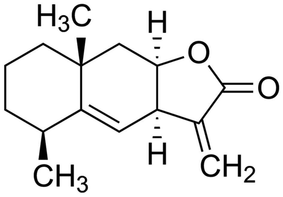
The chemical structure of alantolactone (purity of
98% calculated by high-performance liquid chromatography) is shown
in Fig. 1. The anticancer effects of
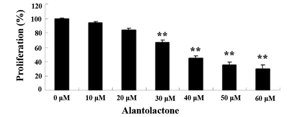
alantolactone on the growth of HeLa cells were measured using an
MTT assay. Administration of alantolactone (0, 10, 20, 30, 40, 50
and 60 µM) for 12 h inhibited the growth of HeLa cells in a
dose-dependent manner (Fig. 2).
Therefore, 30 µM of alantolactone was used in the subsequent
experiments.
Alantolactone induces the apoptosis of
HeLa cells
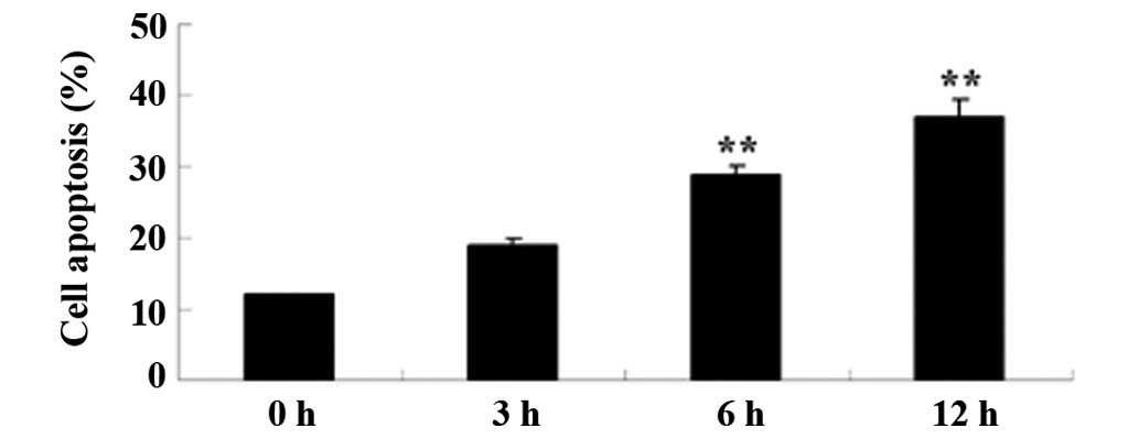
To examine the anticancer effects of alantolactone
on apoptosis, HeLa cells were treated with 30 µM of alantolactone
for 0, 3, 6 and 12 h, and apoptosis was measured with flow
cytometry. As shown in Fig. 3,
treatment with alantolactone induced apoptosis of HeLa cells in a
time-dependent manner. The results of the present study suggested
that alantolactone-induced cell apoptosis indicated early apoptosis
prior to the 6 h time point.
Alantolactone induces ROS generation
in HeLa cells
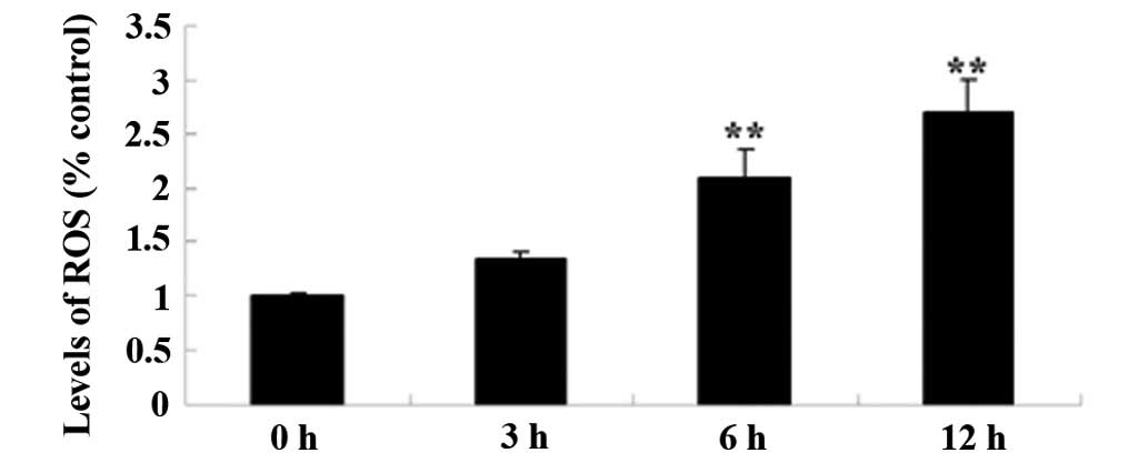
It was verified that the levels of ROS generation
have a significant role in the anticancer effects of alantolactone
on HeLa cells. Levels of ROS were measured using the H2DCFDA kit.
Pretreatment of HeLa cells with 30 µM of alantolactone for 0, 3, 6
and 12 h induced ROS generation in HeLa cells in a time-dependent
manner (Fig. 4). The results of the
present study showed that alantolactone effectively induced ROS
generation prior to the 6 h time point.
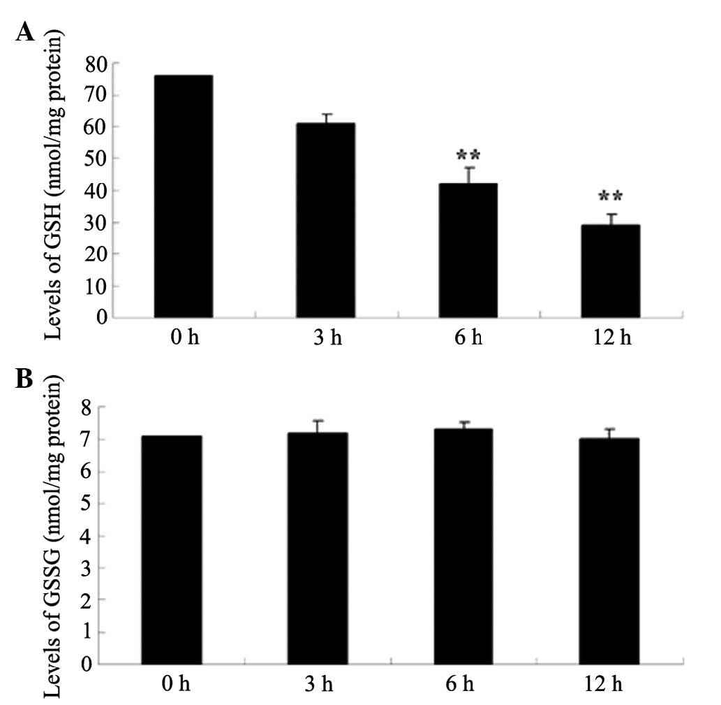
Alantolactone inhibits GSH and GSSG
production in HeLa cells
To investigate whether the levels of GSH and GSSG
generation had significant roles in the anticancer effects of
alantolactone on HeLa cells, the levels of GSH and GSSG were
measured using commercial kits. The results of the present study
showed that alantolactone inhibited GSH generation in a
time-dependent manner (Fig. 5).
Following treatment with alantolactone for 6 h, GSH generation was
effectively inhibited. However, the levels of GSSG generation did
not demonstrate a statistically significant difference in any of
the experimental groups.
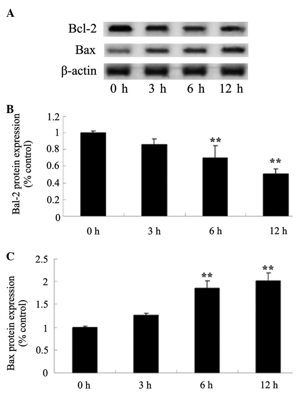
Alantolactone inhibits the Bcl-2/Bax
signaling pathway in HeLa cells
To investigate the Bcl-2/Bax signaling pathway in
alantolactone-induced apoptosis, Bcl-2 and Bax protein expression
was determined using western blot analysis. As shown in Fig. 6, Bcl-2 and Bax protein expression were
inhibited and increased, respectively, in a time-dependent manner
following treatment with 30 µM of alantolactone for 0, 3, 6 and 12
h. Following treatment with alantolactone for 6 h, the Bcl-2/Bax
signaling pathway was effectively inhibited.
Discussion
Cervical carcinoma is one of the primary diseases
that present a serious threat to women's health. In terms of global
malignant tumor occurrence, the incidence of cervical carcinoma is
second only to breast cancer. Cervical carcinoma is one of the
leading causes of cancer in developing countries, whereas in
developed countries, it is ranked below breast cancer and third
overall (1). According to the World
Health Organization, there are 500,000 new cases of cervical
carcinoma worldwide each year, and 80% of these cases occur in
developing countries (17). There are
a total of 140,000 new cases of cervical carcinoma each year in
China (18). The results of the
present study indicated that administration of alantolactone was
able to inhibit growth and induce the apoptosis of HeLa cells in a
dose or time-dependent manner. In addition, Jarrett et al
and Shi et al demonstrated clearly that alantolactone
inhibited the proliferation of HCT-8 human colon adenocarcinoma
cells (16,19). Khan et al (20) reported that alantolactone induced
apoptosis in HepG2 hepatoma cells.
Chronic inflammation has long been considered to be
a risk factor for a variety of human malignant tumors, particularly
cervical carcinoma. The chronic inflammation hypothesis emphasizes
that the ROS produced by phagocytes leads to cytotoxicity and
mutation (21). In the event that a
chronic inflammatory tissue contains large amounts of nitric oxide
(NO) and derivative ROS, NO and ROS may cause direct and indirect
damage to DNA and other genetic material (22). The balance and interaction between NO
and ROS has a significant role in the etiology of a tumor (23). The results of the present study
regarding the underlying mechanism of alantolactone action on HeLa
cells have implications for the promotion of ROS-mediated curative
effects. Khan et al (15)
demonstrated that alantolactone induced cell apoptosis via GSH
depletion and ROS generation in glioblastoma cells. Zhang et
al (24) reported that
alantolactone induced cell apoptosis via ROS generation in RKO
cells.
The onset of cervical carcinoma may be associated
with long-term estrogen stimulation, as cervical carcinoma is a
type of hormone dependent tumor (25). It has been proposed that androgen
transfer into estrogen in fat tissue is involved in an increase in
the levels of estrogen synthesis (26). Previous studies identified that
Japanese fat consumption and cervical carcinoma incidence was lower
compared with Finland. Finnish hydrogen peroxide levels in normal
endometrium and GSH-peroxidase (Px) activity were lower compared
with that of Japanese individuals. However, in Japan and Finland,
patients exhibiting cervical carcinoma had increased endometrial
lipid peroxide (LPO) levels compared with a healthy group, while
superoxide dismutase and GSH-Px activity was reduced compared with
a healthy group. This result demonstrated that cervical carcinoma
and multiple quantity increased the intake of fat, resulting in the
lipid peroxidation and antioxidant system being weakened (27,28). In
addition, a previous study demonstrated a significant increase in
GSH-Px activity in cervical carcinoma tissue, and in
well-differentiated adenocarcinomas this association was more
marked compared with moderate or poorly differentiated
adenocarcinomas (29). In the present
study, the levels of GSH generation had significant roles in the
anticancer effects of alantolactone on HeLa cells. Khan et
al (15,20) demonstrated that alantolactone was able
to induce apoptosis of HepG2 cells and glioblastoma cells via GSH
depletion.
The present study demonstrated that of the various
genes involved in the regulation of cell apoptosis, Bcl-2 is
particularly important, as it is regarded as one of the final
common pathways of apoptosis regulation (30). Bcl-2 exhibits increased expression in
a number of tumors, and is able to inhibit the natural apoptosis of
cells, as well as suppress the apoptosis induced by many antitumor
drugs, reducing their cytotoxicity. By contrast, Bax promotes
apoptosis (31). The underlying
mechanism of Bax activity does not block apoptosis directly, but
inhibits the anti-apoptotic role of Bcl-2 (32). The results of the present study
demonstrate that the anticancer effects of alantolactone on HeLa
cells were associated with the Bcl-2/Bax signaling pathway.
Administration of alantolactone may be capable of inhibiting the
Bcl-2/Bax signaling pathway in HeLa cells. Recent studies have
suggested that alantolactone likely increases the ratio of Bcl-2 to
Bax, therefore inducing apoptosis in cancer cells (33,34).
In conclusion, the results of the present study
provide evidence that alantolactone is capable of inducing
apoptosis in human cervical cancer cells. Consequently, promotion
of ROS generation, and inhibition of GSH generation and the
Bcl-2/Bax signaling pathway induced the anticancer effects of
alantolactone on apoptosis in HeLa cells. The results suggest that
alantolactone is a promising Traditional Chinese Medicine that may
be utilized for the treatment of cervical cancer. Additional
investigation is required to verify the contribution of
alantolactone to anticancer therapy in vitro and in
vivo.
References
|
1.
|
Yau Hsiung W and Abdul Kadir H: Leea
indica ethyl acetate fraction induces growth-inhibitory effect
in various cancer cell lines and apoptosis in ca ski human cervical
epidermoid carcinoma cells. Evid Based Complement Alternat Med.
2011:2930602011.PubMed/NCBI
|
|
2.
|
Einck JP, Hudson A, Shulman AC, Yashar CM,
Dieng MM, Diagne M, Gueye L, Gningue F, Gaye PM, Fisher BJ, et al:
Implementation of a high-dose-rate brachytherapy program for
carcinoma of the cervix in Senegal: A pragmatic model for the
developing world. Int J Radiat Oncol Biol Phys. 89:462–467. 2014.
View Article : Google Scholar : PubMed/NCBI
|
|
3.
|
Misra JS, Srivastava AN and Das V: Single
life time cytological screening in high risk women as an economical
and feasible approach to control cervical cancer in developing
countries like India. Asian Pac J Cancer Prev. 16:859–862. 2015.
View Article : Google Scholar : PubMed/NCBI
|
|
4.
|
Di Gao Z, Pan Q, Lv H, Sun Y, Ma X, Qin Z
and Sun YP: HPV genotypes in paraffin sections of non-cervical
squamous cell carcinoma in Qingdao of China. Oncol Lett.
5:1219–1222. 2013.PubMed/NCBI
|
|
5.
|
Bigoni J, Gundar M, Tebeu PM, Bongoe A,
Schäfer S, Fokom-Domgue J, Catarino R, Tincho EF, Bougel S,
Vassilakos P and Petignat P: Cervical cancer screening in
sub-Saharan Africa: A randomized trial of VIA versus cytology for
triage of HPV-positive women. Int J Cancer. 137:127–134. 2015.
View Article : Google Scholar : PubMed/NCBI
|
|
6.
|
Gerli S, Bavetta F and Di Renzo GC:
Antisepsis regimen in the surgical treatment of HPV generated
cervical lesions: Polyhexamethylene biguanide vs chlorhexidine. A
randomized, double blind study. Eur Rev Med Pharmacol Sci.
16:1994–1998. 2012.PubMed/NCBI
|
|
7.
|
Rodriguez EF, Reynolds JP, Jenkins SM,
Winter SM, Henry MR and Nassar A: Atypical squamous cells of
undetermined significance in patients with HPV positive DNA testing
and correlation with disease progression by age group: An
institutional experience. Int J Clin Exp Pathol. 5:428–435.
2012.PubMed/NCBI
|
|
8.
|
Xiang T, Du L, Pham P, Zhu B and Jiang S:
Nelfinavir, an HIV protease inhibitor, induces apoptosis and cell
cycle arrest in human cervical cancer cells via the ROS-dependent
mitochondrial pathway. Cancer Lett. 364:79–88. 2015. View Article : Google Scholar : PubMed/NCBI
|
|
9.
|
Singh M, Bhui K, Singh R and Shukla Y: Tea
polyphenols enhance cisplatin chemosensitivity in cervical cancer
cells via induction of apoptosis. Life Sci. 93:7–16. 2013.
View Article : Google Scholar : PubMed/NCBI
|
|
10.
|
Dupuis G, Mitchell JC and Towers GH:
Reaction of alantolactone, an allergenic sesquiterpene lactone,
with some amino acids. Resultant loss of immunologic reactivity.
Can J Biochem. 52:575–581. 1974. View
Article : Google Scholar : PubMed/NCBI
|
|
11.
|
Yao Y, Xia D, Bian Y, Sun Y, Zhu F, Pan B,
Niu M, Zhao K, Wu Q, Qiao J, et al: Alantolactone induces G1 phase
arrest and apoptosis of multiple myeloma cells and overcomes
bortezomib resistance. Apoptosis. 20:1122–1133. 2015. View Article : Google Scholar : PubMed/NCBI
|
|
12.
|
Zhang J, Li Y, Duan D, Yao J, Gao K and
Fang J: Inhibition of thioredoxin reductase by alantolactone
prompts oxidative stress-mediated apoptosis of HeLa cells. Biochem
Pharmacol. 2015.(In press).
|
|
13.
|
Lim HS, Jin SE, Kim OS, Shin HK and Jeong
SJ: Alantolactone from Saussurea lappa exerts
antiinflammatory effects by inhibiting chemokine production and
STAT1 phosphorylation in TNF-α and IFN-γ-induced in HaCaT cells.
Phytother Res. 29:1088–1096. 2015. View
Article : Google Scholar : PubMed/NCBI
|
|
14.
|
Ketai W, Huitao L, Yunkun Z, Xingguo C,
Zhide H, Yucheng S and Xiao M: Separation and determination of
alantolactone and isoalantolactone in traditional Chinese herbs by
capillary electrophoresis. Talanta. 52:1001–1005. 2000. View Article : Google Scholar : PubMed/NCBI
|
|
15.
|
Khan M, Yi F, Rasul A, Li T, Wang N, Gao
H, Gao R and Ma T: Alantolactone induces apoptosis in glioblastoma
cells via GSH depletion, ROS generation, and mitochondrial
dysfunction. IUBMB Life. 64:783–794. 2012. View Article : Google Scholar : PubMed/NCBI
|
|
16.
|
Jarrett SG, Albon J and Boulton M: The
contribution of DNA repair and antioxidants in determining cell
type-specific resistance to oxidative stress. Free Radic Res.
40:1155–1165. 2006. View Article : Google Scholar : PubMed/NCBI
|
|
17.
|
Liao S, Xiao S, Zhu G, Zheng D, He J, Pei
Z, Li G and Zhou Y: CD38 is highly expressed and affects the
PI3K/Akt signaling pathway in cervical cancer. Oncol Rep.
32:2703–2709. 2014.PubMed/NCBI
|
|
18.
|
Levin CE, Sharma M, Olson Z, Verguet S,
Shi JF, Wang SM, Qiao YL, Jamison DT and Kim JJ: An extended
cost-effectiveness analysis of publicly financed HPV vaccination to
prevent cervical cancer in China. Vaccine. 33:2830–2841. 2015.
View Article : Google Scholar : PubMed/NCBI
|
|
19.
|
Shi Y, Bao YL, Wu Y, Yu CL, Huang YX, Sun
Y, Zheng LH and Li YX: Alantolactone inhibits cell proliferation by
interrupting the interaction between Cripto-1 and activin receptor
type II A in activin signaling pathway. J Biomol Screen.
16:525–535. 2011. View Article : Google Scholar : PubMed/NCBI
|
|
20.
|
Khan M, Li T, Khan Ahmad MK, Rasul A,
Nawaz F, Sun M, Zheng Y and Ma T: Alantolactone induces apoptosis
in HepG2 cells through GSH depletion, inhibition of STAT3
activation, and mitochondrial dysfunction. BioMed Res Int.
2013:7198582013. View Article : Google Scholar : PubMed/NCBI
|
|
21.
|
Yang J, Xiao YL, He XR, Qiu GF and Hu XM:
Aesculetin-induced apoptosis through a ROS-mediated mitochondrial
dysfunction pathway in human cervical cancer cells. J Asian Nat
Prod Res. 12:185–193. 2010. View Article : Google Scholar : PubMed/NCBI
|
|
22.
|
He L, Nan MH, Oh HC, Kim YH, Jang JH,
Erikson RL, Ahn JS and Kim BY: Asperlin induces G2/M
arrest through ROS generation and ATM pathway in human cervical
carcinoma cells. Biochem Biophys Res Commun. 409:489–493. 2011.
View Article : Google Scholar : PubMed/NCBI
|
|
23.
|
Agostinelli E and Seiler N:
Non-irradiation-derived reactive oxygen species (ROS) and cancer:
Therapeutic implications. Amino Acids. 31:341–355. 2006. View Article : Google Scholar : PubMed/NCBI
|
|
24.
|
Zhang Y, Bao YL, Wu Y, Yu CL, Huang YX,
Sun Y, Zheng LH and Li YX: Alantolactone induces apoptosis in RKO
cells through the generation of reactive oxygen species and the
mitochondrial pathway. Mol Med Rep. 8:967–972. 2013.PubMed/NCBI
|
|
25.
|
Kim YT, Kim SW, Yoon BS, Cho HJ, Nahm EJ,
Kim SH, Kim JH and Kim JW: Effect of intravenously administered
iron sucrose on the prevention of anemia in the cervical cancer
patients treated with concurrent chemoradiotherapy. Gynecol Oncol.
105:199–204. 2007. View Article : Google Scholar : PubMed/NCBI
|
|
26.
|
Srivastava K, Paul S, Chufal KS,
Shamsundar SD, Lal P, Pant MC, Bhatt M, Singh S and Gupta R:
Concurrent chemoradiation versus radiotherapy alone in cervical
carcinoma: A randomized phase III trial. Asia Pac J Clin Oncol.
9:349–356. 2013. View Article : Google Scholar : PubMed/NCBI
|
|
27.
|
Fan S, Yu Y, Qi M, Sun Z, Li L, Yao G,
Tashiro S, Onodera S and Ikejima T: P53-mediated GSH depletion
enhanced the cytotoxicity of NO in silibinin-treated human cervical
carcinoma HeLa cells. Free Radic Res. 46:1082–1092. 2012.
View Article : Google Scholar : PubMed/NCBI
|
|
28.
|
Kwaśniewska A, Tukendorf A and Semczuk M:
Frequency of HPV infection and GSH levels in plasma of women with
cervical dysplasia. Eur J Gynaecol Oncol. 18:196–199.
1997.PubMed/NCBI
|
|
29.
|
Moon HJ and Park WH: Butylated
hydroxyanisole inhibits the growth of HeLa cervical cancer cells
via caspase-dependent apoptosis and GSH depletion. Mol Cell
Biochem. 349:179–186. 2011. View Article : Google Scholar : PubMed/NCBI
|
|
30.
|
Chan HW, Liu T, Verdile G, Bishop G, Haasl
RJ, Smith MA, Perry G, Martins RN and Atwood CS: Copper induces
apoptosis of neuroblastoma cells via post-translational regulation
of the expression of Bcl-2-family proteins and the tx mouse is a
better model of hepatic than brain Cu toxicity. Int J Clin Exp Med.
1:76–88. 2008.PubMed/NCBI
|
|
31.
|
Li J, Wang H, Ma Z, Fan W, Li Y, Han B,
Zhang Z and Wang J: TAT-Apoptin induces apoptosis in the human
bladder cancer EJ cell line and regulates Bax, Bcl-2, caspase-3 and
survivin expression. Exp Ther Med. 3:1033–1038. 2012.PubMed/NCBI
|
|
32.
|
Ji M, Yuan L, Lv X, Dong W and Peng X:
EBP50 regulates the apoptosis of pancreatic cancer cells by
decreasing the expression levels of Bcl-2. Exp Ther Med. 8:919–924.
2014.PubMed/NCBI
|
|
33.
|
Mi XG, Song ZB, Wu P, Zhang YW, Sun LG,
Bao YL, Zhang Y, Zheng LH, Sun Y, Yu CL, et al: Alantolactone
induces cell apoptosis partially through down-regulation of
testes-specific protease 50 expression. Toxicol Lett. 224:349–355.
2014. View Article : Google Scholar : PubMed/NCBI
|
|
34.
|
Zhao P, Pan Z, Luo Y, Zhang L, Li X, Zhang
G, Zhang Y, Cui R, Sun M and Zhang X: Alantolactone induces
apoptosis and cell cycle arrest on lung squamous cancer SK-MES-1
cells. J Biochem Mol Toxicol. 29:199–206. 2015. View Article : Google Scholar : PubMed/NCBI
|