Introduction
In the Western world, colorectal cancer (CRC) is the
fourth leading cause of cancer-associated mortality (1), and >95% of colorectal cancers are
adenocarcinomas. The main risk factors are genetic factors (family
history), a low fiber and high fat diet, and smoking (2). Following curative surgery alone, the
percentage of patients that subsequently relapse and succumb to
metastatic disease ranges from 40 to 50%. This percentage falls to
33% when patients receive postoperative adjuvant treatment with
5-fluorouracil (5-FU) and leucovorin, and to 23% when the
platinum-containing compound oxaliplatin is added to this treatment
[FOLFOX: folinic acid (leucovorin), 5-FU and oxaliplatin] (2,3).
Unfortunately, despite the positive results obtained
by the combination protocols of oxaliplatin with fluoropyrimidines
(e.g. 5-FU) and folinic acid (leucovorin) in disease-free survival
of stage II or III colon cancer (3),
unwanted side effects develop that can affect, to various degrees,
the quality of life of the patients. Several side effects have been
reported for oxaliplatin, which include gastrointestinal toxicity,
moderate hematological toxicity, hypersensitivity and neurological
toxicity. This unpredictable neural toxicity has quite unique
features and determines the dose-limiting toxicity of oxaliplatin
(4).
The identification of biomarkers that could predict
the onset of these secondary effects would be of great value in
preventing long-term toxicity or permanent damage in patients at
risk. Recent research from our laboratory has led to the
identification of several genes that are significantly up- or
downregulated in peripheral white cells (PWCs) of CRC patients
following oxaliplatin-based chemotherapy treatment (5). In our screening, the expression levels
of the gene encoding for the immunophilin FK506-binding protein 5
(FKBP51) were 2.76-times lower [3,812 (pre) vs. 1,383 (post)] in
PWCs after 3 cycles of oxaliplatin-based chemotherapy (5).
FKBP51 protein plays multiple roles in the
regulation of a variety of signaling pathways, and has altered
expression levels in many different tumor types. By regulating
steroid receptor maturation, as well as the Akt and nuclear factor
κB signaling pathways, FKBP51 is important in tumorigenesis and in
the response to chemotherapy (6–13). FKBP51
belongs to a superfamily of peptidyl-prolyl isomerases (PPIs),
which also includes FKBP52 and the cyclosporine A-binding protein
cyclophilin-40. FKBP51 is a 51 kD FK506-binding protein with a
C-terminal tetratricopeptide repeat (TPR) domain, and an N-terminal
FK1 domain responsible for PPIase activity, which catalyzes the
cis-trans conversion of prolyl peptide bonds within target proteins
(14). Through the TPR domain, FKBP51
binds to heat shock protein 90 (HSP90) complexes, such as those
associated with steroid hormone receptors. The mechanisms
underlying the regulation of steroid hormone receptor signaling by
this immunophilin and its physiological roles in endocrine-related
processes are very well studied and will be further discussed in
the discussion section.
Research in this field has led to the identification
of FKBP51 as a potential therapeutic target for several
endocrine-related diseases, such as metabolic and stress-related
diseases, prostate cancer and breast cancer (13). Diseases associated with this protein
include major depressive disorder and glucocorticoid resistance
(Gene Ontology annotations). FKBP51 is a ubiquitous protein
expressed in the cytoplasm, nucleus and mitochondria (14). Nuclear-mitochondrial shuttling is
triggered by oxidative stress: The protein translocates to the
nucleus upon the onset of oxidative stress to protect the cells
from stress damage (7).
Recently, a role for the immunophilins FKBP51 and
FKBP52 in regulating microtubules has been suggested, acting via
their interaction with τ proteins (15). Regulation of microtubule dynamics by
FKBPs has been associated with neurite outgrowth (16). Furthermore, FKBP51 has been identified
as a regulator of cell death in response to gemcitabine and
cytarabine treatment: High levels of FKBP51 expression were
associated with sensitivity, while low levels of expression were
associated with resistance to these drugs (17).
Several studies also link FKBP51 to cell
proliferation and cancer. It has been demonstrated, for example,
that it plays a role in negatively regulating the Akt pathway.
Acting as a scaffold protein, FKBP51 promotes the interaction of
Akt and PHLPP, a phosphatase that specifically dephosphorylates Akt
at Ser473 and inhibits its activity (9). Recently it has been demonstrated that
FKBP51 is key in promoting the activation of genes involved in
melanoma progression (11), and
modulates the transforming growth factor β (TGF-β) signal in
malignant melanocytes, increasing the tumor-promoter potential of
TGF-β (12). FKBP51 expression is
also decreased in pancreatic cancer tissues and in numerous cancer
cell lines.
In the current study, an immunohistochemical
(IHC)analysis of FKBP51 in CRC tissue sections (namely before
antineoplastic therapy) and primary metastases (resected after
oxaliplatin-based chemotherapy) was performed in order to determine
whether the alteration in FKBP51 gene expression observed in PWCs
can be detected at the protein level, the nature of their role in
tumoral physiopathology, and whether these alterations have any
prognostic implications.
Materials and methods
Patients
The study was approved by the Ethics Committee of La
Laguna University (La Laguna, Spain) and the Ethics Committee of
Nuestra Señora de Candelaria University Hospital (HUNSC; Santa Cruz
de Tenerife, Spain). All patients signed informed consent for
diagnosis and research on tissue specimens, prior to entering the
study. All subjects were treated with FOLFOX chemotherapy as
follows: Day 1, oxaliplatin [100 mg/m2 intravenous
(i.v.) over 2 h], leucovorin calcium (400 mg/m2 i.v.
over 2 h); followed by 5-FU (400 mg/m2 i.v. bolus) and
by 5-FU (2,400 mg/m2 i.v. over 46 h), every 14 days,
days, and no patient underwent previous CT scans or received
radiation therapy. The patients were treated between October 2010
and July 2015.
Several (between 10 and 15) mounted slides of
paraffin-embedded tissue samples (5 µm thick; colon adenocarcinoma
and metastasized liver and lung from selected patients) obtained
during resection and clinical data were collected from 33 patients
(16 males and 17 females), aged between 38 and 76 years, from the
reference medical areas of HUNSC.
Antibodies
The following antibodies were used: Rabbit
polyclonal antibody (pAb) against FKBP51 (#ab46002; Abcam,
Cambridge, UK; dilution, 1.25:100); mouse monoclonal antibody
against proliferating cell nuclear antigen (PCNA; clone PC10; #1486
772, Roche Diagnostics Deutschland GmbH, Mannheim, Germany;
dilution, 1:100); fluorescein isothiocyanate (FITC)-conjugated goat
pAb against rabbit IgG (#F9887; Sigma-Aldrich, St. Louis, MO, USA;
dilution, 1:200); goat pAb against mouse IgG (DyLight®
650; #ab97018; Abcam, dilution, 1:100); biotin-conjugated goat pAb
against rabbit IgG (H+L) (#31820; Thermo Fisher Scientific, Inc.,
Waltham, MA, USA; dilution, 1:300).
IHC
Immunoperoxidase staining of formalin-fixed,
paraffin-embedded tissue sections was performed using an ordinary
avidin-biotin method. Briefly, 5 µm-thick tissue sections were
deparaffinized in xylene and hydrated in graded alcohol.
Heat-induced epitope retrieval was achieved by heating samples in
sodium citrate buffer pH 6.0 at 120°C for 10 min in an autoclave.
After non-specific sites were blocked with 5% non-fat dry milk in
Tris-buffered saline (TBS) for 1 h at room temperature, endogenous
biotin was blocked using an Avidin/Biotin Blocking Kit (Vector
Laboratories, Inc., Burlingame, CA, USA). Primary antibody against
FKBP51 (1.25:100) was applied to slides overnight at 4°C.
Biotin-conjugated anti-rabbit secondary antibody was incubated for
2 h at 37°C at a dilution of 1:300. To block endogenous peroxidase
activity, slides were incubated with 3% hydrogen peroxidase in
methanol for 15 min. A Pierce ABC Peroxidase Staining Kit (Thermo
Fisher Scientific, Inc.) was used to amplify the specific antibody
staining. Concentrated 3,3′-diaminobenzidine Substrate (#IHC-101F;
Bethyl Laboratories, Inc., Montgomery, TX, USA) was used to
visualize IHC reactions. Samples incubated without primary
antibodies were used as negative controls. Slides were
counterstained with Harris Hematoxylin Solution DC (Panreac Quimica
SLU, Barcelona, Spain) to visualize cell nuclei. Slides were
mounted with Eukitt (Panreac Quimica SLU). An optical light
microscope (BX50, Olympus Corporation, Tokyo, Japan) was used to
visualize immunostaining results.
Image analysis and statistics
For semi-quantitative image analysis, the open
resource digital image analysis software ImageJ was used,
implemented with the IHC Profiler plug-in developed by Varghese
et al (18), which creates a
pixel-by-pixel analysis profile of a digital IHC image, and further
assigns a score in a four tier system: High positive (pixel
intensity range, 0–60), positive (pixel intensity range, 61–120),
low positive (pixel intensity range, 121–180), negative (pixel
intensity range, 181–235). All images were captured at the same
magnification (40x) and with the same levels of contrast and
brightness. Pearson's Correlation Coefficient and Student's t-test
were performed using SPSS version 20 software (IBM SPSS, Madrid,
Spain) in order to estimate the reliability of the study.
Double immunofluorescence simultaneous
staining
Following the deparaffinization, hydration and
heat-induced epitope retrieval procedures (as described), slides
were incubated with 5% bovine serum albumin (catalog no. A9647;
Sigma-Aldrich, St. Louis, MO, USA) and 1% Triton X-100 in TBS to
block non-specific sites. Tissue sections were then incubated
simultaneously with a mixture of two distinct primary antibodies
(rabbit anti-FKBP51 and mouse anti-PCNA) overnight at 4°C, at
concentrations of 1:50 and 1:100, respectively. Slides were then
incubated for 1 h at room temperature with a mixture of two
secondary antibodies (FITC-conjugated anti-rabbit and
DyLight® 650-conjugated anti-mouse). Slides were mounted
with ProLong® Diamond Anti-fade Mountant with DAPI
(Molecular Probes; Thermo Fisher Scientific, Inc., Eugene, Oregon,
USA) to visualize cell nuclei. Slides were analyzed using a
confocal microscope (FV1000, Olympus Corporation).
Results
IHC analysis of FKBP51 expression in
colon tissue samples from CRC patients
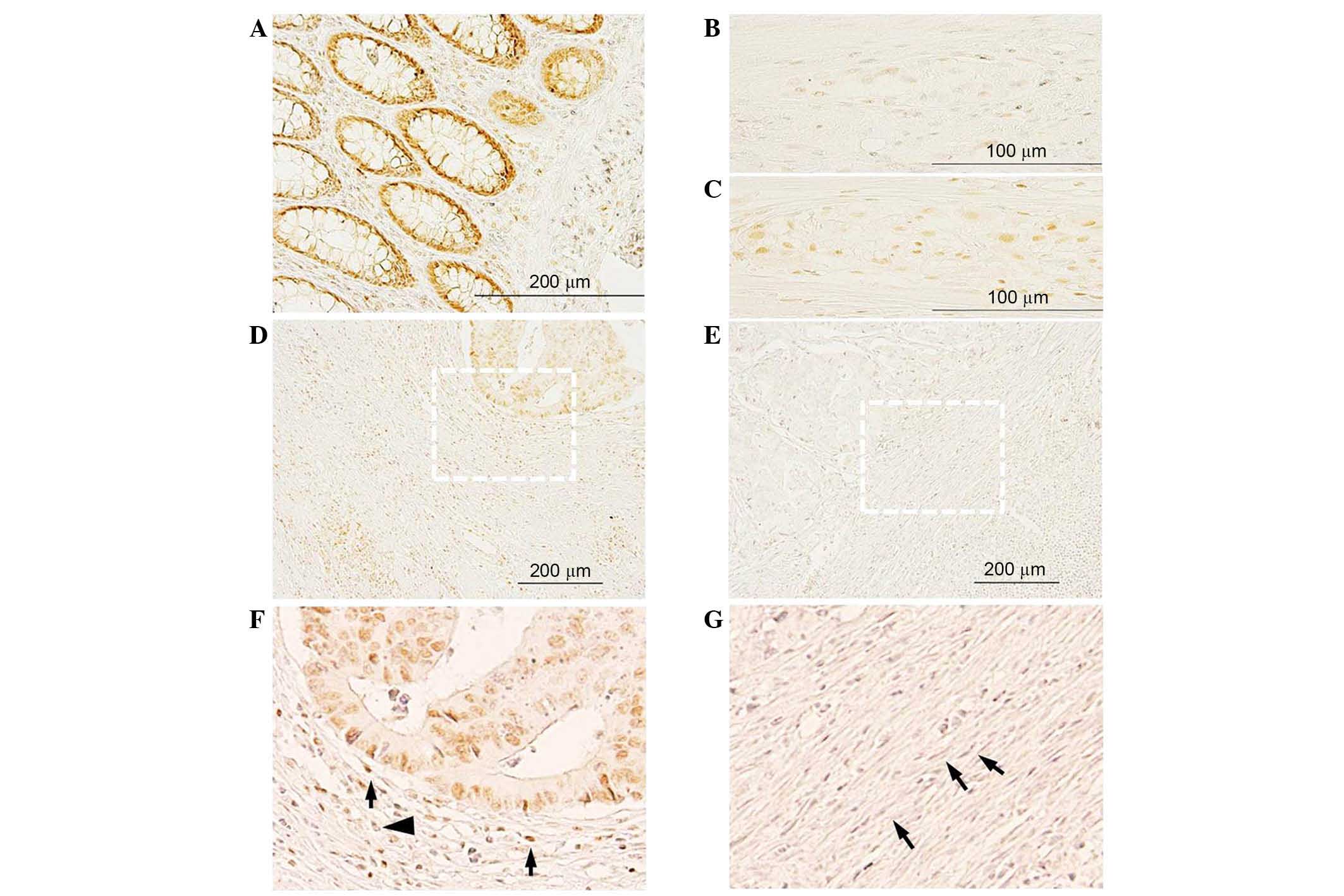
In healthy colon and in the apparently healthy
region of the CRC tissue sections (Fig.
1), intestinal glands exhibited intense positive FKBP51 nuclear
staining in enterocytes and in cells of the lamina propria
(Fig. 1A). In healthy colon, few
cells in the myenteric plexus exhibited a weak signal (Fig. 1B), while in CRC tissue sections,
several cells in the plexus were strongly positive (Fig. 1C).
In colon adenocarcinoma tissue sections, FKBP51
protein was localized in the cytoplasm and/or nucleus of tumor
cells, as well as in inflammatory and fibrous stromal cells
surrounding the lesions (Fig. 1D-G).
In certain areas of the section, a variable positive signal could
be observed in tumor cells, while in other areas, no staining was
detected. Notably, the phenotype of the connective tissue
surrounding the lesions appeared variable: In those areas where no
immunophilin expression was observed in tumor and stromal cells,
stromal fibroblasts exhibited a mature phenotype, with thin, wavy
and small spindle cell morphology (Fig.
1E and G, arrows); by contrast, in those areas where positive
FKBP51 immunostaining could be observed in tumor and stromal cells,
fibroblasts exhibited an immature phenotype, with large, puffy,
spindle-shaped morphology (Fig. 1F).
An increased microvessel density and enhanced infiltration of
tumor-associated macrophages was also observed in the connective
tissue surrounding FKBP51-positive lesions (Fig. 1F).
In the stroma surrounding tumor nests, the
expression of FKBP51 was variable, with cells exhibiting a strong
positive signal (Fig. 1F, arrow) and
others completely negative (Fig. 1F,
arrowhead).
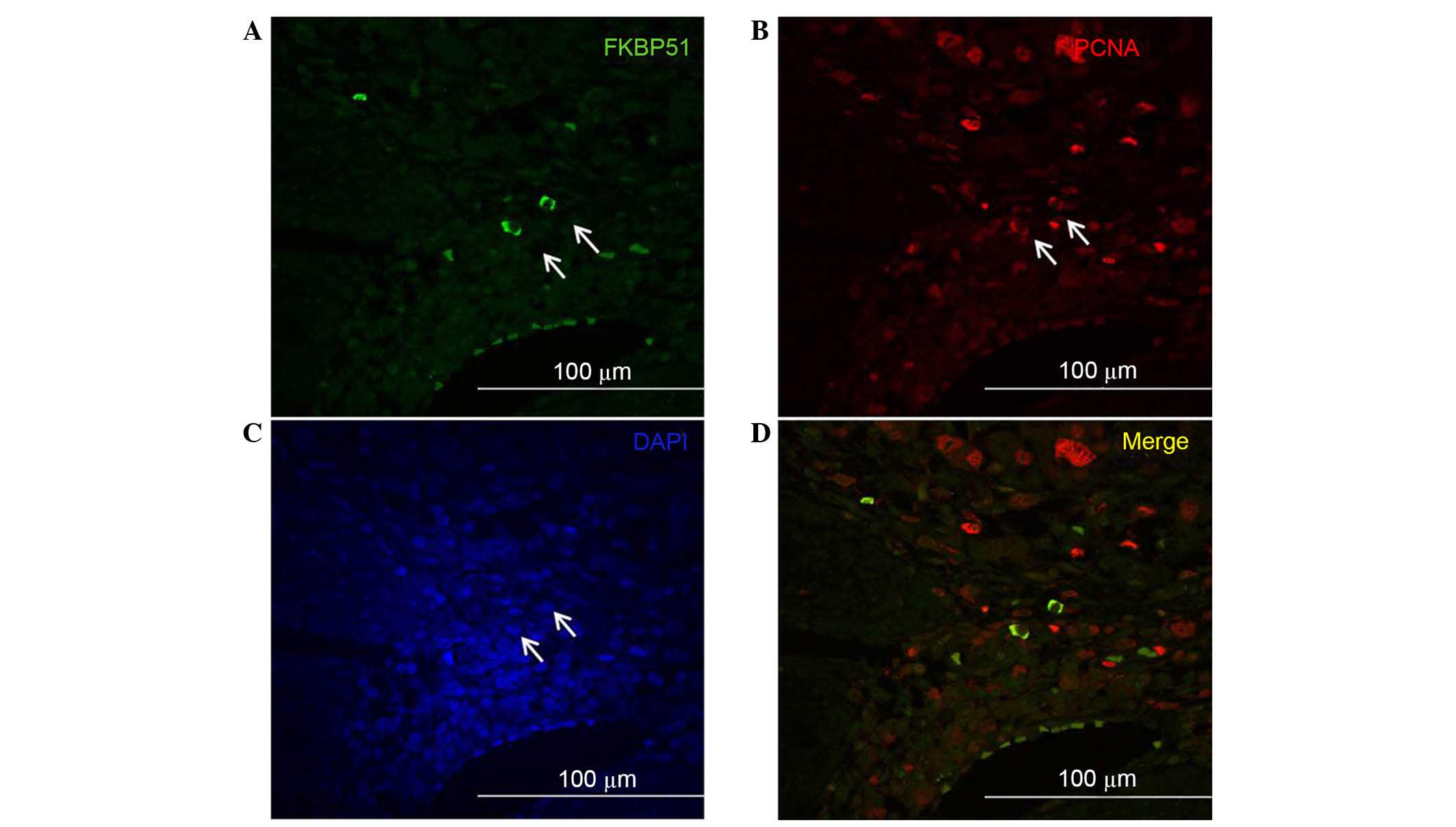
Double immunofluorescence experiments to detect
FKBP51 and PCNA, the clamp subunit of DNA polymerase and marker of
S phase of cell cycle (19), revealed
that, among the stromal cells expressing PCNA, only a few
coexpressed FKBP51 (Fig. 2). This
suggests a potential role for this immunophilin protein as a marker
of specific subtypes of stromal cells; however, further studies are
needed to assess this hypothesis.
IHC analysis of FKBP51 expression in
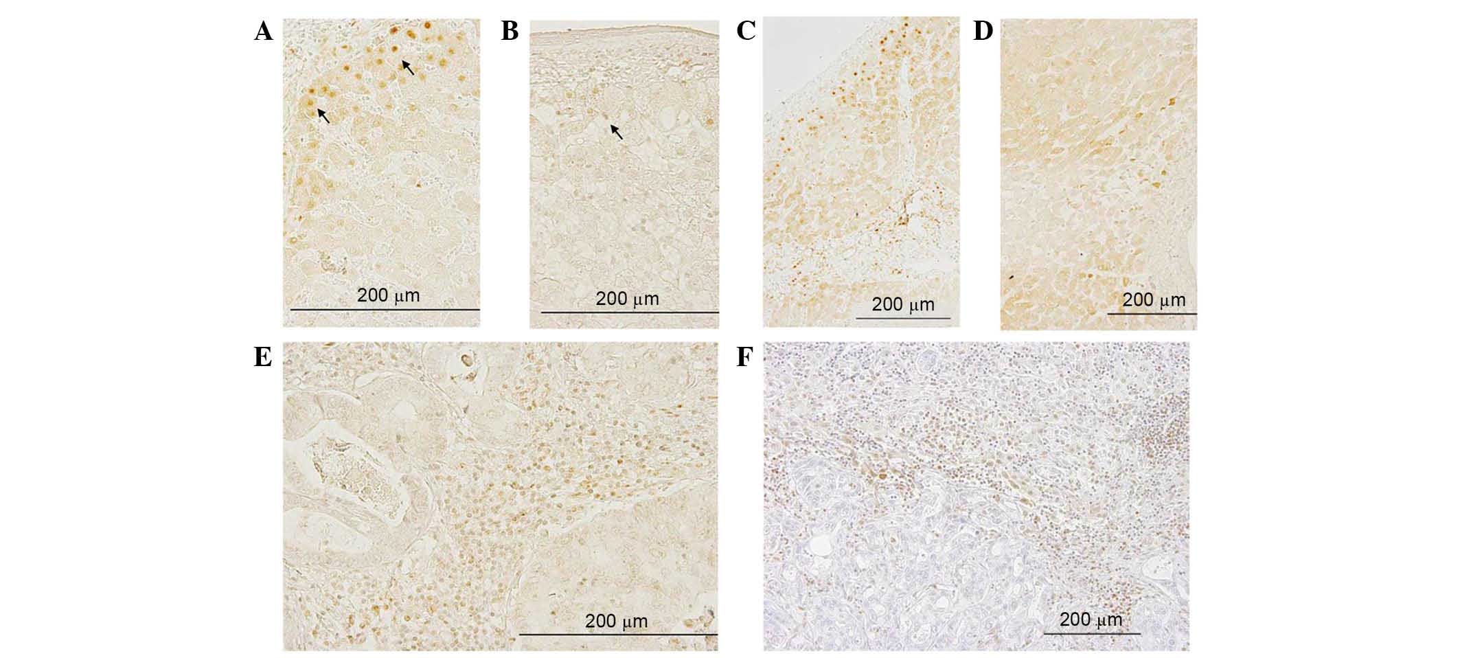
metastasized liver tissue samples from CRC patients
In the overall sections of healthy liver (Fig. 3C) and in the apparently healthy part
of metastasized liver (Fig. 3D),
there were areas with a more intense signal and well-delimited
areas in which FKBP51 protein expression was weak or absent.
Intense staining could be observed in the nuclei of hepatocytes
lining the edge of the connective tissue capsule (Glisson's
capsule) (Fig. 3A, arrows). By
contrast, in metastasized liver, this nuclear signal was fainter
(Fig. 3B, arrow). In metastases, the
signal appeared faint or absent, while several of the inflammatory
fibrous stroma cells exhibited a strong signal (Fig. 3E).
IHC analysis of FKBP51 expression in
metastasized lung tissue samples from CRC patients
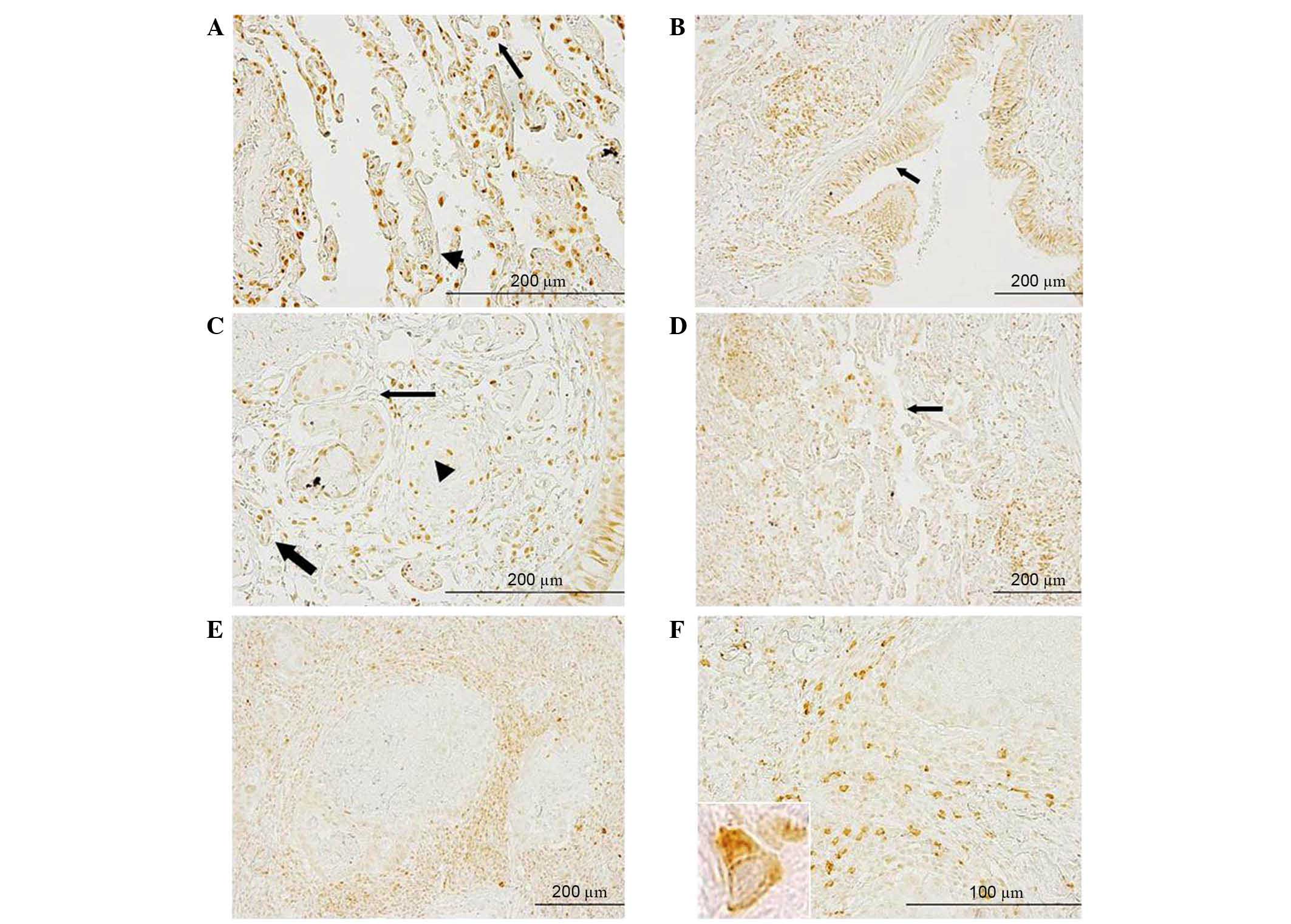
In healthy lung tissue, positive FKBP51 staining was
observed in macrophages and endothelial cells. In the respiratory
mucosa, a strong signal was present in the nuclei and cytoplasm of
ciliated cells (data not shown). In metastasized lung, strong
positive staining could be observed in macrophages (Fig. 4A, black arrow), in cells of the
lamina propria, in endothelial cells and, to a lesser
extent, in the nuclei of bronchial gland cells (Fig. 4C, arrowhead, thick arrow and thin
arrow, respectively). In the bronchial epithelial cells, FKBP51
protein was localized to the nuclei and/or cytoplasm of ciliated
cells; strong staining in the basal bodies of these cells was
present (Fig. 4B, arrow). In the
malignant area of the section, weakly positive or no staining was
observed in tumor cells, while the signal appeared stronger in
inflammatory and fibrous stromal cells surrounding the lesions
(Fig. 4E and F). Fig. 4F shows the positive protein staining
in several stromal cells between two metastases; the intracellular
distribution pattern suggested mitochondrial localization of the
protein in these cells.
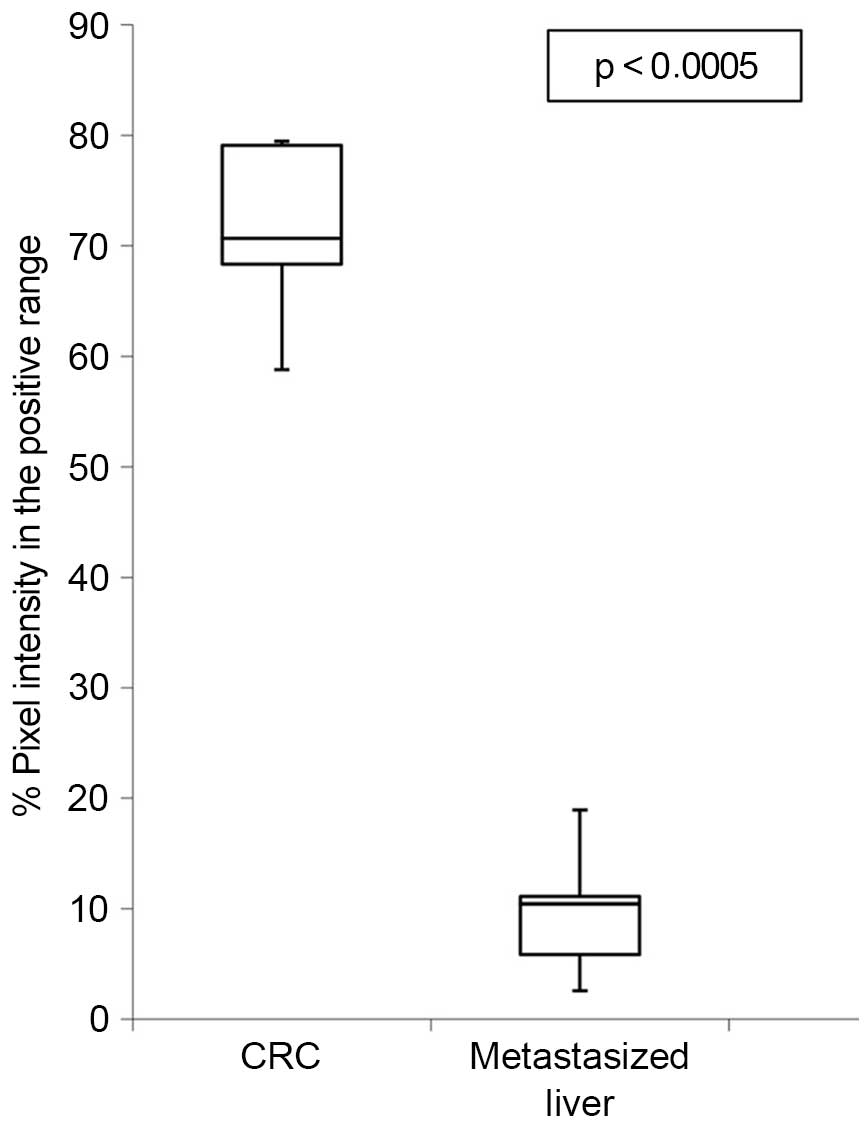
FKBP51 protein is downregulated in
metastasized liver tissue samples
Specimens were evaluated by two independent
observers (a biologist and a pathologist) who were blinded to the
conditions. In addition, ImageJ software and the open source
plug-in IHC Profiler developed by Varghese et al (18) were used to compare the visual human
interpretation to that of the computer-aided vision. Fig. 5 shows a box-and-whisker plot
illustrating the results obtained using IHC Profiler to compare the
percentage of positive pixels (pixel intensity range, 61–120) in
the tissue samples. This clearly demonstrates the downregulation of
FKBP51 protein in malignant liver specimens vs. CRC tissue samples
(7.5±4.3% in liver vs. 71.3±7.6% in CRC; P<0.003).
No differences in distribution or in staining
intensity were detected between samples from male or female
patients, or among patients of different ages (data not shown).
Discussion
In a previous screening for biomarkers involved in
oxaliplatin toxicity, FKBP5 was the gene whose transcriptional
expression level was most downregulated quantitatively (5). In the current study, IHC analysis of
FKBP51 protein expression and localization allowed observation of
strong staining in the nuclei of enterocytes in healthy colon,
whereas in colonic adenocarcinoma cells, the staining was localized
in nuclei and cytoplasm. However, lesions exhibited variable
staining, ranging from tumor nests with malignant cells strongly
expressing FKBP51, to the surrounding stroma cells and lesions
where no positive signal could be detected (Fig. 1E and F).
The observation that the expression of FKBP51 in
tumor and stromal cells is associated with an immature phenotype of
the surrounding stromal fibroblasts and with an increased
microvessel density, as well as augmented tumor-associated
macrophage infiltration, suggests a role for this protein in the
epithelial-to-mesenchymal transition (EMT) process in CRC (20).
Patients in the current study received
oxaliplatin-based chemotherapy prior to resection of the
metastases. IHC analyses allowed the observation of changes in
FKBP51 expression levels and localization in malignant liver
compared with CRC. While in healthy liver FKBP51 protein exhibited
strong staining in the nuclei of hepatocytes, in healthy regions of
metastatic liver, the nuclear signal was fainter. The observed
alterations in the liver tissue surrounding the metastases could be
related to hepatic sinusoidal injury elicited by oxaliplatin
therapy (21).
In liver metastases, the signal appeared faint or
absent, while in the inflammatory fibrous stroma, several cells
exhibited a strong signal (Fig. 3E).
These structural changes could be associated with the effect of
chemotherapy on tumor cells rather than with intrinsic changes of
transformation of cells. This phenomenon is further supported by
the fact that a patient with a predominantly negative
immunostaining signal had a tumor (metastasis) that was completely
resistant to chemotherapy. Lung metastases exhibited a similar
expression pattern to liver metastases, with weak staining in tumor
cells and a strong signal in inflammatory and fibrous stromal cells
surrounding the metastases. Whether this weaker level of expression
in the metastatic cells is related to chemotherapy or to their cell
biology is to be determined by further studies.
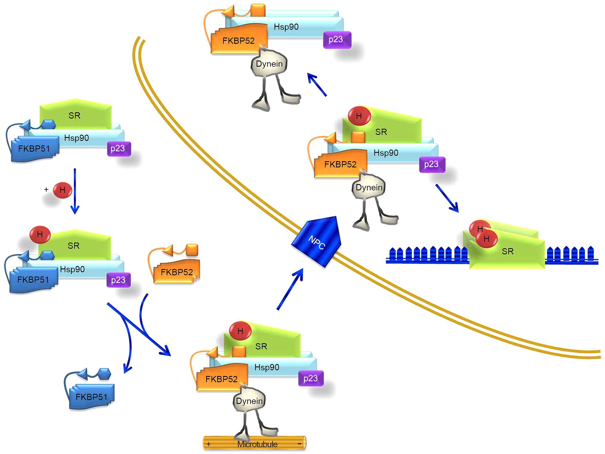
FKBP51 and its related protein FKBP52 are HSP90
co-chaperones that influence steroid hormone receptor activity.
These two immunophilins share a similar structure but act
divergently due to differences in the FK1 domain and the
proline-rich loop (13). Fig. 6 illustrates their coordinated
functions. Due to differences in the FK1 domain, repression of
hormone binding occurs in the presence of FKBP51, and potentiation
in the presence of FKBP52. In the absence of a ligand, certain
steroid hormone receptors reside primarily in the cytoplasm,
whereas others are nuclear. Regardless of their primary
localization, these receptors are not confined to any particular
cell compartment, and instead shuttle continuously between the
cytoplasm and nucleus (13). It is
assumed that these signaling molecules move across the cell by
simple diffusion. However, the fact that proteins of the
HSP90-FKBP52 complex co-immunoprecipitate with the glucocorticoid
and mineralocorticoid receptors, and with the dynein-dynactin
complex (22–24), suggests that these motor proteins
could power the retrograde movement of these steroid receptors.
FKBP51 is generally considered to be a negative regulator of
receptor function. Evidence suggests that it displays tissue-
and/or cell-type-specific effects on receptor signaling (13). Furthermore, it has been reported that
colorectal tumors produce glucocorticoids; these glucocorticoids
would have immunosuppressive functions, leading to an increase in
tumor survival and growth (25).
In transformed cells, the current results indicated
a decrease in the expression levels of FKBP51, as observed in
leukocytes. FKBP52 was not selected for analysis as no change in
this gene was detected in the transcriptome analysis (5) or in the preliminary immunolocalization
experiments. Fig. 6, which is based
on the model of Storer et al (13), illustrates how a decrease in FKBP51
expression brings about the predominance or easier driving of the
steroid receptor-hormone-HSP90-p23 complex to the nucleus, making
the transformed cell (at least after FOLFOX chemotherapy), somehow,
more sensitive to steroid hormones. This fact needs to be further
confirmed experimentally.
Baughman et al (26) reported in 1997 that FKBP51 is
expressed in various tissues, but not in the colon, lung and
spleen. In that study, the expression of FKBP51 was only analyzed
by western blotting of protein lysate. However, in another study,
in which different techniques were used (western blot, reverse
transcription-polymerase chain reaction and IHC analyses), Mukaide
et al (27) demonstrated that
FKBP51 is expressed in normal epithelial cells and in
adenocarcinoma cells in the human colon, and that there are no
significant differences in the expression of FKBP51 between these
cell types. Furthermore, the authors suggested that FKBP51 may
suppress the proliferation of colorectal adenocarcinoma, possibly
due to the suppression of function of the glucocorticoid receptors
(27). The current findings only
agree partially with these facts.
Previously, the RNA interference technique has been
used to knock down the expression of FKBP5 in the A174 glioma cell
line, revealing that FKBP5 expression aids in the regulation of
glioma cell growth. By contrast, overexpression of FKBP5 markedly
enhanced growth in this cell line (28). This fact agrees with the current
results and explains a complementary effect of chemotherapy through
FKBP pathways.
In summary, the present study supports the
fundamental role of cell-by-cell IHC analysis in molecular data
interpretation. The findings have demonstrated that the changes in
FKBP51 gene expression elicited by FOLFOX chemotherapy in PWCs of
CRC patients can be confirmed at the protein level in tissue
samples of colon adenocarcinoma prior to chemotherapy, compared
with tissue sections of metastasized liver resected after
oxaliplatin-based chemotherapy. Furthermore, the results indicated
that, in CRC tissue sections, the expression of FKBP51 in tumor and
stroma cells is associated with the immature phenotype of stromal
fibroblasts and with the EMT phenotype, suggesting a role for this
protein in the EMT process in CRC. The expression of FKBP51 in
neural cells of the Auerbach's and Meissner's plexus could explain
the development of oxaliplatin-induced autonomic neuropathy
(29).
Finally, the observation that only certain cells in
the tumor-associated stroma express FKBP51 must be further
investigated to assess the hypothesis of a potential role for this
immunophilin as a stromal cell subtype marker (30–32).
Acknowledgements
The authors would like to thank Dr Sonja Kennington
for English language supervision and critical reading of the
manuscript. This study was supported by Health Research Fund (FIS)
PI11/00114 and FIS PI12/00729 grants, Spain. The study was also
supported partially by the Insular Council of Tenerife and
MCT-FEDER 2003/2004 (Olympus FV1000), and by an Improving
Biomedical Research in the Canary Islands-7th Framework Program
Research Potential (IMBRAIN-FP7-REGPOT)-2012-31637 grant.
References
|
1
|
Ferlay J, Shin HR, Bray F, Forman D,
Mathers C and Parkin DM: Estimates of worldwide burden of cancer in
2008: GLOBOCAN 2008. Int J Cancer. 127:2893–2917. 2010. View Article : Google Scholar : PubMed/NCBI
|
|
2
|
Xu R, Zhou B, Fung PC and Li X: Recent
advances in the treatment of colon cancer. Histol Histopathol.
21:867–872. 2006.PubMed/NCBI
|
|
3
|
André T, Boni C, Mounedji-Boudiaf L,
Navarro M, Tabernero J, Hickish T, Topham C, Zaninelli M, Clingan
P, Bridgewater J, et al: Oxaliplatin, fluorouracil, and leucovorin
as adjuvant treatment for colon cancer. N Engl J Med.
350:2343–2351. 2004. View Article : Google Scholar : PubMed/NCBI
|
|
4
|
Raymond E, Chaney SG, Taamma A and
Cvitkovic E: Oxaliplatin: A review of preclinical and clinical
studies. Ann Oncol. 9:1053–1071. 1998. View Article : Google Scholar : PubMed/NCBI
|
|
5
|
Morales M, Ávila J, González-Fernández R,
Boronat L, Soriano ML and Martín-Vasallo P: Differential
transcriptome profile of peripheral white cells to identify
biomarkers involved in oxaliplatin induced neuropathy. J Pers Med.
4:282–296. 2014. View Article : Google Scholar : PubMed/NCBI
|
|
6
|
Erlejman AG, De Leo SA, Mazaira GI,
Molinari AM, Camisay MF, Fontana V, Cox MB, Piwien-Pilipuk G and
Galigniana MD: NF-kB transcriptional activity is modulated by
FK506-binding proteins FKBP51 and FKBP52: A role for
peptidyl-prolyl isomerase activity. J Biol Chem. 289:26263–26276.
2014. View Article : Google Scholar : PubMed/NCBI
|
|
7
|
Gallo LI, Lagadari M, Piwien-Pilipuk G and
Galigniana MD: The 90-kDa heat-shock protein (Hsp90)-binding
immunophilin FKBP51 is a mitochondrial protein that translocates to
the nucleus to protect cells against oxidative stress. J Biol Chem.
286:30152–30160. 2011. View Article : Google Scholar : PubMed/NCBI
|
|
8
|
Li L, Lou Z and Wang L: The role of FKBP5
in cancer aetiology and chemoresistance. Br J Cancer. 104:19–23.
2011. View Article : Google Scholar : PubMed/NCBI
|
|
9
|
Pei H, Li L, Fridley BL, Jenkins GD,
Kalari KR, Lingle W, Petersen G, Lou Z and Wang L: FKBP51 affects
cancer cell response to chemotherapy by negatively regulating Akt.
Cancer Cell. 16:259–266. 2009. View Article : Google Scholar : PubMed/NCBI
|
|
10
|
Romano S, D'Angelillo A, Pacelli R,
Staibano S, De Luna E, Bisogni R, Eskelinen EL, Mascolo M, Cali G,
Arra C and Romano MF: Role of FK506-binding protein 51 in the
control of apoptosis of irradiated melanoma cells. Cell Death
Differ. 17:145–157. 2010. View Article : Google Scholar : PubMed/NCBI
|
|
11
|
Romano S, Staibano S, Greco A, Brunetti A,
Nappo G, Ilardi G, Martinelli R, Sorrentino A, Di Pace A, Mascolo
M, et al: FK506 binding protein 51 positively regulates melanoma
stemness and metastatic potential. Cell Death Dis. 4:e5782013.
View Article : Google Scholar : PubMed/NCBI
|
|
12
|
Romano S, D'Angelillo A, D'Arrigo P,
Staibano S, Greco A, Brunetti A, Scalvenzi M, Bisogni R, Scala I
and Romano MF: FKBP51 increases the tumour-promoter potential of
TGF-beta. Clin Transl Med. 3:12014. View Article : Google Scholar : PubMed/NCBI
|
|
13
|
Storer CL, Dickey CA, Galigniana MD, Rein
T and Cox MB: FKBP51 and FKBP52 in signaling and disease. Trends
Endocrinol Metab. 22:481–490. 2011. View Article : Google Scholar : PubMed/NCBI
|
|
14
|
Hubler TR, Denny WB, Valentine DL,
Cheung-Flynn J, Smith DF and Scammell JG: The FK506-binding
immunophilin FKBP51 is transcriptionally regulated by progestin and
attenuates progestin responsiveness. Endocrinology. 144:2380–2387.
2003. View Article : Google Scholar : PubMed/NCBI
|
|
15
|
Jinwal UK, Koren J III, Borysov SI, Schmid
AB, Abisambra JF, Blair LJ, Johnson AG, Jones JR, Shults CL,
O'Leary JC III, et al: The Hsp90 cochaperone, FKBP51, increases Tau
stability and polymerizes microtubules. J Neurosci. 30:591–599.
2010. View Article : Google Scholar : PubMed/NCBI
|
|
16
|
Chambraud B, Sardin E, Giustiniani J,
Dounane O, Schumacher M, Goedert M and Baulieu EE: A role for
FKBP52 in Tau protein function. Proc Natl Acad Sci USA.
107:2658–2663. 2010. View Article : Google Scholar : PubMed/NCBI
|
|
17
|
Li L, Fridley B, Kalari K, Jenkins G,
Batzler A, Safgren S, Hildebrandt M, Ames M, Schaid D and Wang L:
Gemcitabine and cytosine arabinoside cytotoxicity: Association with
lymphoblastoid cell expression. Cancer Res. 68:7050–7058. 2008.
View Article : Google Scholar : PubMed/NCBI
|
|
18
|
Varghese F, Bukhari AB, Malhotra R and De
A: IHC Profiler: An open source plugin for the quantitative
evaluation and automated scoring of immunohistochemistry images of
human tissue samples. PLoS One. 9:e968012014. View Article : Google Scholar : PubMed/NCBI
|
|
19
|
Bleau AM, Agliano A, Larzabal L, de
Aberasturi AL and Calvo A: Metastatic dormancy: A complex network
between cancer stem cells and their microenvironment. Histol
Histopathol. 29:1499–1510. 2014.PubMed/NCBI
|
|
20
|
Ha SY, Yeo SY, Xuan Y and Kim SH: The
prognostic significance of cancer-associated fibroblasts in
esophageal squamous cell carcinoma. PLoS One. 9:e999552014.
View Article : Google Scholar : PubMed/NCBI
|
|
21
|
Nalbantoglu IL, Tan BR Jr, Linehan DC, Gao
F and Brunt EM: Histological features and severity of
oxaliplatin-induced liver injury and clinical associations. J Dig
Dis. 15:553–560. 2014. View Article : Google Scholar : PubMed/NCBI
|
|
22
|
Davies TH, Ning YM and Sánchez ER: A new
first step in activation of steroid receptors: Hormone-induced
switching of FKBP51 and FKBP52 immunophilins. J Biol Chem.
277:4597–4600. 2002. View Article : Google Scholar : PubMed/NCBI
|
|
23
|
Galigniana MD, Radanyi C, Renoir JM,
Housley PR and Pratt WB: Evidence that the peptidylprolyl isomerase
domain of the hsp90-binding immunophilin FKBP52 is involved in both
dynein interaction and glucocorticoid receptor movement to the
nucleus. J Biol Chem. 276:14884–14889. 2001. View Article : Google Scholar : PubMed/NCBI
|
|
24
|
Wochnik GM, Rüegg J, Abel GA, Schmidt U,
Holsboer F and Rein T: FK506-binding proteins 51 and 52
differentially regulate dynein interaction and nuclear
translocation of the glucocorticoid receptor in mammalian cells. J
Biol Chem. 280:4609–4616. 2005. View Article : Google Scholar : PubMed/NCBI
|
|
25
|
Sidler D, Renzulli P, Schnoz C, Berger B,
Schneider-Jakob S, Flück C, Inderbitzin D, Corazza N, Candinas D
and Brunner T: Colon cancer cells produce immunoregulatory
glucocorticoids. Oncogene. 30:2411–2419. 2011. View Article : Google Scholar : PubMed/NCBI
|
|
26
|
Baughman G, Wiederrecht GJ, Chang F,
Martin MM and Bourgeois S: Tissue distribution and abundance of
human FKBP51, and FK506-binding protein that can mediate
calcineurin inhibition. Biochem Biophys Res Commun. 232:437–443.
1997. View Article : Google Scholar : PubMed/NCBI
|
|
27
|
Mukaide H, Adachi Y, Taketani S, Iwasaki
M, Koike-Kiriyama N, Shigematsu A, Shi M, Yanai S, Yoshioka K,
Kamiyama Y and Ikehara S: FKBP51 expressed by both normal
epithelial cells and adenocarcinoma of colon suppresses
proliferation of colorectal adenocarcinoma. Cancer Invest.
26:385–390. 2008. View Article : Google Scholar : PubMed/NCBI
|
|
28
|
Jiang W, Cazacu S, Xiang C, Zenklusen JC,
Fine HA, Berens M, Armstrong B, Brodie C and Mikkelsen T: FK506
binding protein mediates glioma cell growth and sensitivity to
rapamycin treatment by regulating NF-kappaB signaling pathway.
Neoplasia. 10:235–243. 2008. View Article : Google Scholar : PubMed/NCBI
|
|
29
|
Vandamme M, Pauwels W and Bleecker JD: A
case of delayed oxaliplatin-induced pseudo-obstruction: An atypical
presentation of oxaliplatin neurotoxicity. Acta Clin Belg.
70:207–210. 2015. View Article : Google Scholar : PubMed/NCBI
|
|
30
|
Calon A, Lonardo E, Berenguer-Llergo A,
Espinet E, Hernando-Momblona X, Iglesias M, Sevillano M,
Palomo-Ponce S, Tauriello DV, Byrom D, et al: Stromal gene
expression defines poor-prognosis subtypes in colorectal cancer.
Nat Genet. 10:320–329. 2015. View
Article : Google Scholar
|
|
31
|
Kuroda N, Nakayama H, Miyazaki E, Toi M,
Hiroi M and Enzan H: The distribution of CD34-positive stromal
cells and myofibroblasts in colorectal carcinoid tumors. Histol
Histopathol. 20:27–33. 2005.PubMed/NCBI
|
|
32
|
Sugimoto H, Mundel TM, Kieran MW and
Kalluri R: Identification of fibroblast heterogeneity in the tumor
microenvironment. Cancer Biol Ther. 5:1640–1646. 2006. View Article : Google Scholar : PubMed/NCBI
|