Introduction
Tuberous sclerosis complex (TSC) is an autosomal
dominant disorder transmitted by two genes: TSC1 and TSC2, which
are located on chromosomes 9q34 and 16p13.3, respectively (1). TSC is a rare genetic disease, which was
first described in depth by Bourneville in 1880, with an incidence
of 1 case per 6,000 individuals worldwide (2,3). TSC may
affect any organ or system (4,5); however,
neurological symptoms are the most common manifestations of the
disease. In total, ~90% of patients experience seizures and ~50% of
those patients experience cognitive impairment, autism or other
behavioral disorders, such as anxiety and depression (6,7). Renal
lesions, including angiomyolipomas (AMLs), renal cysts, renal cell
carcinoma and oncocytomas, are the second most common presentation
associated with TSC, which occur in 50–80% of patients (8–10).
TSC was originally identified as a neurological and
dermatological disorder; however, renal disease is the leading
cause of mortality in adults with TSC (11), as AMLs exhibit a high risk of
hemorrhage and may invade adjacent normal renal parenchyma, which
leads to chronic kidney disease and end stage renal disease
(12). The etiologies of AMLs with
and without TSC are different. Compared with sporadic
angiomyolipomas, TSC-associated angiomyolipomas tend to arise in
multiples and bilaterally, at a younger age and be predominant in
females, grow more aggressively and be more symptomatic (13). Therefore, the treatment of AMLs in TSC
patients is much more complex than that for sporadic AML. Treatment
strategies include active surveillance, embolization, and nephron
sparing surgeries (NSS), such as partial nephrectomy (PN) or
ablative therapy. In the present study, the treatment of 17
patients with TSC-associated renal AMLs, who underwent treatment at
the Department of Urology, Zhongshan Hospital (Shanghai, China),
was retrospectively analyzed, to identify a feasible strategy for
surgical management.
Patients and methods
Patients
A total of 17 patients diagnosed with TSC-associated
renal AMLs in the bilateral kidney, that were admitted to the
Department of Urology, Zhongshan Hospital (Shanghai, China) between
January 1998 and December 2012 were retrospectively analyzed.
Ethical approval was obtained from the Clinical Research Ethics
Committee of Zhongshan Hospital of Fudan University. All patients
provided written informed consent for the collection and use of
their tissue samples and clinical data. The patient cohort included
7 males and 10 females with a mean age of 37.6 years (range, 18–62
years). Renal AMLs were identified during physical examination (PE)
in 12 patients, while 5 cases were identified due to spontaneous
hemorrhage. A total of 10 patients presented with tumor-associated
symptoms, including a palpable mass in 8 patients and flank pain in
3 patients. TSC was confirmed in all cases following comprehensive
examination, which was performed according to the 2012 diagnostic
criteria of the International TSC Consensus Group (14).
All cases underwent ultrasound and contrast-enhanced
computed tomography (CT) as the initial diagnostic modality. The
AML was diagnosed when a highly echogenic lesion was identified on
the ultrasound and when masses of dense fat were found on the
kidneys using CT scans. MR was required in cases where diagnosis
proved difficult. A total of 7 patients underwent brain CT
examination. Additional symptoms, including multiple fibroids of
skin, facial angiofibromas, subependymal nodules and shagreen
patches were examined. Epilepsy status of the patients and family
history were also considered. AML size was defined as the maximum
diameter of the largest tumor identified on CT scan.
Treatments
A total of 12 surgeries were performed on 9
patients. Of these, 4 nephrectomies and 8 PN/tumor enucleations
were performed. None of the tumors were considered to be malignant.
Nephrectomy and PN were performed as described previously (15). Simple enucleation of renal AML was
performed for cases with large AML or AML with hematoma. The tumors
were enucleated along the capsule.
Statistical analysis
IBM SPSS version 19.0 (IBM SPSS, Armonk, NY, USA)
was used for the statistical analysis. Comparisons of categorical
data were analyzed using Fisher's exact test. P-values were
two-tailed, and differences were considered significant at values
of P<0.05.
Results
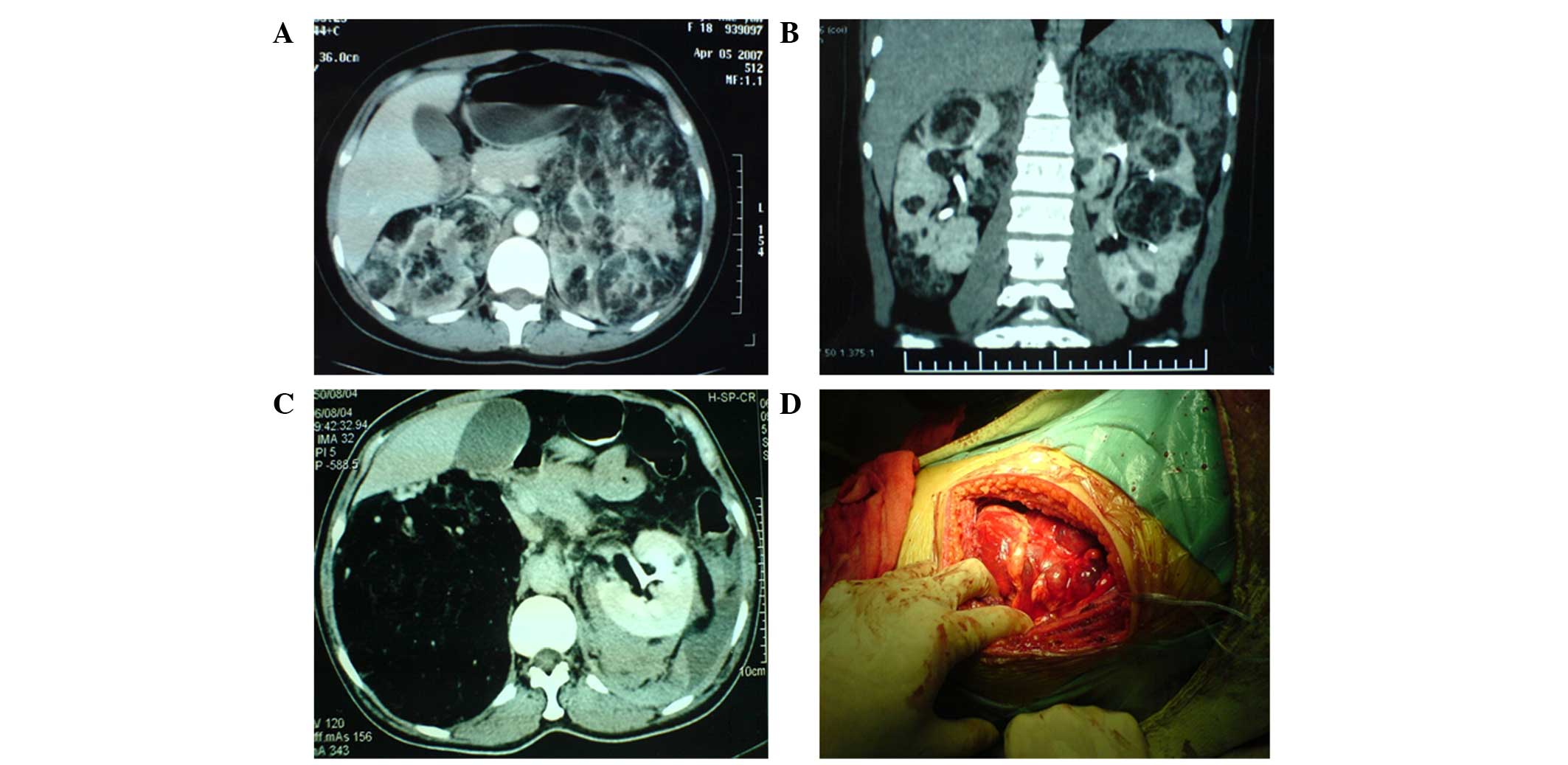
Ultrasonography revealed highly echogenic lesions in
both kidneys in all patients and mixed echogenic masses in patients
with spontaneous ruptures of the kidney. Abdominal CT scans
revealed masses of dense fat within the tumor in 16 patients
(Fig. 1). One patient exhibited no
significant intratumoral fat density on the CT and was subsequently
diagnosed using MR. The mean tumor size was 10.0±4.0 cm (range,
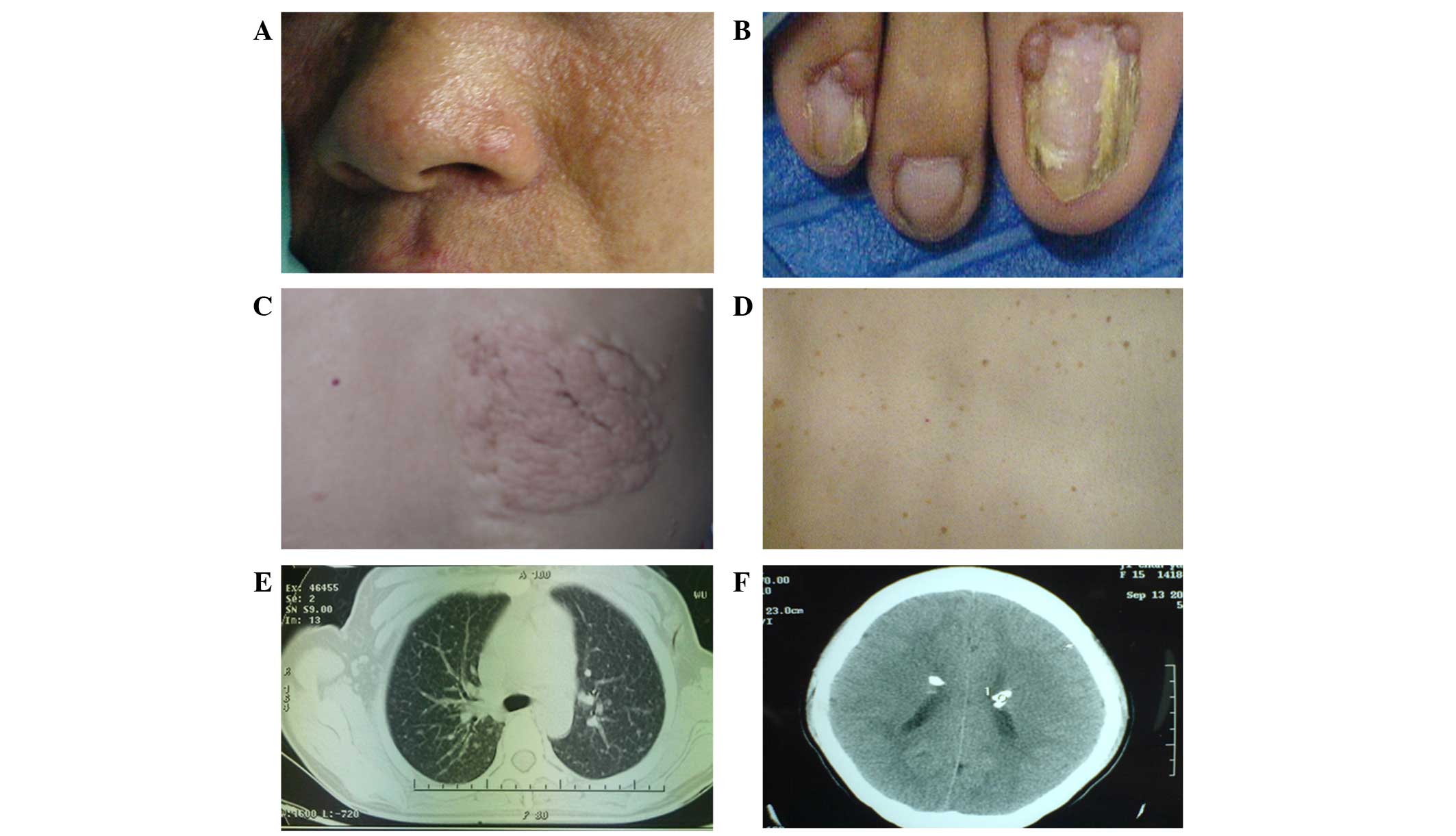
3.0–17.5 cm) In addition to renal AMLs, multiple fibroids and
facial angiofibromas were also identified in all 17 patients,
periungual fibroma in 9 patients, shagreen patches in 3 patients
and subependymal nodules with calcification in 5 patients, which
were identified by brain CT (Fig. 2).
A total of 5 patients had epilepsy and 8 patients reported a family
history of epilepsy.
Unilateral nephrectomy was performed in 4 patients,
of which 3 cases required emergency treatment due to
life-threatening hemorrhage following spontaneous rupture of AMLs,
while one case underwent the procedure due to large lesions (>15
cm). Of these 4 cases, 2 patients underwent prophylactic
contralateral PN/tumor enucleation 3 and 6 months following initial
surgery for spontaneous rupture of AML. One patient underwent
unilateral tumor enucleation in the right side of the kidney with
contralateral tumor enucleation 4 months after initial surgery due
to a large tumor (8 cm) in the right side and hematoma following
spontaneous rupture of an AML lesion, 17.5 cm in size, in the left
side of the kidney. Unilateral PN/tumor enucleation was performed
in 4 patients that exhibited large lesions (≥5 cm) as a precaution.
The other 8 patients received symptomatic medical treatment, for
example controlling blood pressure for hypertension and pain relief
for abdominal pain. The mean follow-up time for the patients was
44±17 months (range, 10–67 months) during which 1 patient succumbed
due to multiple organ dysfunction caused by hypovolemic shock that
had resulted from spontaneous rupture of left renal AML. Notably,
this patient had undergone right side nephrectomy at the First
People's Hospital of Jiashan, Jiaxing, China, 6 years previously,
due to the spontaneous rupture of AML. The patient was transfered
to our hospital due to spontaneous rupture of the AML on June 2010.
Patient demographics and outcomes are shown in Tables I and II.
 | Table I.Spontaneous rupture group patient
demographics and outcomes. |
Table I.
Spontaneous rupture group patient
demographics and outcomes.
|
|
|
|
| Treatment |
|
|---|
|
|
|
|
|
|
|
|---|
| Gender | Age, years | AML size, cm | Features of TSC | Right Kidney | Left Kidney | Outcome |
|---|
| F | 21 | 12.0 | Facial angiofibromas,
renal AML |
Nephrectomya nephrectomyb | Partial | Alive |
| M | 32 | 12.0 | Facial angiofibromas,
periungual fibroma, renal AML |
Nephrectomyc | Follow-up | Dead |
| F | 56 | 17.5 | Facial angiofibromas,
renal AML | Tumor
enucleationa | Tumor
enucleationb | Alive |
| M | 55 | 14.0 | Periungual fibroma,
subependymal nodule, renal AML |
Nephrectomya | Follow-up | Alive |
| F | 52 | 12.0 | Facial angiofibromas,
shagreen patch, periungual fibroma, renal AML | Follow-up |
Nephrectomya | Alive |
| Table II.Physical examination group patient
demographics and outcomes. |
Table II.
Physical examination group patient
demographics and outcomes.
|
|
|
|
| Treatment |
|
|---|
|
|
|
|
|
|
|
|---|
| Gender | Age, years | AML size, cm | Features of TSC | Right Kidney | Left Kidney | Outcome |
|---|
| F | 26 | 5.0 | Periungual fibroma,
renal AML | Follow-up | Partial
neprectomya | Alive |
| M | 25 | 13.0 | Facial angiofibromas,
periungual fibroma, subependymal nodule, renal AML | Follow-up | Follow-up | Alive |
| F | 37 | 9.0 | Facial angiofibromas,
renal AML | Follow-up | Follow-up | Alive |
| M | 49 | 10.0 | Facial angiofibromas,
renal AML | Follow-up | Follow-up | Alive |
| F | 18 | 15.0 | Facial angiofibromas,
subependymal nodule, renal AML | Tumor enucleation 6
months latera,b |
Nephrectomya | Alive |
| M | 19 |
5.5 | Facial
angiofibromas, renal AML | Follow-up | Follow-up | Alive |
| F | 62 |
3.0 | Confetti skin
lesion, hepatic hamartoma, renal AML | Follow-up | Partial
neprectomya | Alive |
| M | 32 |
7.0 | Facial
angiofibromas, shagreen patch, periungual fibroma, renal AML | Follow-up | Partial
neprectomya | Alive |
| F | 29 |
9.0 | Facial
angiofibromas, renal AML | Follow-up | Tumor
enucleationa | Alive |
| F | 51 | 10.0 | Facial
angiofibromas, subependymal nodule, periungual fibroma, renal
AML | Follow-up | Follow-up | Alive |
| F | 22 |
9.0 | Facial
angiofibromas, shagreen patch, lung periungual fibroma, renal
AML | Follow-up | Follow-up | Alive |
| M | 18 |
8.0 | Facial
angiofibromas, subependymal nodule, periungual fibroma, renal
AML | Follow-up | Follow-up | Alive |
The kidney reservation rate during surgery was 87.5%
(7/8) in the PE group and 25% (1/4) in the spontaneous rupture
group. A study with additional cases is required to verify the
differences between the rupture group and PE group, since the
P-value was 0.067 using Fisher's exact test, which is close to
0.05. The surgery demographics of each group are listed in Table III. AMLs with or without hemorrhage
were confirmed by pathological examination in 9 cases following
surgery.
| Table III.Surgery demographics of the
spontaneous rupture and PE groups. |
Table III.
Surgery demographics of the
spontaneous rupture and PE groups.
| Surgery | Rupture group,
n | PE group, n | P-value |
|---|
| Nephrectomy | 3 | 1 | 0.067 |
| Partial
nephrectomy | 1 | 7 |
|
Discussion
TSC is a rare disease, which may affect any organ or
system, and patients most commonly present with skin and
neurological symptoms (4–6). Renal manifestations are the second most
common symptom associated with TSC, occurring in 50–80% of patients
with TSC (6,10,16). The
renal lesions that commonly occur in TSC patients include AMLs,
renal cysts and renal cell carcinoma (RCC) (8–10). The
incidence of RCC in TSC patients is extremely low, which is similar
to that in healthy individuals, and can be easily diagnosed by CT
or MR scan. Renal cysts occur in 20% of patients with TSC and tend
to stabilize with increasing age (16). AML is the most common renal
manifestation and 66.6% of TSC patients develop multiple renal AML
(17–19). Treatment for diseases of the nervous
system has markedly improved; however, long-term survival depends
on the effective control of kidney disease in TSC patients.
Spontaneous rupture of renal AML is the primary cause of mortality
in adult TSC patients (11). In the
present study, 29.4% (5/17) of patients were admitted to hospital
due to spontaneous rupture of the affected kidney, of which 20%
(1/5) of patients succumbed due to blood loss. Therefore, the
management of renal AMLs is extremely important in treating TSC
patients.
At present, the management of renal AMLs in TSC
patients is the same as that for sporadic AMLs. Surgery is
considered essential if the tumor diameter is >4 cm, as 50% of
tumors spontaneously rupture, leading to life-threatening shock in
33.3% of patients. However, AMLs in TSC patients exhibit different
features when compared with sporadic AMLs. Firstly, AMLs in TSC
patients are always bilateral and multiple and thus, surgery is not
curative. Secondly, the tumor volume of TSC-associated AMLs are
often larger than sporadic AMLs and thus, avoiding nephrectomy is
difficult, particularly when spontaneous ruptures occur. In the
present study, only 25% (1/4) of kidneys were preserved
successfully during surgery for spontaneous ruptured AMLs and one
patient succumbed due to blood loss. However, the kidney
reservation rate was 87.5% (7/8) in the PE group. Thirdly, both
sides of TSC-associated AMLs exhibit a risk of spontaneous rupture
and hemorrhage, even following nephrectomy, and the remaining
lesions in the contralateral kidney may continue to grow, leading
to life-threatening complications. Furthermore, removal of one
kidney may significantly increase the probability of renal failure
if spontaneous rupture occurs in the contralateral kidney. At
present, standard guidelines for the management of TSC-associated
AMLs remain to be established.
We hypothesize that the management of renal AMLs in
TSC patients should be different from patients with sporadic AMLs,
particularly with regard to surgical treatment. As TSC-associated
AMLs usually arise in multiples and bilaterally, growth faster and
are more likely to spontaneously hemorrhage compared with sporadic
AMLs, surgical therapies should be performed actively following a
cautious risk-benefit analysis. The risk of hemorrhage must be
evaluated and a comprehensive treatment plan should be established
according to tumor location and size, which aims to preserve the
kidney. It is impossible to cure multiple AMLs without nephrectomy
and thus, treatment should focus on kidney reservation and
prevention of life-threatening hemorrhage. The tumor size must be
monitored closely during follow-up, as it may increase rapidly.
Surgery is the first line treatment and PN/tumor enucleation should
be performed immediately, even if the tumor size is <4 cm. Tumor
enucleation is useful in surgical treatment, as it involves the
removal of the large tumor in addition to the capsule or sub
capsule. In the present study, 3 patients received 4 tumor
enucleations, including 1 patient with a tumor size of >10 cm.
Although the control rate (rate of the prevention of spontaneous
AML rupture) is lower in TSC-associated AMLs compared with sporadic
AMLs (40 vs. 100%), endovascular embolization may be a feasible
alternative in TSC patients with bilateral large renal AMLs,
solitary kidney with huge AML or AML associated with a higher risk
of severe hemorrhage during surgery (20). Additionally, ablative therapies,
including radiofrequency ablation (RFA) or cryoablation, present
alternatives to NSS and embolization for renal AML. RFA may be used
safely and effectively for the treatment of small (<4 cm) and
symptomatic renal AMLs, as shown previously (21). Conservative treatment and follow-up
must not be performed blindly, as it may lead to kidney loss and
life-threatening hemorrhage following the spontaneous rupture of
AMLs. In the present study, 1 patient succumbed due to spontaneous
rupture of AML in the left kidney. Notably, this patient had
received nephrectomy due to spontaneous rupture of AML in the right
kidney 6 years previously. Furthermore, the maximum size of the
endogenous tumor in the left kidney had been >10 cm prior to
rupture; however, surgery was postponed due to the hesitation of
the patient and to allow the doctor to make the right decision.
In addition, a number of novel medical therapies for
renal AMLs in TSC have been identified (12,22–24). After
the identification of TSC1 and TSC2 and their encoded proteins
(hamartin and tuberin, respectively), the mammalian target of
rapamycin (mTOR) complex1 was established as a downstream target of
the hamartin/tuberin complex. Therefore, the use of mTOR inhibitors
(mTORis) may present a potential targeted therapy as they have been
demonstrated to affect the AML volume (12,22–24). A
multicentre randomized double-blind placebo-controlled trial by
Bissler et al (25)
demonstrated that everolimus, an mTORi, was more effective than a
placebo in AML response rate (42 vs. 0%), skin lesion response rate
(26 vs. 0%), and the median time to AML progression was 11.4 months
for the placebo and was not reached for everolimus. Furthermore, at
week 24, >50% everolimus patients exhibited a ≥50% reduction
from baseline in target AML volume. Two other trials demonstrated
that sirolimus, an associated mTORi, exhibited similar effects on
AML volume (24,26). Therefore, mTORis present a novel
treatment for TSC patients that allows more nephrons to be spared,
avoiding further AML growth.
In conclusion, AMLs occur with high frequency in
patients with TSC and may lead to considerable hemorrhage, renal
failure and mortality. The management of TSC-associated renal AMLs
is different from that of solitary sporadic AMLs. Treatment must
not only focus on treating the tumors, but should fully evaluate
the risk of hemorrhage and actively avoid nephrectomy. Surgical
therapies should be performed actively following comprehensive
risk-benefit analysis, and should be considered for fast-growing
tumors even if the maximum tumor size is <4 cm. Furthermore,
mTORis present a potential pharmacological treatment modality for
TSC-associated renal AMLs. The results of the present study
indicate that active surgical treatment is useful to prevent
spontaneous hemorrhage.
Glossary
Abbreviations
Abbreviations:
|
AMLs
|
angiomyolipomas
|
|
TSC
|
tuberous sclerosis complex
|
|
CT
|
computed tomography
|
|
NSS
|
nephron sparing surgeries
|
|
PN
|
partial nephrectomy
|
|
RCC
|
renal cell carcinoma
|
|
MR
|
magnetic resonance
|
|
mTORi
|
mammalian target of rapamycin
inhibitor
|
References
|
1
|
Winterkorn EB, Daouk GH, Anupindi S and
Thiele EA: Tuberous sclerosis complex and renal angiomyolipoma:
Case report and review of the literature. Pediatr Nephrol.
21:1189–1193. 2006. View Article : Google Scholar : PubMed/NCBI
|
|
2
|
Crino PB, Nathanson KL and Henske EP: The
tuberous sclerosis complex. N Engl J Med. 355:1345–1356. 2006.
View Article : Google Scholar : PubMed/NCBI
|
|
3
|
Bourneville DM: Tuberous sclerosis of the
cerebral cortex: Intellectual disability and hemiplegic epilepsy.
Arch Neurol (Paris). 1:81–91. 1880.(In French).
|
|
4
|
Borkowska J, Schwartz RA, Kotulska K and
Jozwiak S: Tuberous sclerosis complex: Tumors and tumorigenesis.
Int J Dermatol. 50:13–20. 2011. View Article : Google Scholar : PubMed/NCBI
|
|
5
|
Yates JR: Tuberous sclerosis. Eur J Hum
Genet. 14:1065–1073. 2006. View Article : Google Scholar : PubMed/NCBI
|
|
6
|
Franz DN, Bissler JJ and McCormack FX:
Tuberous sclerosis complex: Neurological, renal and pulmonary
manifestations. Neuropediatrics. 41:199–208. 2010. View Article : Google Scholar : PubMed/NCBI
|
|
7
|
de Vries PJ, Whittemore VH, Leclezio L,
Byars AW, Dunn D, Ess KC, Hook D, King BH, Sahin M and Jansen A:
Tuberous sclerosis associated neuropsychiatric disorders (TAND) and
the TAND Checklist. Pediatr Neurol. 52:25–35. 2015. View Article : Google Scholar : PubMed/NCBI
|
|
8
|
Rakowski SK, Winterkorn EB, Paul E, Steele
DJ, Halpern EF and Thiele EA: Renal manifestations of tuberous
sclerosis complex: Incidence, prognosis, and predictive factors.
Kidney Int. 70:1777–1782. 2006. View Article : Google Scholar : PubMed/NCBI
|
|
9
|
Henske EP: Tuberous sclerosis and the
kidney: From mesenchyme to epithelium and beyond. Pediatr Nephrol.
20:854–857. 2005. View Article : Google Scholar : PubMed/NCBI
|
|
10
|
O'Callaghan FJ, Noakes MJ, Martyn CN and
Osborne JP: An epidemiological study of renal pathology in tuberous
sclerosis complex. BJU Int. 94:853–857. 2004. View Article : Google Scholar : PubMed/NCBI
|
|
11
|
Shepherd CW, Gomez MR, Lie JT and Crowson
CS: Causes of death in patients with tuberous sclerosis. Mayo Clin
Proc. 66:792–796. 1991. View Article : Google Scholar : PubMed/NCBI
|
|
12
|
Dixon BP, Hulbert JC and Bissler JJ:
Tuberous sclerosis complex renal disease. Nephron Exp Nephrol.
118:e15–e20. 2011. View Article : Google Scholar : PubMed/NCBI
|
|
13
|
Rabenou RA and Charles HW: Differentiation
of sporadic versus tuberous sclerosis complex-associated
angiomyolipoma. AJR Am J Roentgenol. 205:292–301. 2015. View Article : Google Scholar : PubMed/NCBI
|
|
14
|
Northrup H and Krueger DA: International
Tuberous Sclerosis Complex Consensus Group: Tuberous sclerosis
complex diagnostic criteria update: Recommendations of the 2012
Iinternational tuberous sclerosis complex consensus conference.
Pediatr Neurol. 49:243–254. 2013. View Article : Google Scholar : PubMed/NCBI
|
|
15
|
Wang H, Zhou L, Guo J, Sun L, Long Q, Ma
Y, Zhang L, Lin Z, Zhu T and Wang G: Mini-flank supra-12th rib
incision for open partial nephrectomy compared with laparoscopic
partial nephrectomy and traditional open partial nephrectomy. PloS
One. 9:e891552014. View Article : Google Scholar : PubMed/NCBI
|
|
16
|
Ewalt DH, Sheffield E, Sparagana SP,
Delgado MR and Roach ES: Renal lesion growth in children with
tuberous sclerosis complex. J Urol. 160:141–145. 1998. View Article : Google Scholar : PubMed/NCBI
|
|
17
|
Steiner MS, Goldman SM, Fishman EK and
Marshall FF: The natural history of renal angiomyolipoma. J Urol.
150:1782–1786. 1993.PubMed/NCBI
|
|
18
|
Malone MJ, Johnson PR, Jumper BM, Howard
PJ, Hopkins TB and Libertino JA: Renal angiomyolipoma: 6 case
reports and literature review. J Urol. 135:349–353. 1986.PubMed/NCBI
|
|
19
|
Curatolo P, Bombardieri R and Jozwiak S:
Tuberous sclerosis. Lancet. 372:657–668. 2008. View Article : Google Scholar : PubMed/NCBI
|
|
20
|
Kothary N, Soulen MC, Clark TW, Wein AJ,
Shlansky-Goldberg RD, Crino PB and Stavropoulos SW: Renal
angiomyolipoma: Long-term results after arterial embolization. J
Vasc Interv Radiol. 16:45–50. 2005. View Article : Google Scholar : PubMed/NCBI
|
|
21
|
Castle SM, Gorbatiy V, Ekwenna O, Young E
and Leveillee RJ: Radiofrequency ablation (RFA) therapy for renal
angiomyolipoma (AML): An alternative to angio-embolization and
nephron-sparing surgery. BJU Int. 109:384–387. 2012. View Article : Google Scholar : PubMed/NCBI
|
|
22
|
Curatolo P and Moavero R: mTOR inhibitors
in tuberous sclerosis complex. Curr Neuropharmacol. 10:404–415.
2012. View Article : Google Scholar : PubMed/NCBI
|
|
23
|
Budde K and Gaedeke J: Tuberous sclerosis
complex-associated angiomyolipomas: Focus on mTOR inhibition. Am J
Kidney Dis. 59:276–283. 2012. View Article : Google Scholar : PubMed/NCBI
|
|
24
|
Bissler JJ, McCormack FX, Young LR, Elwing
JM, Chuck G, Leonard JM, Schmithorst VJ, Laor T, Brody AS, Bean J,
et al: Sirolimus for angiomyolipoma in tuberous sclerosis complex
or lymphangioleiomyomatosis. N Engl J Med. 358:140–151. 2008.
View Article : Google Scholar : PubMed/NCBI
|
|
25
|
Bissler JJ, Kingswood JC, Radzikowska E,
Zonnenberg BA, Frost M, Belousova E, Sauter M, Nonomura N,
Brakemeier S, de Vries PJ, et al: Everolimus for angiomyolipoma
associated with tuberous sclerosis complex or sporadic
lymphangioleiomyomatosis (EXIST-2): A multicentre, randomised,
double-blind, placebo-controlled trial. Lancet. 381:817–824. 2013.
View Article : Google Scholar : PubMed/NCBI
|
|
26
|
Davies DM, de Vries PJ, Johnson SR,
McCartney DL, Cox JA, Serra AL, Watson PC, Howe CJ, Doyle T,
Pointon K, et al: Sirolimus therapy for angiomyolipoma in tuberous
sclerosis and sporadic lymphangioleiomyomatosis: A phase 2 trial.
Clin Cancer Res. 17:4071–4081. 2011. View Article : Google Scholar : PubMed/NCBI
|