Introduction
Osteosarcoma (OSA) is the most common malignant bone
cancer in dogs, representing 85% of canine skeletal neoplasms
(1). OSA is a highly aggressive and
painful tumor that occurs primarily in the appendicular skeleton of
large and giant breed dogs (1).
Treatment of OSA is difficult due to its high metastasis rate and
aggressive local behavior (2). The
current standard treatment for OSA in dogs is amputation or
limb-sparing surgery combined with chemotherapy (2). Amputation or limb-sparing surgery is not
always the choice of treatment for dogs with OSA due to neurologic
or orthopedic disease (1).
Alternative treatments to suppress tumor growth and alleviate the
pain of the primary tumor would have significant clinical relevance
in these cases. Coarsely fractionated, or ‘palliative’ radiation
therapy using several fractioned doses (for example, 8–10 Gy per
fraction) may be used for palliation of pain, but is not able to
achieve local tumor control (3–5). High dose
per fraction radiation therapy has been described as a method for
local tumor control using stereotactic radiosurgery, particularly
when combined with chemotherapy (5,6).
Stereotactic radiosurgery is able to deliver a single very high
dose of radiation (for example, 30 Gy) using multiple beams with a
linear accelerator to a designated target, while sparing the
surrounding tissues (6).
Charged particle radiation therapy, including proton
and heavy ion therapy, has gained interest in human radiation
oncology as a novel therapeutic modality (7). Clinical application of protons and
carbon ions for human cancer treatment has been expanding and has
achieved significantly improved clinical outcomes for a number of
tumor types, including OSA in non-resectable tumors (8). Unlike conventional photon radiation,
protons and carbon ions may be manipulated to release the majority
of their energy only when they reach their target (7,9). This
property is known as the Bragg peak. Particle radiation therapy
offers the possibility of a significantly increased dose of
ionizing radiation to the tumor with very little dose deposited in
the normal tissue (7–9). Particle radiation therapy has the
potential to be considered as an alternative modality for the
treatment of canine and human OSA in the future. Carbon ions have
increased ionization density [high linear energy transfer (LET)
properties], and heavy ions differ from protons or photons with low
LET due to their radiobiological properties. A number of in
vitro studies have demonstrated that high LET radiation is more
effective than low LET radiation in terms of cell-killing effects
(10–13). Additionally, the extent of cell
killing by heavy ions is not cell cycle dependent and is not
decreased due to the presence of hypoxia (14–16). In
human oncology, the relative biological effectiveness (RBE) is used
to describe the increase in effectiveness of particle radiation
(10,12). RBE is defined as the ratio of doses of
photons and charged particles inducing the same biological
endpoints, including cell killing, mutation and cytogenetic
aberrations. The RBE values of cell killing for heavy ions
generally increases up to a LET near 200 keV/µm and decreases
afterwards (17,18).
Particle radiation therapy may be expensive to
implement, and the application of heavy ions within veterinary
clinics requires additional background information to determine its
effectiveness. To the best of our knowledge, there are only limited
reports of the use of protons for treating dogs with brain tumors
and currently there have been no reports of using heavy ions to
treat tumors in veterinary medicine (19,20). RBE
values of 1.96–2.50 in canine squamous cell carcinoma, fibrosarcoma
and hemangiopericytoma cell lines for carbon ions (LET=108 keV/µm)
have been previously reported (21).
Our previous study described canine OSA cell lines that were either
radioresistant or highly radiosensitive to photon radiation, along
with their basic cellular characteristics (22). In the present study, an in
vitro comparison of the cell-killing effects of photons,
protons and heavy ions on canine OSA cells was performed. For heavy
ion irradiation, SOBP (spread out Bragg peak) carbon ions (LET at
50 keV/µm), which are used in radiotherapy, and iron ions (LET at
200 keV/µm), as above are expected to have maximum biological
effects of heavy ions (23).
Materials and methods
Cell culture
The canine OSA cell lines Abrams, D17, Grey and
Moresco were a gift from the Animal Cancer Center of Colorado State
University (Fort Collins, CO, USA) (22,24). Cells
were grown in minimal essential media (Invitrogen; Thermo Fisher
Scientific, Inc., Waltham, MA, USA) supplemented with 10% (v/v)
fetal bovine serum (FBS; Sigma-Aldrich, St. Louis, MO, USA) and 1%
(v/v) Pen/Strep and fungizone solution (Invitrogen; Thermo Fisher
Scientific, Inc.), and they were maintained in a tissue culture
incubator at 37°C in a 100% humidified atmosphere of 5%
CO2. The cell doubling times of these cells were 19 h
for Abrams, 22 h for D17, 18 h for Grey and 22 h for Moresco
(22).
Radiation conditions
Particle-based irradiation experiments were
performed at the National Institute of Radiological Sciences (NIRS)
in Chiba, Japan. For heavy ion exposure, accelerated ions were
irradiated using the Heavy Ion Medical Accelerator in Chiba (HIMAC;
Chiba, Japan) at room temperature. The details concerning the beam
characteristics of the particle radiation, biological irradiation
procedures and dosimetry have been described previously (12,25,26).
Accelerated monoenergetic iron ions have 500 MeV/nucleon of initial
energy and 200 keV/µm of LET at the irradiated position. Carbon
ions were accelerated at 290 MeV/nucleon of initial energy and
spread out with a ridge filter for 6 cm width of SOBP. The
monolayer cell culture was irradiated at the center (50 keV/µm of
average LET) within the SOBP at a distance of 119 mm from the
entrance (27). Monoenergetic protons
that were accelerated to 70 MeV using the NIRS-930 cyclotron have a
LET value of 1.0 at the irradiated position. Dose rates for heavy
ions and protons were set at 1 and 5 Gy/min respectively. Gamma-ray
irradiations were performed at the Colorado State University (Fort
Collins, CO, USA) with 137Cs gamma-rays delivered at a dose rate of
~2.5 Gy/min at room temperature (using a J.L. Shepherd Model Mark
I-68, nominal 6000 Ci 137Cs irradiator).
Cell survival assays
Exponentially growing cells cultured in T12.5 flasks
(BD Biosciences, Franklin Lakes, NJ, USA) were irradiated, then
trypsinized and plated onto 100 mm cell culture dishes at an
density of 100 colonies. Following incubation for 7–10 days to
allow colony formation, surviving colonies were rinsed with 0.9%
NaCl, fixed with 100% ethanol and stained by 0.1% crystal violet.
Each colony consisting of >50 cells was scored as a survivor. At
least three independent experiments were performed.
RBE was calculated, which is defined as the ratio of
dose of photons and charged particles inducing identical biological
effects, based on D10 values. The D10 values,
which represent doses required to achieve 10% survival, were
obtained from each survival curve using Prism 5 software (GraphPad
Software, Inc., La Jolla, CA, USA).
Statistical analysis
Data were analyzed using Prism 5 software. Data are
presented as the mean ± standard error. Differences with a
P<0.05 were considered statistically significant. Statistical
comparison of mean values in the RBE was performed using unpaired
two tailed t-test.
Results
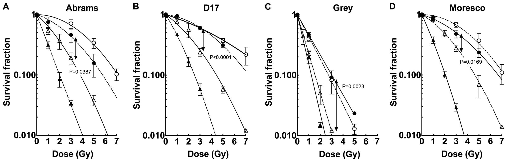
The present study used canine OSA cell lines with
various radiation sensitivities as reported previously (22). Abrams, D17 and Moresco were fairly
resistant to gamma-rays (D10=7.12–9.33 Gy), while Grey
was relatively radiosensitive (D10=2.72 Gy). Fig. 1 shows the dose-response curves for
cell-killing effect on the four canine OSA cell lines irradiated
with various radiation sources. It was observed that the clinically
relevant carbon ion beams (LET at 50 keV/µm) decreased cell
survival fractions of the four canine OSA cell lines compared with
gamma-rays (P<0.05). Iron ion beams decreased cell survival more
than carbon ions in the three radioresistant cell lines (Abrams,
D17 and Moresco). However, the radiosensitive cell line Grey
demonstrated similar cell survival for both carbon and iron ion
irradiation. The proton cell survival curves demonstrated similar
profiles to those of gamma-rays for the four cell lines. In order
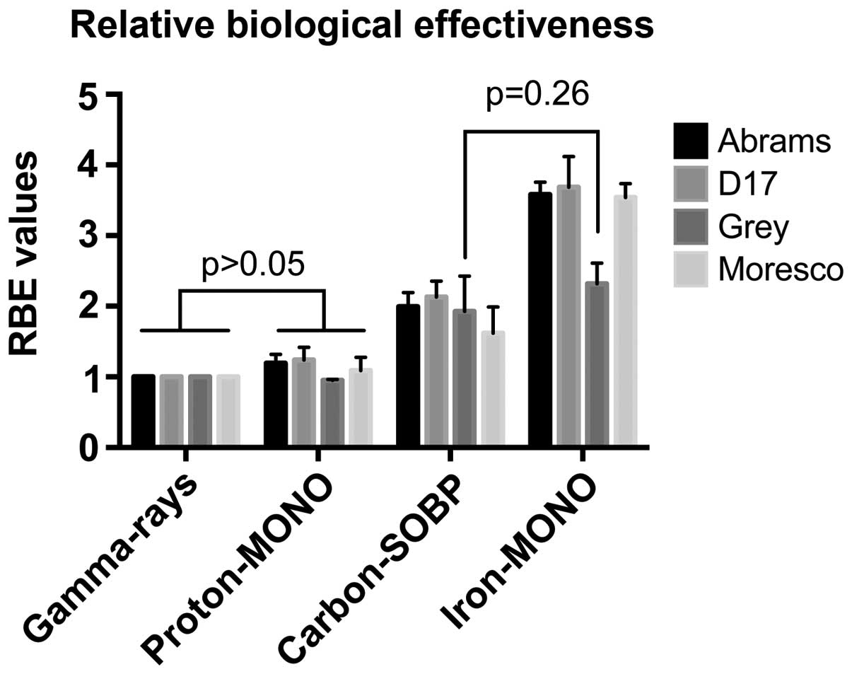
to describe the increased effects of particle radiation, RBE was
calculated based on the D10 values relative to
gamma-rays (Fig. 2 and Table I). The RBE values ranged from
0.90–1.26 for protons, 1.56–2.10 for carbon-ions and 2.22–3.69 for
iron ions among the cell lines. The RBEs for iron ions in the
radiosensitive Grey cell line (2.22) was reduced compared with
those in the three radioresistant cell lines.
 | Table I.Relative biological effectiveness of
particle radiation types in canine osteosarcoma cell lines. |
Table I.
Relative biological effectiveness of
particle radiation types in canine osteosarcoma cell lines.
|
| γ-rays, 0.2
keV/µm | Proton, 1
keV/µm | Carbon SOBP, 50
keV/µm | Iron, 200
keV/µm |
|---|
|
|
|
|
|
|
|---|
| Cell lines | D10,
Gy | D10,
Gy | RBE | D10,
Gy | RBE | D10,
Gy | RBE |
|---|
| Abrams | 7.12 | 3.96 | 1.80 | 3.81 | 1.87 | 2.01 | 3.55 |
| D17 | 9.33 | 7.26 | 1.29 | 4.44 | 2.10 | 2.53 | 3.69 |
| Grey | 2.72 | 3.02 | 0.90 | 1.55 | 1.75 | 1.23 | 2.22 |
| Moresco | 7.24 | 6.68 | 1.08 | 4.64 | 1.56 | 2.62 | 3.51 |
Discussion
The present study indicated that heavy ions may be a
superior method to induce canine OSA cell killing in vitro
compared with protons and gamma-rays, based on using four cell
lines with various radiosensitivities. The results of the present
study were consistent with a previous canine cell line study
(21). Additionally, several other
previous studies using human and rodent cells have demonstrated
that high LET heavy ions are more effective for cell killing at
identical physical absorbed doses as low LET radiation types
(10–12,21,26). In
general, a large number of complex clusters of DNA damage generated
by high LET radiation would be more difficult to repair, resulting
in more severe biological damage than that induced by low LET
radiation (28,29).
In the present study, the RBE values for the human
clinical setting carbon ions ranged from 1.56–2.10 using
D10 doses relative to that of gamma-rays. The RBE values
of canine OSA cells for the carbon ions were close to the
previously published values: ~1.5 for human salivary gland tumor
origin HSG cells and 1.8 for HeLa cells, using identical carbon-ion
beams (30). For a proton beam, which
is another type of particle radiation with low LET, the cell
killing effects did not differ substantially from gamma-rays,
providing the RBE values of almost 1.0 for all four cell lines. In
human oncology, the RBE of protons has been reported to exhibit
experimental variations, but in general a constant RBE of either
1.0 or 1.1 is estimated for clinical applications (31–34).
However not only LET, but also DNA repair capacity and cellular
radiosensitivity affect RBE values (7,26). The
results of the present study suggest that the effectiveness of
particle radiation on human and canine tumor cell killing is likely
comparable.
Furthermore, the present results with regard to iron
ion radiation clearly revealed that heavy ions with high LET (200
keV/µm) better enhanced the cell killing effects in radioresistant
canine OSA cells compared with 50 keV/µm carbon ions. It should be
noted that this is consistent with previous human and rodent
studies, which observed the peak RBE is approximately LET 200
keV/µm (17,18). In the case of the radiosensitive cell
line Grey, carbon and iron ion radiation demonstrated more similar
cell killing effects and smaller RBE values compared to the
radioresistant canine OSA cells. The response of Grey to high LET
was similar to DNA-repair deficient cell lines in previous human
and rodent studies (26,35). Previous studies have suggested that
high LET is effective to cells with normal cellular DNA repair
capacity (36,37). Furthermore, in a recent investigation,
a variable response to radiation was detected clinically in dogs
affected by OSA (38). Although it
was beyond the scope of the present study, investigation into the
DNA repair deficiencies of the radiosensitive OSA cell line may
provide an insight into whether tumor cell radiosensitivity can be
used as an indicator of clinical response to radiation therapy.
Canine OSA cells are considered to be resistant to
conventional radiotherapy (1). In
addition to improved dose distribution conferred by particle
irradiation, a markedly increased efficiency of cell killing is
another attractive reason for investigating heavy ions (15). Furthermore, hypoxic cells naturally
occur within cancerous tissues. Hypoxic cells are resistant to
low-LET photons, but may be effectively killed by high-LET heavy
ions (14,39). According to previous reports, it has
been suggested that heavy ions may suppress the metastatic
capabilities of cancer cells; thus, the effects of heavy ions on
the control of lung metastasis of canine OSA should be investigated
for clinical application (40,41).
Additional investigations are required to determine whether heavy
ion radiotherapy is able to confer clinical advantages over photon
irradiation in veterinary clinics, including in the treatment of
dogs with OSA.
In conclusion, the results of the present study
provide basic insights into the application of heavy ions in
veterinary clinics. With the use of radioresistant and
radiosensitive canine OSA cells, the present study demonstrated
that high LET charged particles, particularly iron ions, are
effective at killing canine OSA cells independent of gamma-ray
radiosensitivity. Radioresistant and radiosensitive cells exhibited
significantly higher RBE values for carbon and iron ion radiation
(P<0.05; Fig. 2). These findings
support the future investigation of heavy ion application within
veterinary clinics to study their effectiveness compared with
currently available radiation therapy.
Acknowledgements
The present research was funded by the College
Research Council at Colorado State University College of Veterinary
Medicine and Biosciences (TAK), the Dr. Akiko M. Ueno Radiobiology
Fund (TAK) and the International Open Laboratory at the National
Institute of Radiological Sciences (AF). The authors acknowledge
support from NIRS-HIMAC and Cyclotron (Chiba, Japan).
References
|
1
|
Ehrhart NP, Ryan SD and Fan TM: Tumors of
the skeletal system. Withrow and MacEwen's Small Animal Clinical
Oncology. Withrow SJ, Vail DM and Page RL: (5th). W.B. Saunders.
(St. Louis, MO). 463–503. 2013. View Article : Google Scholar
|
|
2
|
Mueller F, Fuchs B and Kaser-Hotz B:
Comparative biology of human and canine osteosarcoma. Anticancer
Res. 27:155–164. 2007.PubMed/NCBI
|
|
3
|
Ramirez O III, Dodge RK, Page RL, Price
GS, Hauck ML, LaDue TA, Nutter F and Thrall DE: Palliative
radiotherapy of appendicular osteosarcoma in 95 dogs. Vet Radiol
Ultrasound. 40:517–522. 1999. View Article : Google Scholar : PubMed/NCBI
|
|
4
|
Green EM, Adams WM and Forrest LJ: Four
fraction palliative radiotherapy for osteosarcoma in 24 dogs. J Am
Anim Hosp Assoc. 38:445–451. 2002. View
Article : Google Scholar : PubMed/NCBI
|
|
5
|
Coomer A, Farese J, Milner R, Liptak J,
Bacon N and Lurie D: Radiation therapy for canine appendicular
osteosarcoma. Vet Comp Oncol. 7:15–27. 2009. View Article : Google Scholar : PubMed/NCBI
|
|
6
|
Farese JP, Ashton J, Milner R, Ambrose LL
and Van Gilder J: The effect of the bisphosphonate alendronate on
viability of canine osteosarcoma cells in vitro. In Vitro Cell Dev
Biol Anim. 40:113–117. 2004. View Article : Google Scholar : PubMed/NCBI
|
|
7
|
Schulz-Ertner D, Jäkel O and Schlegel W:
Radiation therapy with charged particles. Semin Radiat Oncol.
16:249–259. 2006. View Article : Google Scholar : PubMed/NCBI
|
|
8
|
Schulz-Ertner D and Tsujii H: Particle
radiation therapy using proton and heavier ion beams. J Clin Oncol.
25:953–964. 2007. View Article : Google Scholar : PubMed/NCBI
|
|
9
|
Tobias CA, Lyman JT, Chatterjee A, Howard
J, Maccabee HD, Raju MR, Smith AR, Sperinde JM and Welch GP:
Radiological physics characteristics of the extracted heavy ion
beams of the bevatron. Science. 174:1131–1134. 1971. View Article : Google Scholar : PubMed/NCBI
|
|
10
|
Cox R, Thacker J, Goodhead DT and Munson
RJ: Mutation and inactivation of mammalian cells by various
ionising radiations. Nature. 267:425–427. 1977. View Article : Google Scholar : PubMed/NCBI
|
|
11
|
Eguchi-Kasai K, Murakami M, Itsukaichi H,
Fukutsu K, Yatagai F, Kanai T, Ohara H and Sato K: Repair of DNA
double-strand breaks and cell killing by charged particles. Adv
Space Res. 22:543–549. 1998. View Article : Google Scholar : PubMed/NCBI
|
|
12
|
Suzuki M, Kase Y, Yamaguchi H, Kanai T and
Ando K: Relative biological effectiveness for cell-killing effect
on various human cell lines irradiated with heavy-ion medical
accelerator in Chiba (HIMAC) carbon-ion beams. Int J Radiat Oncol
Biol Phys. 48:241–250. 2000. View Article : Google Scholar : PubMed/NCBI
|
|
13
|
Fujisawa H, Genik PC, Kitamura H, Fujimori
A, Uesaka M and Kato TA: Comparison of human chordoma cell-kill for
290 MeV/n carbon ions versus 70 MeV protons in vitro. Radiat Oncol.
8:912013. View Article : Google Scholar : PubMed/NCBI
|
|
14
|
Tobias CA, Blakely EA, Alpen EL, Castro
JR, Ainsworth EJ, Curtis SB, Ngo FQ, Rodriguez A, Roots RJ,
Tenforde T and Yang TC: Molecular and cellular radiobiology of
heavy ions. Int J Radiat Oncol Biol Phys. 8:2109–2120. 1982.
View Article : Google Scholar : PubMed/NCBI
|
|
15
|
Hamada N, Imaoka T, Masunaga S, Ogata T,
Okayasu R, Takahashi A, Kato TA, Kobayashi Y, Ohnishi T, Ono K, et
al: Recent advances in the biology of heavy-ion cancer therapy. J
Radiat Res. 51:365–383. 2010. View Article : Google Scholar : PubMed/NCBI
|
|
16
|
Blakely EA, Tobias CA, Yang TC, Smith KC
and Lyman JT: Inactivation of human kidney cells by high-energy
monoenergetic heavy-ion beams. Radiat Res. 80:122–160. 1979.
View Article : Google Scholar : PubMed/NCBI
|
|
17
|
Tsuruoka C, Suzuki M, Kanai T and Fujitaka
K: LET and ion species dependence for cell killing in normal human
skin fibroblasts. Radiat Res. 163:494–500. 2005. View Article : Google Scholar : PubMed/NCBI
|
|
18
|
Kato TA, Tsuda A, Uesaka M, Fujimori A,
Kamada T, Tsujii H and Okayasu R: In vitro characterization of
cells derived from chordoma cell line U-CH1 following treatment
with X-rays, heavy ions and chemotherapeutic drugs. Radiat Oncol.
6:1162011. View Article : Google Scholar : PubMed/NCBI
|
|
19
|
Kaser-Hotz B, Sumova A, Lomax A, Schneider
U, Klink B, Fidel J and Blattmann H: A comparison of normal tissue
complication probability of brain for proton and photon therapy of
canine nasal tumors. Vet Radiol Ultrasound. 43:480–486. 2002.
View Article : Google Scholar : PubMed/NCBI
|
|
20
|
Bley CR, Sumova A, Roos M and Kaser-Hotz
B: Irradiation of brain tumors in dogs with neurologic disease. J
Vet Intern Med. 19:849–854. 2005. View Article : Google Scholar : PubMed/NCBI
|
|
21
|
Wada S, Van Khoa T, Kobayashi Y, Funayama
T, Ogihara K, Ueno S and Ito N: Prediction of cellular
radiosensitivity from DNA damage induced by gamma-rays and carbon
ion irradiation in canine tumor cells. J Vet Med Sci. 67:1089–1095.
2005. View Article : Google Scholar : PubMed/NCBI
|
|
22
|
Maeda J, Yurkon CR, Fujisawa H, Kaneko M,
Genet SC, Roybal EJ, Rota GW, Saffer ER, Rose BJ, Hanneman WH, et
al: Genomic instability and telomere fusion of canine osteosarcoma
cells. PLoS One. 7:e433552012. View Article : Google Scholar : PubMed/NCBI
|
|
23
|
Tsujii H, Mizoe J, Kamada T, Baba M, Tsuji
H, Kato H, Kato S, Yamada S, Yasuda S, Ohno T, et al: Clinical
Results of Carbon Ion Radiotherapy at NIRS. J Radiat Res.
48:S1–S13. 2007. View Article : Google Scholar
|
|
24
|
Legare ME, Bush J, Ashley AK, Kato T and
Hanneman WH: Cellular and phenotypic characterization of canine
osteosarcoma cell lines. J Cancer. 2:262–270. 2011. View Article : Google Scholar : PubMed/NCBI
|
|
25
|
Kamada T, Tsujii H, Tsuji H, Yanagi T,
Mizoe JE, Miyamoto T, Kato H, Yamada S, Morita S, Yoshikawa K, et
al: Working Group for the Bone and Soft Tissue Sarcomas: Efficacy
and safety of carbon ion radiotherapy in bone and soft tissue
sarcomas. J Clin Oncol. 20:4466–4471. 2002. View Article : Google Scholar : PubMed/NCBI
|
|
26
|
Cartwright IM, Bell JJ, Maeda J, Genet MD,
Romero A, Fujii Y, Fujimori A, Kitamuta H, Kamada T, Chen DJ and
Kato TA: Effects of targeted phosphorylation site mutations in the
DNA-PKcs phosphorylation domain on low and high LET radiation
sensitivity. Oncol Lett. 9:1621–1627. 2015.PubMed/NCBI
|
|
27
|
McMillan DD, Maeda J, Bell JJ, Genet MD,
Phoonswadi G, Mann KA, Kraft SL, Kitamura H, Fujimori A and Yoshii
Y: Validation of 64Cu-ATSM damaging DNA via high-LET Auger electron
emission. J Radiat Res. 56:784–791. 2015. View Article : Google Scholar : PubMed/NCBI
|
|
28
|
Fakir H, Sachs RK, Stenerlöw B and Hofmann
W: Clusters of DNA double-strand breaks induced by different doses
of nitrogen ions for various LETs: Experimental measurements and
theoretical analyses. Radiat Res. 166:917–927. 2006. View Article : Google Scholar : PubMed/NCBI
|
|
29
|
Hada M and Georgakilas AG: Formation of
clustered DNA damage after high-LET irradiation: A review. J Radiat
Res. 49:203–210. 2008. View Article : Google Scholar : PubMed/NCBI
|
|
30
|
Kanai T, Endo M, Minohara S, Miyahara N,
Koyama-ito H, Tomura H, Matsufuji N, Futami Y, Fukumura A and
Hiraoka T: Biophysical characteristics of HIMAC clinical
irradiation system for heavy-ion radiation therapy. Int J Radiat
Oncol Biol Phys. 44:201–210. 1999. View Article : Google Scholar : PubMed/NCBI
|
|
31
|
Paganetti H, Niemierko A, Ancukiewicz M,
Gerweck LE, Goitein M, Loeffler JS and Suit HD: Relative biological
effectiveness (RBE) values for proton beam therapy. Int J Radiat
Oncol Biol Phys. 53:407–421. 2002. View Article : Google Scholar : PubMed/NCBI
|
|
32
|
Carabe A, Moteabbed M, Depauw N, Schuemann
J and Paganetti H: Range uncertainty in proton therapy due to
variable biological effectiveness. Phys Med Biol. 57:1159–1172.
2012. View Article : Google Scholar : PubMed/NCBI
|
|
33
|
Tsunemoto H, Morita S, Ishikawa T,
Furukawa S, Kawachi K, Kanai T, Ohara H, Kitagawa T and Inada T:
Proton therapy in Japan. Radiat Res Suppl. 8:S235–S243. 1985.
View Article : Google Scholar : PubMed/NCBI
|
|
34
|
Wouters BG, Lam GK, Oelfke U, Gardey K,
Durand RE and Skarsgard LD: Measurements of relative biological
effectiveness of the 70 MeV proton beam at TRIUMF using Chinese
hamster V79 cells and the high-precision cell sorter assay. Radiat
Res. 146:159–170. 1996. View
Article : Google Scholar : PubMed/NCBI
|
|
35
|
Tobias CA, Blakely EA, Chang PY, Lommel L
and Roots R: Response of sensitive human ataxia and resistant T-1
cell lines to accelerated heavy ions. Br J Cancer Suppl. 6:175–185.
1984.PubMed/NCBI
|
|
36
|
Loucas BD and Geard CR: Kinetics of
chromosome rejoining in normal human fibroblasts after exposure to
low- and high-LET radiations. Radiat Res. 138:352–360. 1994.
View Article : Google Scholar : PubMed/NCBI
|
|
37
|
George K, Wu H, Willingham V, Furusawa Y,
Kawata T and Cucinotta FA: High- and low-LET induced chromosome
damage in human lymphocytes: A time-course of aberrations in
metaphase and interphase. Int J Radiat Biol. 77:175–183. 2001.
View Article : Google Scholar : PubMed/NCBI
|
|
38
|
Walter CU, Dernell WS, LaRue SM, Lana SE,
Lafferty MH, LaDue TA and Withrow SJ: Curative-intent radiation
therapy as a treatment modality for appendicular and axial
osteosarcoma: A preliminary retrospective evaluation of 14 dogs
with the disease. Vet Comp Oncol. 3:1–7. 2005. View Article : Google Scholar : PubMed/NCBI
|
|
39
|
Brurberg KG, Skogmo HK, Graff BA, Olsen DR
and Rofstad EK: Fluctuations in pO2 in poorly and well-oxygenated
spontaneous canine tumors before and during fractionated radiation
therapy. Radiother Oncol. 77:220–226. 2005. View Article : Google Scholar : PubMed/NCBI
|
|
40
|
Ogata T, Teshima T, Kagawa K, Hishikawa Y,
Takahashi Y, Kawaguchi A, Suzumoto Y, Nojima K, Furusawa Y and
Matsuura N: Particle irradiation suppresses metastatic potential of
cancer cells. Cancer Res. 65:113–120. 2005.PubMed/NCBI
|
|
41
|
Akino Y, Teshima T, Kihara A,
Kodera-Suzumoto Y, Inaoka M, Higashiyama S, Furusawa Y and Matsuura
N: Carbon-ion beam irradiation effectively suppresses migration and
invasion of human non-small-cell lung cancer cells. Int J Radiat
Oncol Biol Phys. 75:475–481. 2009. View Article : Google Scholar : PubMed/NCBI
|