Introduction
Cancer is the biggest killer threatening human
health globally (1). At present, the
main treatment approaches include radiotherapy, chemotherapy and
combination therapy (2). However,
radiotherapy has significant toxicity and side effects on healthy
tissues of the body, and the toxicity and side effects of
chemotherapy adversely affect the impact of the treatment to a
certain degree (3). Therefore, the
choice of drugs for chemotherapy has become key in tumor treatment.
In recent years, an emphasis has been placed upon individualized
medication, and improvement of drug sensitivity and treatment
effect through combination therapy in tumor treatment (4).
Dihydroartemisinin (DHA) is an active derivative of
the first-line anti-malarial drug artemisinin, which has been
extracted from the goldenrod Artemisia annua in China for
the first time (5,6). Due to the quick activity, effectiveness
and safety of artemisinin and its derivative, the World Health
Organization promotes their application in the treatment of severe
and drug-resistant malaria (7). It
has been demonstrated that artemisinin and its derivative show an
antitumor effect and that they act on a series of cell biochemical
processes, inhibiting proliferation, inducing apoptosis and
oxidative stress, and acting in anti-angiogenesis (8). Doxorubicin (DOX), also a currently-used
antitumor antibiotic, can inhibit the synthesis of RNA and DNA,
with the strongest inhibitory effect on RNA. The drug is
efficacious against multiple types of tumor, with a wide antitumor
spectrum (9,10).
In the present study, the natural drug DHA and the
chemical drug DOX were combined to act on HeLa tumor cells, and the
survival rate and the mechanism of death were detected. The
activity of the DHA + DOX combination in the OVCAR-3, MCF-7, PC-3
and A549 cells was also investigated in the study. In addition, the
in vivo treatment effect of DHA + DOX was studied with an
intratumoral method.
Materials and methods
Experimental materials and
reagents
The cervical cancer (HeLa), ovarian cancer
(OVCAR-3), breast cancer (MCF-7), lung cancer (A549) and prostate
cancer (PC-3) cells were purchased from the American Type Culture
Collection (Manassas, VA, USA). Flasks and 96-well plates for cell
culture were purchased from Corning Inc. (Corning, NY, USA). The
Dulbecco's modified Eagle's medium (DMEM) was obtained from Thermo
Fisher Scientific Inc., (Gibco; Waltham, MA, USA) and the new-born
calf serum was purchased from Hangzhou Sijiqing Biological
Engineering Materials Co., Ltd. (Hangzhou, China). Cell Counting
kit-8 (CCK-8) was bought from Dojindo Molecular Technologies Inc.
(Kumamoto, Japan). The DHA, DOX, Hoechst 33258 dye, broad-spectrum
caspase inhibitors Z-IETD-fmk, Z-LEHD-fmk and Z-DQMD-fmk, and
staurosporin (STS) were purchased from Sigma-Aldrich (St. Louis,
MO, USA). The fluoroscein isothiocyanate (FITC)-Annexin V/propidium
iodide (PI) apoptosis kit was purchased from eBioscience Inc. (San
Diego, CA, USA).
Experimental instruments
The flow cytometer (FACSCanto™ II) was obtained from
BD Biosciences (Franklin Lakes, NJ, USA). The laser scanning
confocal microscope (LSM510/ConfoCor2) was purchased from Zeiss
GmbH (Jena, Germany). The microplate reader (Infinite M200) was
purchased from Tecan Austria GmbH (Grödig, Austria).
Cell culture
The HeLa, OVCAR-3, MCF-7, A549 and PC-3 cells were
cultivated in the DMEM liquid medium containing 10% new-born calf
serum. Once 80–90% confluence was reached, 0.25% trypsin was used
to digest the cells for passaging and then the cells were placed
into a 5% CO2 incubator at 37°C for culture, ready for
use in subsequent experiments.
Determination of cell viability with
CCK-8
Cell suspension (100 µl; 4,000–5,000 cells/well) was
inoculated into the 96-well plate and then placed into a 37°C, 5%
CO2, saturated humidity incubator for 24-h culture.
Next, the medium was replaced by fresh medium, and the samples were
assigned into the control group and the treatment groups, with 4
parallel wells in each group. Cells in the treatment groups were
treated with 0.5, 1, 2.5, 5, 10 or 20 µg/ml DHA and/or DOX. After
treatment, the medium in the 96-well plate was replaced with fresh
medium, and 10% CCK-8 working fluid was added to each well. After a
30-min culture in the incubator, a microplate reader was used to
determine absorbance [optical density at a wavelength of 450 nm
(OD450)], and the OD450 value was positively
correlated with the cell survival rate.
Hoechst 33258 staining
After the HeLa cells underwent serial subcultivation
for 24 h, and were treated with DHA alone (10 µg/ml), DOX alone (10
µg/ml) or DHA (10 µg/ml) + DOX (10 µg/ml) for 24 h, PBS was used
for washing three times. Next, Hoechst 33258 dye at a final
concentration of 1 µM was added for 20 min for staining, PBS was
used for washing three times and the cell karyotype was observed
under a laser scanning confocal microscope. The excitation light
source was a mercury lamp, and the amplification factor of the
fluorescence microscope was 400-fold.
Flow cytometry with FITC-Annexin V/PI
double staining
Flow cytometry with FITC-Annexin V/PI double
staining is considered ideal for the quantitative determination of
cell apoptosis. The cells were inoculated in a six-well plate,
ensuring that the number of cells was 1×10⁶/well. After
the cells were treated with DHA alone (10 µg/ml), DOX alone (10
µg/ml) or DHA (10 µg/ml) + DOX (10 µg/ml) for 24 h, 5 µl
FITC-Annexin V (at a concentration of 10 µM) was added and then the
cells were incubated for 30 min in the dark. PI (5 µl; at a
concentration of 10 µg/ml) was then added and the cells were
incubated again for 10 min in the dark. A 300-mesh sieve was used
to filter the cells and analysis was performed on a flow cytometer.
The light source was a 488-nm argon ion laser.
Tumor inoculation and treatment
A total of 35 male BALB/c nude mice (age, 6–8 weeks;
weight, ~25 g) were obtained from the Experimental Animal Center of
Southern Medical University (Guangzhou, China). The present study
was approved by the institutional review board. The mice were
maintained at 25°C and 70% relative humidity, under a 12-h
light/dark cycle, with ad libitum access to food and water.
To establish an animal HeLa tumor model, 100 µl PBS containing
2×10⁶ HeLa cells was injected subcutaneously into the
back of each mouse. The tumor volume was measured once every 2 days
and calculated using the formula: Tumor volume = 0.5 × long axis ×
short axis2. The weight of the mice was also measured
once every 2 days. When the tumor volume was ~100 mm3,
the mice were divided into five groups (7 mice/group), as follows:
i) The control group (no injection); ii) the saline group, in which
the mice were intratumorally injected with 100 µl normal saline on
day 1; iii) the DHA group, in which mice received intratumoral
injection with 15 mg/kg DHA; iv) the DOX group, in which mice
received intratumoral injection with 15 mg/k DOX; and v) the DHA +
DOX group, in which mice received intratumoral injection with 15
mg/kg DHA and 15 mg/kg DOX.
Hematoxylin and eosin (HE) staining of
vital organs in mice
After 35 days, the mice were anesthetized with 10%
chloral hydrate (300 mg/kg; Chengdu Kelong Chemicals, Co., Ltd.,
Chengdu, China) and sacrificed by cervical dislocation. The organs
of the mice were isolated and placed into 4% formaldehyde solution
for fixation, then gradually dehydrated and finally embedded in
paraffin. Sections (5-µm) were cut for HE staining and organ
toxicity analysis.
Statistical analysis
Data are presented as the mean ± standard deviation.
One-way analysis of variance followed by the Holm-Šídák test was
used to determine differences in cell viability and the ratios of
apoptotic cells among the different groups. Each experiment was
performed in triplicate, and statistical analysis was performed
with the SPSS 13.0 software (SPSS Inc., Chicago, IL, USA). P≤0.05
was used to indicate a statistically significant difference.
Results
Effect of the combination of DHA and
DOX at different concentrations on cell viability
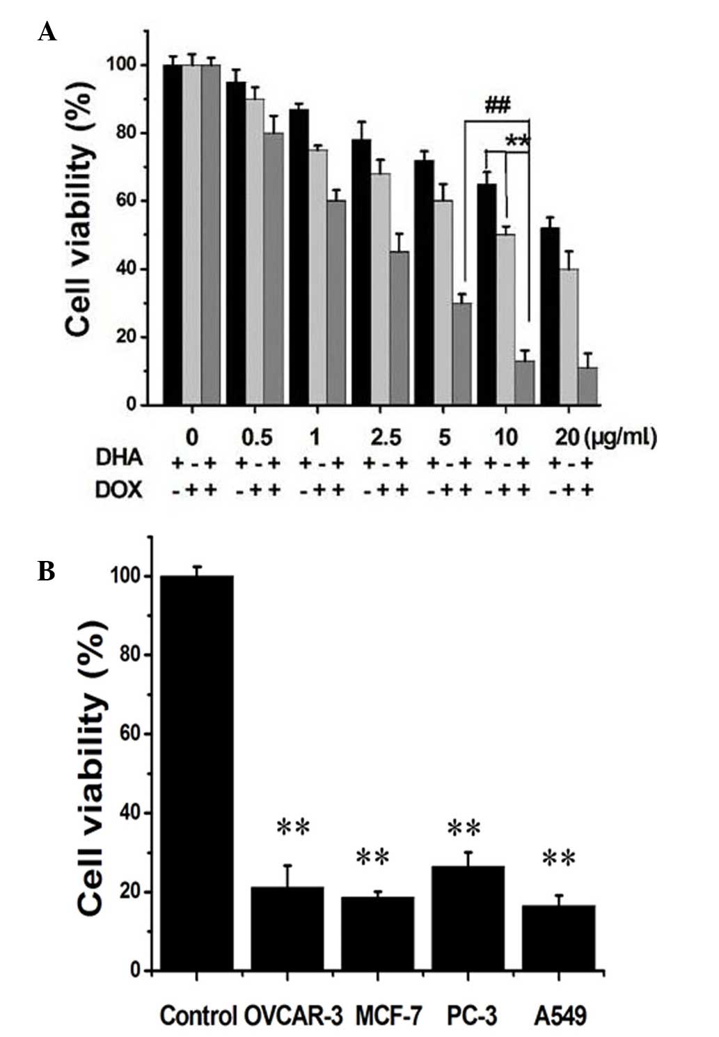
CCK-8 is commonly used to detect cell viability. As
shown in Fig. 1A, the viability of
the HeLa cells had a tendency to decrease as the concentrations of
DHA and DOX increased. After the cells were treated with DHA alone
at 0.5, 1, 2.5, 5, 10 and 20 µg/ml for 24 h, the viability ranged
between 45 and 95%, and after treatment with DOX alone at 0.5, 1,
2.5, 5, 10 and 20 µg/ml for 24 h, the viability ranged between 42
and 94%. After treatment with DHA + DOX, the cell viability
decreased up to 90%. The combination of 10 µg/ml DHA and 10 µg/ml
DOX resulted in a decrease of 89% in cell viability, which was
significantly different from that caused by the combination of 5
µg/ml DHA and 5 µg/ml DOX (P=0.008; Fig.
1), and was comparable with that caused by the combination of
20 µg/ml DHA and 20 µg/ml DOX (P=0.643). Therefore 10 µg/ml DHA
plus 10 µg/ml DOX (DHA + DOX) was the optimal concentration
combination. In Fig. 1B, combination
of the drugs at the optimal concentrations was used to treat the
OVCAR-3, MCF-7, PC-3 and A549 cells for 24 h, and a good inhibitory
effect was shown for these tumor cells, with statistically
significant differences compared with the control group (P=0.002,
0.001, 0.003 and 0.001, respectively).
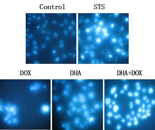
Pattern of DHA + DOX-induced cell
death
In order to verify the pattern of cell death induced
by the combination of the drugs, Hoechst 33258 staining was used
first to observe the morphological changes in the cell nuclei.
Under confocal fluorescence microscope, it was observed that the
cells treated with DHA + DOX and DHA or DOX alone showed marked
apoptosis, with chromatin agglutination and karyopyknosis (Fig. 2). In the blank control group, the cell
fluorescence was relatively superficial and uniform, and the
structure of the cell nuclei was normal. When compared with the
cells treated with the apoptosis-positive control reagent, STS, the
results shown with the combination of the drugs was similar to
those of the positive control. These results indicated that DHA +
DOX may have induced a type of programmed cell death, which may
have been apoptosis induced by the synergistic action of DHA and
DOX.
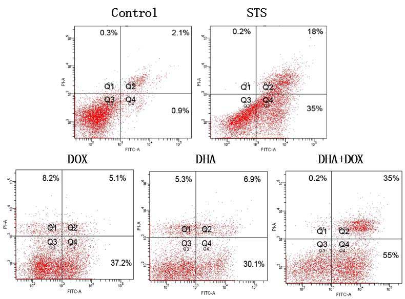
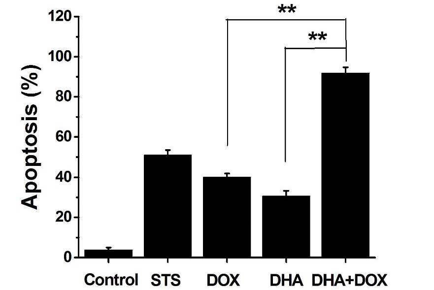
In order to further confirm that the cell death
induced by DHA + DOX was apoptosis, Annexin V/PI double staining
was used to detect the extroversion of phosphatidyl serine on the
cell membrane and the PI-specific stained cell nuclei after rupture
of the membrane, indicating apoptosis (11,12). As
shown in Fig. 3, after treatment with
DHA + DOX, the percentage of apoptotic cells (often considered to
be the total of cells of Q2 and Q4) increased from 3% (control) to
90% (24 h), but the percentage of necrotic cells increased
inconspicuously. The positive control indicated that Q2 + Q4 did
represent apoptosis. Among the cells treated with DHA or DOX alone,
there were also apoptotic cells, but the apoptotic rate was
significantly lower than that of the combination group (P=0.005 and
0.006, respectively; Fig. 4). These
results indicated that the DHA-induced cell death was
apoptosis.
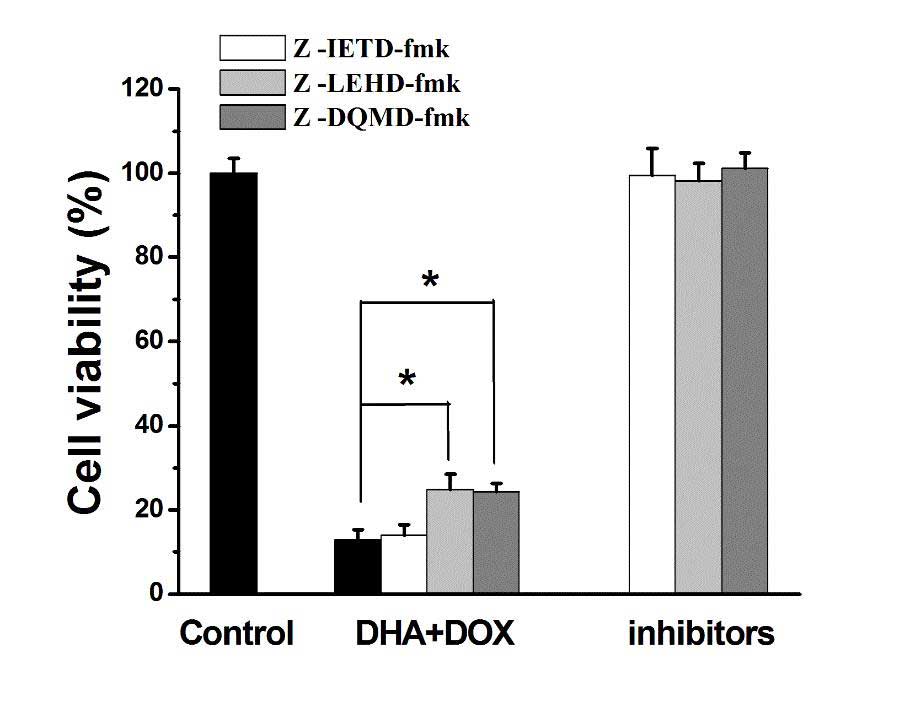
Intrinsic and extrinsic pathways
through which DHA + DOX causes cell apoptosis
The specific inhibitors of caspase-3, −8 and −9,
namely, z-DQMD-fmk, z-IETD-fmk and z-LEHD-fmk, respectively, were
used to determine whether caspase-3, −8 or −9 were involved in the
induction of cell apoptosis by DHA + DOX. Caspase-8 is an essential
apoptotic factor in the extrinsic apoptotic pathway, and caspase-3
and −9 are factors in the intrinsic apoptotic pathway (13,14). As
shown in Fig. 5, unlike the caspase-8
inhibitor z-IETD-fmk, treatment with z-DQMD-fmk and z-LEHD-fmk
increased the viability of the cells treated with DHA + DOX
(P=0.036 and 0.039, respectively; Fig.
5), suggesting that caspase-3 and −9 were involved in the
induction of cell apoptosis by DHA + DOX. Based on this, it was
speculated that DHA + DOX acted through the intrinsic apoptotic
pathway.
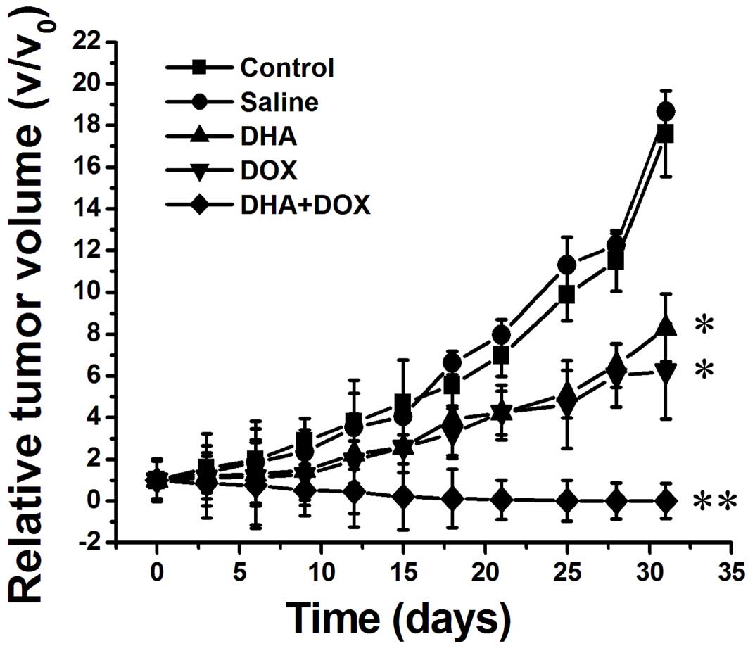
In vivo treatment effect of DHA +
DOX
The heterologous HeLa tumor model was established to
investigate whether DHA + DOX had the effect of tumor elimination
in vivo. DHA + DOX (15 mg/kg) was administered by
intratumoral injection, and the treatment effect was evaluated
through the measurement of tumor volume. As shown in Fig. 6, there was a significant inhibitory
effect after 6 days of intratumoral injections with DHA + DOX
(n=7). After injection of DHA or DOX alone, the tumor size was
significantly inhibited, as compared with the control (P=0.042 and
0.043, respectively) and saline groups (P=0.040 and 0.042,
respectively), although tumor size was inhibited to the greatest
extent in the DHA + DOX group (P=0.031, 0.033, 0.035 and 0.036 vs.
the control, saline, DHA and DOX groups, respectively).
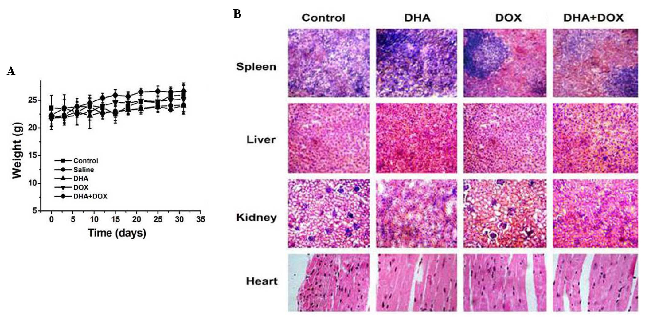
In vivo toxicity detection after
intratumoral injection of DHA + DOX
The in vivo toxicity of drugs has always been
a clinical concern. In the present study, it was determined through
monitoring changes in the weight of the mice after drug
administration and through pathological analysis of the sections of
vital organs after 30 days of administration. As shown in Fig. 7A, compared with the control group,
after injection of DHA or DOX alone, or DHA + DOX, the weight of
the mice did not significantly decrease, but was actually
increasing slowly. The HE staining images (Fig. 7B) showed that after intratumoral
injection in all treatment groups, no toxicity or side effects were
observed in the liver, kidneys, spleen and heart.
Discussion
DHA, extracted from goldenrod Artemisia annua
in China for the first time, is a novel first-line anti-malarial
drug that has been also been revealed to exhibit antitumor effects
(15–17). Park et al (18) found that DHA exhibited no clear
cytotoxicity on normal human breast cells, but that it exhibited
significant cytotoxicity on human breast cancer cells. DOX is a
wide-spectrum antitumor antibiotic and a cell cyclic non-specific
agent that is mostly combined with other anticancer drugs (19). In the present study, four experiments
were designed to investigate the lethal effect of DHA + DOX on
human cervical cancer cells (HeLa) and the corresponding mechanism.
The first investigation studied the effect of DHA + DOX at
different concentrations on the viability of the HeLa cells to
determine the optimal concentrations and synergistic lethal effect
of the combination. The lethal effect of DHA + DOX at optimal
concentrations on the OVCAR-3, MCF-7, PC-3 and A549 cells was also
assessed. These results indicated that, as compared with either
drug alone, combined DHA and DOX treatment showed an enhanced
lethal effect on HeLa cells. Furthermore, this combination
inhibited the viability of OVCAR-3, MCF-7, PC-3 and A549 cells.
Apoptosis has important biological significance in
the development and progression of tumors, and numerous
chemotherapy drugs exert an antitumor effect by inducing the
apoptosis of tumor cells. A previous study revealed that DHA could
induce apoptosis through the intrinsic pathway (8). The second experiment in the present
study investigated whether DHA + DOX also induced tumor cell
apoptosis. The nuclei-specific Hoechst 33258 dye was used to check
the changes in cell nuclei after administration of DHA + DOX, and
flow cytometry was employed to determine the mode of cell death.
The results indicated that after administration of DHA + DOX,
karyopyknosis of the HeLa cells occurred, inducing apoptosis, with
an apoptotic rate of 90%. This indicated that the administration of
DHA + DOX synergistically improved the rate of apoptosis in the
cells.
Caspase proteins are the primary factor that induces
changes in the intracellular biochemical and morphological
properties, with caspase-8 as a typical apoptotic factor in the
extrinsic pathway, and caspase-3 and −9 as typical apoptotic
factors in the intrinsic pathway (20). The third experiment in the present
study used specific inhibitors of caspase-3, −8 and −9 to detect
whether these caspases were involved in the apoptosis induced by
the administration of DHA + DOX. The results indicated that
caspase-8 was not involved in the apoptosis, but that caspase-3 and
−9 were involved in the apoptosis of the HeLa cells, suggesting
that the administration of DHA + DOX induced apoptosis through the
intrinsic pathway.
The present study also investigated the in
vivo treatment effect of DHA + DOX on mice with HeLa tumors.
Intratumoral injection is increasingly being applied in the
treatment of tumors (21). In the
present study, intratumoral injection was employed to treat the
tumor-bearing mice, and it was observed that the tumor elimination
effect of the combined medication was significantly better than
that of separate administration. The results of the in vivo
experiment indicated that combined medication exhibited no clear
toxicity in the mice. Previous studies have demonstrated a high
toxicity for DOX at high concentrations in the hearts of mice.
However, the present study showed that DHA + DOX could not only
reduce drug toxicity shortly after administration, but that it
could also considerably increase the treatment effect of the
drugs.
In conclusion, DHA + DOX exhibits a significant
synergistic inhibitory effect against a variety of tumor cell
lines. The present study showed that, in Hela cells, the lethal
mechanism behind the combined DHA + DOX medication was apoptosis
induced by the intrinsic pathway, with involvement of caspase-3 and
−9. The in vivo tumor elimination effect of the combined
medication was also significant. The results of this study provide
a novel idea for clinical chemotherapy and the experimental
research into multiple cancers, including human cervical
cancer.
References
|
1
|
Parkin DM, Bray F, Ferlay J and Pisani P:
Estimating the world cancer burden: Globocan 2000. Int J Cancer.
94:153–156. 2001. View
Article : Google Scholar : PubMed/NCBI
|
|
2
|
Chou TC: Theoretical basis, experimental
design, and computerized simulation of synergism and antagonism in
drug combination studies. Pharmacol Rev. 58:621–681. 2006.
View Article : Google Scholar : PubMed/NCBI
|
|
3
|
Roth AD, Maibach R, Martinelli G, Fazio N,
Aapro MS, Pagani O, Morant R, Borner MM, Herrmann R, Honegger H, et
al: Docetaxel (Taxotere)-cisplatin (TC): An effective drug
combination in gastric carcinoma. Swiss group for clinical cancer
research (SAKK), and the European institute of oncology (EIO). Ann
Oncol. 11:301–306. 2000. View Article : Google Scholar : PubMed/NCBI
|
|
4
|
Martello LA, McDaid HM, Regl DL, Yang CP,
Meng D, Pettus TR, Kaufman MD, Arimoto H, Danishefsky SJ, Smith AB
III and Horwitz SB: Taxol and discodermolide represent a
synergistic drug combination in human carcinoma cell lines. Clin
Cancer Res. 6:1978–1987. 2000.PubMed/NCBI
|
|
5
|
Huang FY, Lee TW, Chang CH, Chen LC, Hsu
WH, Chang CW and Lo JM: Evaluation of (188) Re-labeled PEGylated
nanoliposome as a radionuclide therapeutic agent in an orthotopic
glioma-bearing rat model. Int J Nanomedicine. 10:463–473. 2015.
View Article : Google Scholar : PubMed/NCBI
|
|
6
|
Karavitis J and Zhang M: COX2 regulation
of breast cancer bone metastasis. Oncoimmunology. 2:e231292013.
View Article : Google Scholar : PubMed/NCBI
|
|
7
|
Haynes RK: Artemisinin and derivatives:
The future for malaria treatment. Curr Opin Infect Dis. 14:719–726.
2001. View Article : Google Scholar : PubMed/NCBI
|
|
8
|
Guo Y, Shi X and Li L: Effects of
dihydroartemisinin on ovarian cancer HO-8910 cells and expression
of Akt. Zhong Guo Yi Xue Qian Yan Za Zhi. 7:17–18. 2012.
|
|
9
|
Howe LR: Inflammation and breast cancer.
Cyclooxygenase/prostaglandin signaling and breast cancer. Breast
Cancer Res. 9:2102007. View
Article : Google Scholar : PubMed/NCBI
|
|
10
|
Farh KK, Grimson A, Jan C, Lewis BP,
Johnston WK, Lim LP, Burge CB and Bartel DP: The widespread impact
of mammalian MicroRNAs on mRNA repression and evolution. Science.
310:1817–1821. 2005. View Article : Google Scholar : PubMed/NCBI
|
|
11
|
Schutte B, Nuydens R, Geerts H and
Ramaekers F: Annexin V binding assay as a tool to measure apoptosis
in differentiated neuronal cells. J Neurosci Methods. 86:63–69.
1998. View Article : Google Scholar : PubMed/NCBI
|
|
12
|
Cheung JY, Ong RC, Suen YK, Ooi V, Wong
HN, Mak TC, Fung KP, Yu B and Kong SK: Polyphyllin D is a potent
apoptosis inducer in drug-resistant HepG2 cells. Cancer Lett.
217:203–211. 2005. View Article : Google Scholar : PubMed/NCBI
|
|
13
|
Zhang JM, Wang HC, Wang HX, Ruan LH, Zhang
YM, Li JT, Tian S and Zhang YC: Oxidative stress and activities of
caspase-8, −9, and-3 are involved in cryopreservation-induced
apoptosis in granulosa cells. Eur J Obstet Gynecol Reprod Biol.
166:52–55. 2013. View Article : Google Scholar : PubMed/NCBI
|
|
14
|
Hsiao PC, Lee WJ, Yang SF, Tan P, Chen HY,
Lee LM, Chang JL, Lai GM, Chow JM and Chien MH: Nobiletin
suppresses the proliferation and induces apoptosis involving MAPKs
and caspase-8/-9/-3 signals in human acute myeloid leukemia cells.
Tumor Biol. 35:11903–11911. 2014. View Article : Google Scholar
|
|
15
|
Kong R, Jia G, Cheng ZX, Wang YW, Mu M,
Wang SJ, Pan SH, Gao Y, Jiang HC, Dong DL and Sun B:
Dihydroartemisinin enhances Apo2 L/TRAIL-mediated apoptosis in
pancreatic cancer cells via ROS-mediated up-regulation of death
receptor 5. PLoS One. 7:e372222012. View Article : Google Scholar : PubMed/NCBI
|
|
16
|
Sun Q, Teong B, Chen IF, Chang SJ, Gao J
and Kuo SM: Enhanced apoptotic effects of
dihydroartemisinin-aggregated gelatin and hyaluronan nanoparticles
on human lung cancer cells. J Biomed Mater Res B Appl Biomater.
102:455–462. 2014. View Article : Google Scholar : PubMed/NCBI
|
|
17
|
Posobiec LM, Clark RL, Bushdid PB, Laffan
SB, Wang KF and White TE: Dihydroartemisinin (DHA) treatment causes
an arrest of cell division and apoptosis in rat embryonic
erythroblasts in whole embryo culture. Birth Defects Res B Dev
Reprod Toxicol. 98:445–458. 2013. View Article : Google Scholar : PubMed/NCBI
|
|
18
|
Park J, Lai HC, Singh M, Sasaki T and
Singh NP: Development of a dihydroartemisinin-resistant Molt-4
leukemia cell line. Anticancer Res. 34:2807–2810. 2014.PubMed/NCBI
|
|
19
|
Wang Y, Xu L, Duan Z, Cao K, Luo J, Xu Y
and Shi P: Effects of ADM on cell apoptosis and expression of FAK
mRNA in K562. J Trop Med. 12:1195–1198. 2012.
|
|
20
|
Brentnall M, Rodriguez-Menocal L, De
Guevara RL, Cepero E and Boise LH: Caspase-9, caspase-3 and
caspase-7 have distinct roles during intrinsic apoptosis. BMC Cell
Biol. 14:322013. View Article : Google Scholar : PubMed/NCBI
|
|
21
|
Wan L and Zheng X: Expression pattern and
function of SATB1 in the invasiveness of thyroid carcimoma. Chin J
Biochem Pharm. 34:18–20. 2014.
|