Open Access

MicroRNA profile of hepatic epithelioid hemangioendothelioma: A case report

  • Authors:
    • Asahiro Morishita
    • Hisakazu Iwama
    • Hirohito Yoneyama
    • Teppei Sakamoto
    • Koji Fujita
    • Takako Nomura
    • Joji Tani
    • Hisaaki Miyoshi
    • Shintaro Fujihara
    • Noriko Nishiyama
    • Hideki Kobara
    • Hirohito Mori
    • Naoki Yamamoto
    • Keiichi Okano
    • Yasuyuki Suzuki
    • Emi Ibuki
    • Reiji Haba
    • Takashi Himoto
    • Tsutomu Masaki
  • View Affiliations

  • Published online on: January 25, 2017     https://doi.org/10.3892/ol.2017.5643
  • Pages: 1655-1659
  • Copyright: © Morishita et al. This is an open access article distributed under the terms of Creative Commons Attribution License.

Metrics: Total Views: 0 (Spandidos Publications: | PMC Statistics: )
Total PDF Downloads: 0 (Spandidos Publications: | PMC Statistics: )


Abstract

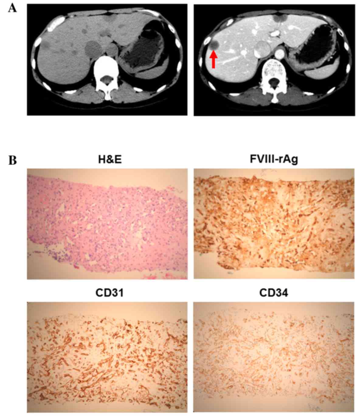
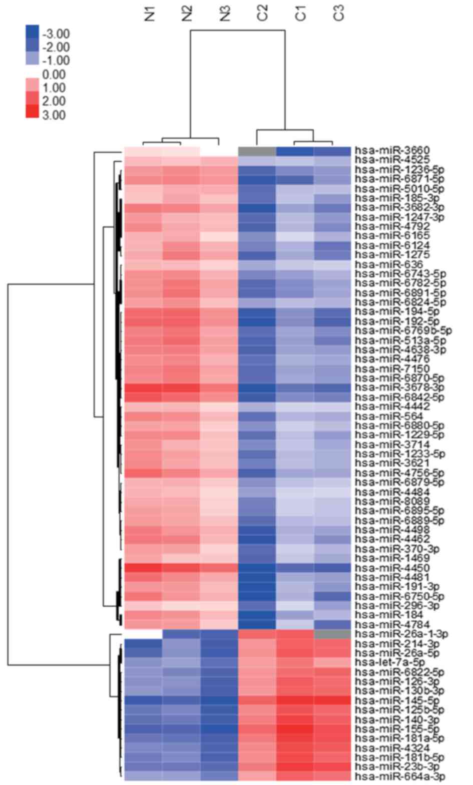
A 72-year-old female was referred for further evaluation of epigastralgia. Abdominal contrast computed tomography revealed numerous tumors in the two lobes of the liver. Liver biopsy and immunohistochemical staining revealed that the tumor cells were positive for factor VIII‑associated antigen, platelet endothelial cell adhesion molecule 1 and human hematopoietic progenitor cell antigen, concordant with a diagnosis of hepatic epithelioid hemangioendothelioma (HEH). To elucidate the etiology of HEH, particularly the microRNA (miRNA) profiles, tissue samples obtained from normal and tumor tissues were analyzed using a miRNA array system. A total of 14 miRNAs were significantly upregulated and 93 miRNAs were downregulated in the tumor tissues (P<0.01). Additionally, unsupervised hierarchical clustering analysis using Pearson's correlation revealed that the tumor tissues clustered separately from the normal tissues. The miRNA expression profile was analyzed in HEH and compared with angiosarcoma, which exhibits histology similar to HEH. Out of a total of 107 miRNAs, only miR‑122‑5p and miR‑1290 demonstrated a differential expression pattern in angiosarcoma. Therefore, these miRNAs may be novel biological markers for the determination of a diagnosis of HEH in primary mesenchymal tumors of the liver. To the best of our knowledge, this study is the first report of a miRNA microarray analysis in HEH.
View Figures
View References

Related Articles

Journal Cover

March-2017
Volume 13 Issue 3

Print ISSN: 1792-1074
Online ISSN:1792-1082

Sign up for eToc alerts

Recommend to Library

Copy and paste a formatted citation
x
Spandidos Publications style
Morishita A, Iwama H, Yoneyama H, Sakamoto T, Fujita K, Nomura T, Tani J, Miyoshi H, Fujihara S, Nishiyama N, Nishiyama N, et al: MicroRNA profile of hepatic epithelioid hemangioendothelioma: A case report. Oncol Lett 13: 1655-1659, 2017
APA
Morishita, A., Iwama, H., Yoneyama, H., Sakamoto, T., Fujita, K., Nomura, T. ... Masaki, T. (2017). MicroRNA profile of hepatic epithelioid hemangioendothelioma: A case report. Oncology Letters, 13, 1655-1659. https://doi.org/10.3892/ol.2017.5643
MLA
Morishita, A., Iwama, H., Yoneyama, H., Sakamoto, T., Fujita, K., Nomura, T., Tani, J., Miyoshi, H., Fujihara, S., Nishiyama, N., Kobara, H., Mori, H., Yamamoto, N., Okano, K., Suzuki, Y., Ibuki, E., Haba, R., Himoto, T., Masaki, T."MicroRNA profile of hepatic epithelioid hemangioendothelioma: A case report". Oncology Letters 13.3 (2017): 1655-1659.
Chicago
Morishita, A., Iwama, H., Yoneyama, H., Sakamoto, T., Fujita, K., Nomura, T., Tani, J., Miyoshi, H., Fujihara, S., Nishiyama, N., Kobara, H., Mori, H., Yamamoto, N., Okano, K., Suzuki, Y., Ibuki, E., Haba, R., Himoto, T., Masaki, T."MicroRNA profile of hepatic epithelioid hemangioendothelioma: A case report". Oncology Letters 13, no. 3 (2017): 1655-1659. https://doi.org/10.3892/ol.2017.5643