Introduction
Colorectal cancer is a common type of malignant
tumor of the digestive tract. In 2012, the morbidity rates of
colorectal cancer in developed countries ranked third and second in
the morbidity rates of malignant tumors in males and females,
respectively (1). During this time,
the morbidity rates of colorectal cancer also increased markedly in
China. In 2013, the morbidity rates of colorectal cancer ranked
fifth amongst the morbidity rates of all types of malignant tumors
in China, whilst in the urban population of China, the morbidity
rates of colorectal cancer were ranked second amongst the morbidity
rates of all types of malignant tumors (2). As surgery is the most effective
therapeutic method for colorectal cancer up to date, the primary
treatment of colorectal cancer is surgery, supplemented by
radiotherapy and chemotherapy. At present, along with the continued
investigation of the pathogenesis of colorectal cancer,
immunotherapy of colorectal cancer has become more acceptable and
has a potentially wide application.
In 1909 Ehrlich first proposed the concept of immune
surveillance (3). In 1959, Thomas
proposed that a low expression of the tumor cell antigen or damaged
cellular immune function was an important factor of tumorigenesis
(3). Subsequently, Burnet developed
the immune surveillance theory, which hypothesized that the immune
system may perform surveillance, recognizing and eliminating novel
antigens expressed, alien components or mutated cells, to maintain
the stability of the internal environment (3). Although the immune system may generate
an immune response to tumor cells and eliminate the tumor, there
remains a certain proportion of primary tumors that may metastasize
and recur. Thus, certain types of tumor may escape the immune
system, which is termed tumor immune evasion. Previous studies have
indicated that the immune evasion mechanism of colorectal cancer
cells may be separated into 3 major categories: Low immunogenicity
of colorectal cancer cells, due to a loss or low expression of
classical human leukocyte antigen (HLA) molecules (4,5) and a loss
of apoptosis antigen 1 (Fas) expression (6,7);
dysfunction of the effector cells due to high expression levels of
the c-c chemokine receptor type 5 (CCR5) from tumor infiltrating
regulatory T-cells (8) and the
expression of immunosuppressive factors by colorectal cancer cells
due to high expression levels of the Fas ligand (FasL) (9–11); and HLA
class I histocompatibility antigen, α chain E (HLA-E) as a type of
nonclassical HLAImolecule (12,13).
At present, studies have revealed that the immune
evasion mechanisms of tumor cells in certain types of advanced
solid malignant tumors, and several types of immune preparations
have been applied as antitumor adjuvant therapy (3). However, in the majority of studies, the
efficacy is limited, which may be associated with the different
immune microenviroments between advanced and early cancers. Thus,
an investigation of the immune evasion mechanisms of early
colorectal cancer cells and a trial of an immune-based intervention
for early colorectal cancer may generate data to improve the
efficacy of immunotherapy in this type of cancer.
To date, previous studies on the immune evasion
mechanisms of early colorectal cancer have not been performed. The
present study aimed to investigate the immune evasion mechanisms in
early colorectal cancer cells and the expression of immune
evasion-associated molecules during the malignant transformation
process of normal colorectal epithelial cells.
Materials and methods
Tissue samples
All tissue specimens were collected between June
2014 and June 2015. A total of 45 colorectal specimens, including
15 adenoma, 15 early cancer and 15 advanced cancer tissues, from
patients undergoing endoscopic procedures such as endoscopic
submucosal dissection, endoscopic mucosal resection, polypectomy
and tumor biopsy in the Huadong Hospital Affiliated to Fudan
University (Shanghai, China) were collected randomly. A total of 15
adenoma tissues were tubulovillous adenomas, as confirmed by
pathology. Amongst the 15 patients with early cancer tissues, 13
were high grade intraepithelial neoplasia and 2 were intramucosal
cancer. In the patients with advanced cancer, 2 were Dukes A stage,
4 were Dukes B stage, 5 were Dukes C1 stage and 4 were Dukes D
stage (14). For a control group, 15
normal colorectal mucosa specimens from 15 consecutive healthy
physical examination persons were collected by colonoscopic biopsy.
All 60 specimens were divided into four groups according to the
type of tissue. All patients and controls enrolled provided
informed consent. The present study was approved by Ethics
Committee of Huadong Hospital (Shanghai, China; trial registration
no. ChiCTR-OOC-15007390).
Immunohistochemical analysis
The formalin-fixed and paraffin-embedded tissue
specimens were assessed by immunohistochemical analysis using the
DouSPTM IHC kit (Fuzhou Maixin Biotech Co., Ltd., Fuzhou, China),
according to the protocol of the manufacturer. Anti-HLA-A (cat. no.
15240-1-AP) and anti-Fas (cat. no. 13098-1-AP) antibodies were
purchased from Wuhan Sanying Biotechnology (Wuhan, China).
Anti-CCR5 (cat. no. ab110103) antibody was purchased from Abcam
(Cambridge, MA, USA). Anti-FasL (cat. no. ARG 53012) and anti-HLA-E
(cat. no. ARG 63017) antibodies were purchased from Arigo (Taiwan,
China). Specimens were incubated with the primary antibody at 4°C
overnight. The secondary antibodies (cat. no. 7074) were purchased
from Cell Signaling Technology (Danvers, MA, USA). Specimens were
incubated with the secondary antibody at room temperature for 2 h.
The secondary antibodies were labeled and visualized using a color
solution (Tiangen Biotech Co., Ltd., Beijing, China). All the
dilutions were 1:100. The expression levels of all the proteins
were determined using a semiquantitative method as previously
described (11) and assessed as
strong positive, positive-staining cells ≥50%; weak positive,
positive-staining cells ≥25 but <50%, and negative, lack of
staining or positive-staining cells <25%. Positive-staining
reactions were measured under a light microscope using ×400
magnification.
Western blot analysis
The expression levels of HLA-A, Fas, CCR5, FasL and
HLA-E in each group were detected by western blot analysis. As
early colorectal cancer lesions were often small and the majority
of such lesions generated from adenoma, the present study did not
obtain fresh colorectal adenoma and early cancer tissues for
western blot analysis to avoiding ethical issues.
A total of 15 patients with advanced colorectal
cancer were enrolled into the present study. Advanced cancer and
paracancer tissues (normal mucosal tissues 3 cm away from the
margin of cancer tissues) were collected from each patient by
colonoscopic biopsy. The specimens were divided into two groups
according to the type of tissue. In the 15 patients with advanced
colorectal cancer, 3 were Dukes A stage, 5 were Dukes B stage, 4
were Dukes C1 stage and 3 were Dukes C2 stage. By location, 4 were
in the ascending colon, 1 was in the transverse colon, 1 was in the
descending colon, 5 were in the sigmoid and 4 were in rectum.
Tissue samples from the patients were extracted
using loading buffer (0.05 M Tris, 10% glycerol, 4%
β-mercaptoethanol, 2% SDS, 0.1% bromophenol blue, pH 6.8) with 1 mM
phenylmethylsulfonyl fluoride (Beyotime Institute of Biotechnology,
Haimen, China), separated by SDS-PAGE (20 µg protein per lane) on a
12% gel and transferred to polyvinylidene fluoride membranes. The
Tanon-5200 Chemi-luminescent Imaging System (Tanon Science and
Technology Co., Ltd., Shanghai, China) was used to observe the
levels of protein expression. The primary antibodies and secondary
antibodies were used as described in the aforementioned
immunohistochemical analysis protocol.
Statistical analysis
All analyses were performed using Statistical
Analysis System 9.13 statistical software package (SAS Institute,
Inc., Cary, NA, USA). The normal distribution of the measurement
data was evaluated by unpaired Student's t-test or analysis of
variance. The enumeration data was evaluated by a χ2
test, and Fisher exact test was used if the frequency was low.
P<0.05 was considered to indicate a statistically significant
difference.
Results
Baseline characteristics
The gender, age and sample location of patients
between the four groups were compared and presented in Table I.
 | Table I.Baseline characteristics of patients
with colorectal cancer. |
Table I.
Baseline characteristics of patients
with colorectal cancer.
|
| Value |
|
|---|
|
|
|
|
|---|
| Characteristic | Normal mucosa
(n=15) | Adenoma (n=15) | Early cancer
(n=15) | Advanced cancer
(n=15) | P-value |
|---|
| Gender
(male/female) | 9/6 | 7/8 | 11/4 | 10/5 | 0.481 |
| Age, years | 58.82±6.82 | 63.47±6.38 | 62.20±9.06 | 68.54±11.53 | 0.060 |
| Location |
|
|
|
| 0.081 |
| Ascending
colon | 3 | 8 | 2 | 2 |
|
|
Transverse colon | 4 | 1 | 1 | 0 |
|
|
Descending colon | 2 | 0 | 3 | 1 |
|
|
Sigmoid | 3 | 4 | 5 | 4 |
|
|
Rectum | 3 | 2 | 4 | 8 |
|
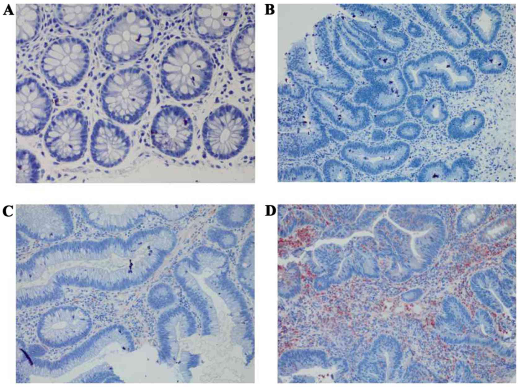
Immunohistochemical analysis
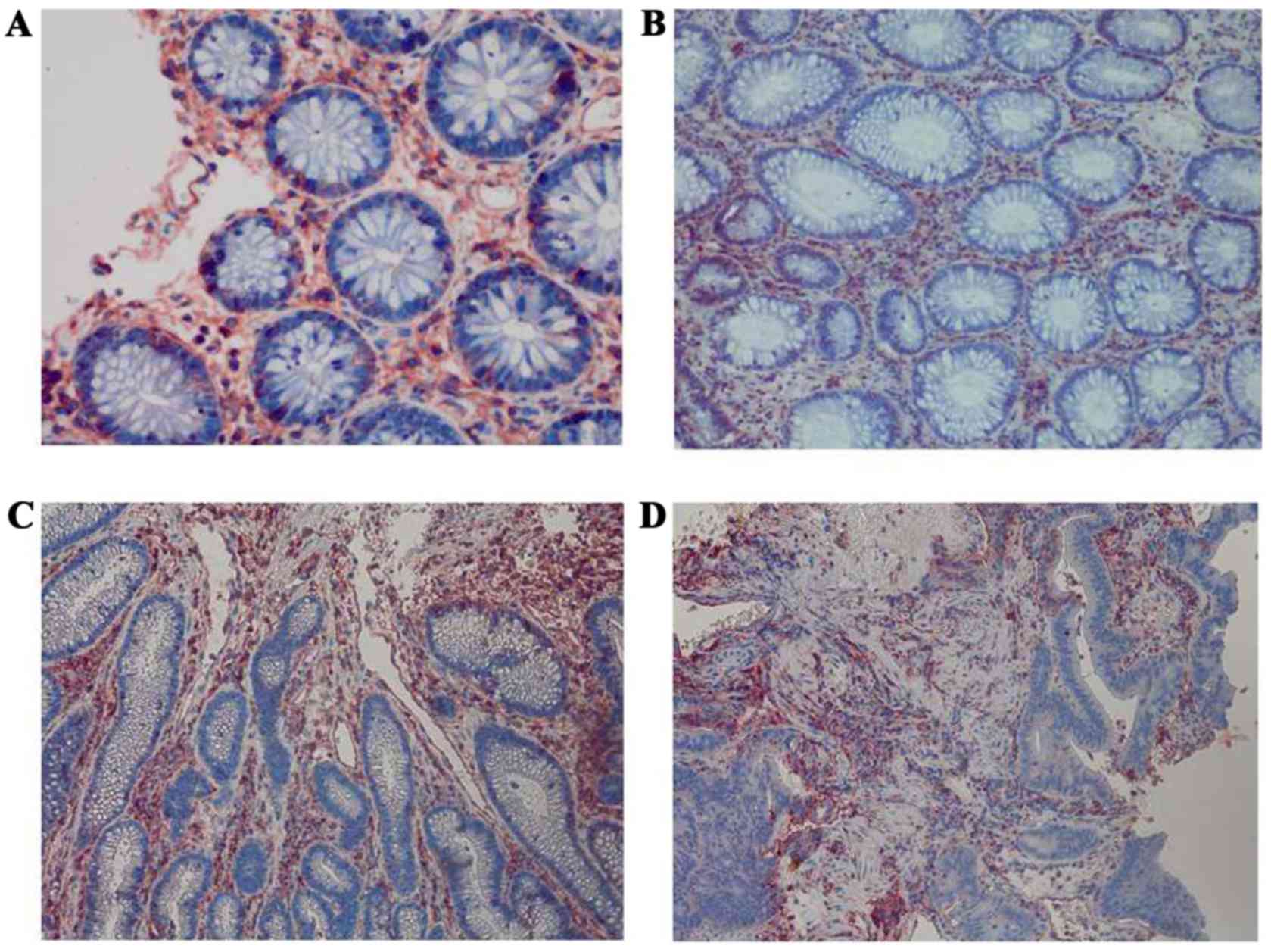
The expression levels of HLA-A, Fas, CCR5, FasL and
HLA-E were analyzed between the four groups: Normal mucosa,
adenoma, early cancer group and advanced cancer groups. It was
observed that the expression levels of HLA-A demonstrated no
significant difference between cancer stages during the malignant
transformation process of normal colorectal epithelial cells
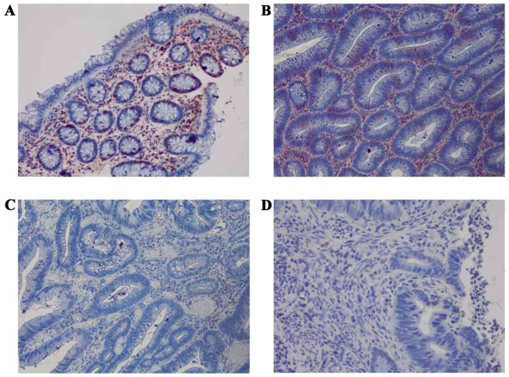
(Fig. 1; Table II). The expression levels of Fas
reduced significantly along with an increased malignancy of the
epithelial cells (P=0.0271). In addition, the expression of Fas
reduced significantly in the early cancer group (P=0.0239) compared
with adenoma group, as demonstrated in Fig. 2 and Table
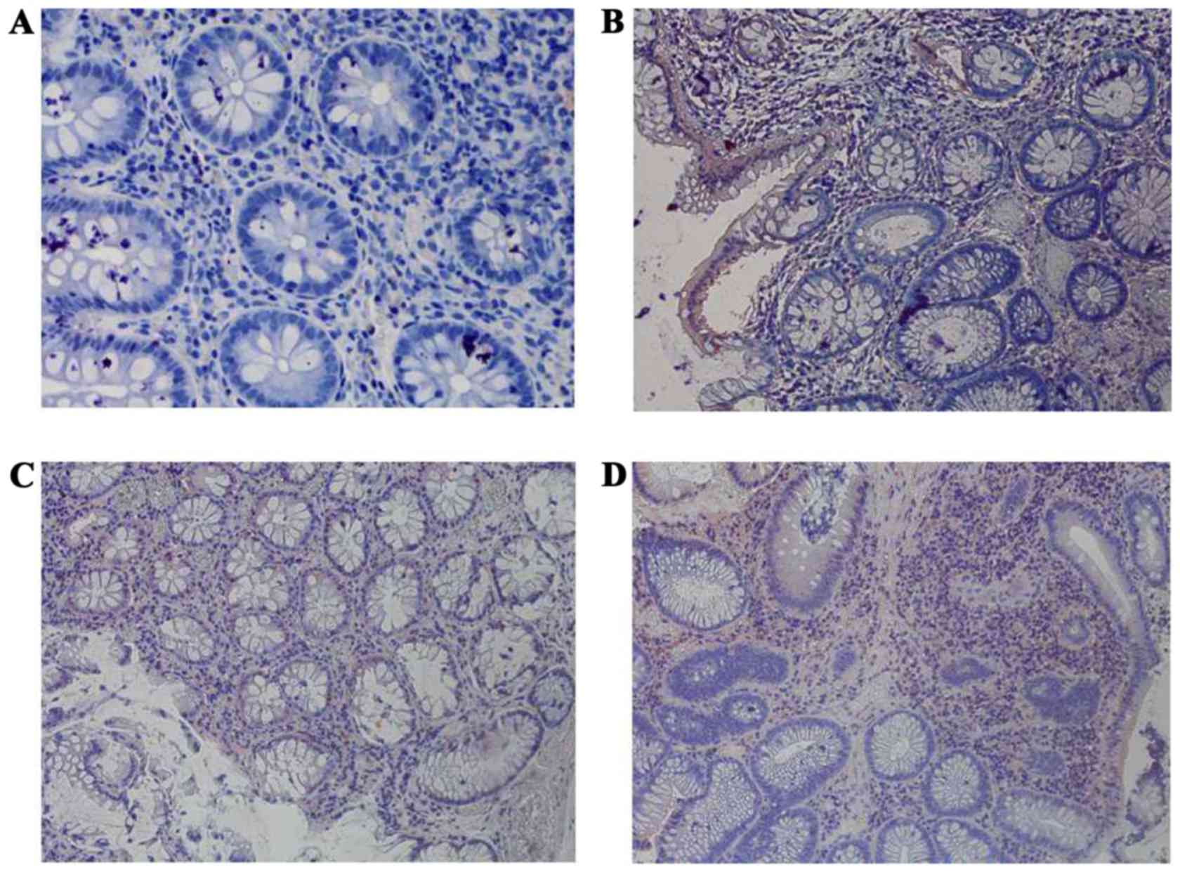
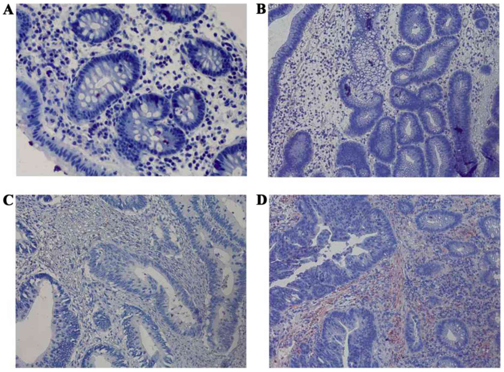
II. The expression of CCR5 and FasL increased significantly
with increased malignancy of the epithelial cells (P<0.001).
However, no significant difference in the expression levels of CCR5
and FasL was observed between the early cancer and adenoma groups
(P>0.05), as demonstrated in Figs.
3 and 4, and Table II. The expression of HLA-E increased
significantly as the malignancy of the epithelial cells increased
(P<0.001). In addition, the expression levels of HLA-E increased
significantly in the early cancer group (P<0.001) compared with
adenoma group, as illustrated in Fig.
5 and Table II.
| Table II.Immunohistochemical analysis: The
expression of HLA-A, Fas, CCR5, FasL and HLA-E among the four
groups. |
Table II.
Immunohistochemical analysis: The
expression of HLA-A, Fas, CCR5, FasL and HLA-E among the four
groups.
|
| No. of patients |
|
|
|---|
|
|
|
|
|
|---|
| Protein
expression | Normal mucosa
(n=15) | Adenoma (n=15) | Early cancer
(n=15) | Advanced cancer
(n=15) | P-value | P'-value |
|---|
| HLA-A |
|
|
|
|
0.460 |
0.682 |
|
Negative | 0 | 0 | 2 | 1 |
|
|
|
Weak | 2 | 4 | 3 | 1 |
|
|
|
Strong | 13 | 11 | 10 | 13 |
|
|
| Fas |
|
|
|
|
0.0271 |
0.0239 |
|
Negative | 3 | 1 | 8 | 8 |
|
|
|
Weak | 4 | 7 | 3 | 5 |
|
|
|
Strong | 8 | 7 | 4 | 2 |
|
|
| CCR5 |
|
|
|
| <0.001 |
0.176 |
|
Negative | 14 | 7 | 2 | 2 |
|
|
|
Weak | 1 | 3 | 5 | 5 |
|
|
|
Strong | 0 | 5 | 8 | 8 |
|
|
| FasL |
|
|
|
| <0.001 |
0.605 |
|
Negative | 15 | 10 | 8 | 4 |
|
|
|
Weak | 0 | 5 | 5 | 8 |
|
|
|
Strong | 0 | 0 | 2 | 3 |
|
|
| HLA-E |
|
|
|
| <0.001 | <0.001 |
|
Negative | 15 | 15 | 5 | 3 |
|
|
|
Weak | 0 | 0 | 9 | 6 |
|
|
|
Strong | 0 | 0 | 1 | 6 |
|
|
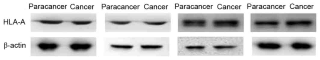
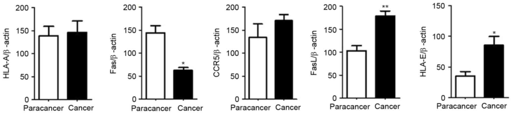
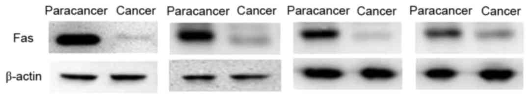
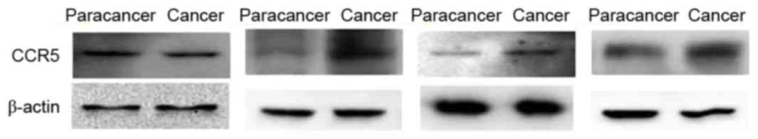
Western blot analysis
The expression of HLA-A, Fas, CCR5, FasL and HLA-E
in the advanced colorectal cancer and paracancer tissues were
analyzed. The results of the western blot analysis were consistent
with the results of the immunohistochemical analysis. No
significant difference was observed in the expression levels of
HLA-A between the advanced colorectal cancer and paracancer groups,
as demonstrated in Figs. 6 and
11, the expression of Fas reduced
significantly in the advanced colorectal cancer group compared with
paracancer group, as illustrated in Figs.
7 and 11, whilst the expression
levels of CCR5, FasL and HLA-E increased significantly in the
advanced colorectal cancer group compared with paracancer group, as
demonstrated in Figs. 8–11.
Discussion
The immune system can perform surveillance,
recognizing and eliminating newly-expressed antigens, alien
components or mutated cells. However, some tumors may escape attack
from immune system, which is termed immune evasion. Previous
studies have reported that the immune evasion mechanisms of
colorectal cancer cells may include a low immunogenicity of
colorectal cancer cells, dysfunction of effector cells and the
expression of immunosuppressive factors by colorectal cancer cells
(4,6,8,11,12). The
present study aimed to investigate the immune evasion mechanisms in
early colorectal cancer cells and the expression of immune
evasion-associated molecules during the malignant transformation
process of normal colorectal epithelial cells, to measure the
effects of immune intervention for early colorectal cancer and to
generate data to improve the efficacy of immunotherapy for this
type of cancer.
The present study indicated that HLA-A was highly
expressed in normal colorectal mucosa, colorectal adenoma, early
colorectal cancer and advanced colorectal cancer tissues, and that
no significant difference was observed between the 4 groups
(P>0.05). According to previous studies, the association between
the expression of HLA class I molecules and the immune evasion of
colorectal cancer cells remains controversial. Hypothetically,
tumor antigens may be presented on the cell surface by HLA class I
molecules and be recognized by cytotoxic T lymphocytes (CTL), which
may kill tumor cells (3). Conversely,
if the expression of HLA class I molecules is lost in tumor cells,
tumor cells may be destroyed by natural killer (NK) cell-mediated
cytotoxicity. Bernal et al (4)
hypothesized that β2-microglobulin is an important component of
major histocompatibility complex (MHC) class I molecules. There is
evidence to demonstrate that a loss of β2-microglobulin may
contribute to tumor immune evasion, particularly in colorectal
cancer and melanoma. An additional study demonstrated that damaging
the β2-microglobulin gene, located at chromosome 15, may result in
a loss of MHC class I molecule expression and tumor immune evasion
(4). As illustrated by Sandel et
al (15), MHC class I molecules
were not expressed in 72% of 88 colorectal cancer specimens, whilst
MHC class I molecules were expressed normally in 28% specimens.
However, NK cells were distributed sparsely in the epithelium of
the primary tumor, where CD8+T cells were distributed
densely instead, indicating that the immune microenviroment was
disordered in primary colorectal cancer. Conversely, Menon et
al (16) revealed that HLA-A was
expressed in up to 98% of 82 primary colorectal cancer specimens.
Other studies demonstrated that the downregulated expression of HLA
class I molecules was deleterious to the prognosis of patients with
colorectal cancer (5,17,18). A
possible explanation is that if the expression of HLA class I
molecules is completely eliminated in tumor cells, the tumor cells
may be destroyed by NK cell-mediated cytotoxicity, whilst a
downregulated expression of HLA class I antigens may protect tumor
cells from immune attack by T cells and NK cells (5,17,18). In summary, although studies
investigating the association between the expression of HLA class I
molecules and the immune evasion of colorectal cancer cells are
common, the conclusions are different. In the present study, HLA-A
was highly expressed in all 4 groups. Thus, investigating the
precise mechanism of HLA-A and immune evasion of colorectal cancer
cells may rely on additional studies of molecular transduction
mechanisms, with larger sample sizes.
Previous studies have demonstrated that Fas is
highly expressed in normal colorectal epithelial cells, whilst a
loss of Fas expression and anti-Fas-mediated apoptosis in
colorectal cancer cells contributed to tumor invasion and
metastasis (6,7,19). The
present study indicated that the expression of Fas reduced
significantly as the malignancy of the epithelial cells increased
(P=0.0271). Furthermore, the expression of Fas reduced
significantly in early cancer group (P'=0.0239) compared with
adenoma group, which suggested that a loss of Fas expression may
serve an important role in the immune evasion in early colorectal
cancer.
Chang et al (8)
revealed that CCR5 was highly expressed in tumor infiltrating
cluster of differentiation (CD)4+ forkhead box
(Fox)p3+ regulatory T cells. C-c chemokine ligand 5
(CCL5) molecules in colorectal cancer cells may bind with CCR5 and
initiate the CCL5/CCR5 signal pathway, which may recruit regulatory
T cells to the tumor and increase the rate of tumor-induced
anti-tumor CD8+T cell death. Thus, the immune escape of
colorectal cancer cells may occur through this pathway. In the
present study, the expression of CCR5 increased significantly with
increased malignancy of the epithelial cells (P<0.001), which
was consistent with the results of Chang et al (8). The expression of CCR5 was not
significantly different between the early cancer and adenoma groups
(P=0.176), which indicated that the expression of CCR5 was not
altered significantly in early colorectal cancer, and that CCR5 may
not be a major factor of the immune evasion in early colorectal
cancer.
Although FasL was expressed highly in active immune
cells such as CTL, studies have revealed that FasL is also
expressed in colorectal cancer cells (9,10). As
active CTL expressed Fas is sensitive to Fas-mediated apoptosis,
several studies have demonstrated that the expression of FasL in
tumor cells may induce the apoptosis of tumor infiltrating
lymphocytes (9,10,20).
Pryczynicz et al (11)
reported that FasL was highly expressed in 70% of patients with
colorectal cancer, and lowly expressed in 30% of patients with
colorectal cancer, whilst all normal colorectal epithelial cells
did not express FasL. Statistical analysis demonstrated that a high
expression of FasL was associated with tumor angiogenesis and
invasion. The present study suggested that the expression of FasL
increased significantly with increased malignancy of the epithelial
cells (P<0.001), which was consistent with the results of
aforementioned research. However, the expression of FasL was not
significantly different between the early cancer and adenoma groups
(P=0.605), which indicated that FasL may not be a major immune
evasion-associated molecule of early colorectal cancer.
HLA-E is a type of atypical HLA class I molecule.
Bossard et al (12) revealed
that the HLA-E expressed in colorectal cancer cells may combine
with the HLA-E receptor CD94/NKG2A expressed on the surface of CTL
and NK cells, which contributed to the inhibition of the activity
of the CTL and NK cells. Zhen et al (21) hypothesized that the overexpression of
HLA-E in colorectal cancer cells was often associated with lower
long-term disease-free survival rate and poor prognosis. Levy et
al (13) demonstrated that HLA-E
expression on the surface of colorectal cancer cell membranes could
inhibit the cetuximab-mediated antibody-dependent cell-mediated
cytotoxicity, which may promote the immune evasion of colorectal
cancer cells. In the present study, the expression of HLA-E
increased significantly as the malignancy of the epithelial cells
increased (P<0.001). In addition, the expression of HLA-E
increased significantly in the early cancer group (P<0.001)
compared with the adenoma group, which suggested that the
expression of HLA-E may serve a key role in the immune evasion in
early colorectal cancer.
The sample size of the present study was relatively
small and it was conducted in a single center. However, the immune
evasion mechanisms of early colorectal cancer cells have been
investigated.
The present study indicated that, the expression of
Fas reduced significantly, whilst the expression of HLA-E increased
significantly in early cancer group compared with adenoma group. It
suggested that a loss of Fas expression and a high expression level
of HLA-E may promote immune evasion in early colorectal cancer
cells, which has not yet been investigated extensively. Thus, on
the basis of the present study, additional studies with larger
sample sizes that investigate the precise mechanisms of immune
evasion in early colorectal cancer cells at the level of molecular
transduction are required.
Acknowledgements
The present study would like to thank the Youth
Scientific Research Project of Shanghai Health Bureau (grant no.
201344073). Furthermore, the present study acknowledges Professor
Xu Fuxing, Dr Ji Danian and Dr Zhou Jun for reviewing the draft
manuscript and providing feedback.
References
|
1
|
Torre LA, Bray F, Siegel RL, Ferlay J,
Lortet-Tieulent J and Jemal A: Global cancer statistics. CA Cancer
J Clin. 65:87–108. 2015. View Article : Google Scholar : PubMed/NCBI
|
|
2
|
Ren JS, Shi JF, Zhang HZ, Liu Q, Zhang YM,
Zou SM, Zhang K and Dai M: Preliminary analysis of the colorectal
cancer screening among urban populations in China, 2012–2013.
Zhonghua Yu Fang Yi Xue Za Zhi. 49:441–443. 2015.(In Chinese).
PubMed/NCBI
|
|
3
|
Ge HL: Tumor immunityPrinciples of
immunology. Zhou GY: Shanghai Scientific and Technical Publishers;
Shanghai: pp. 279–294. 2007
|
|
4
|
Bernal M, Ruiz-Cabello F, Concha A,
Paschen A and Garrido F: Implication of the β2-microglobulin gene
in the generation of tumor escape phenotypes. Cancer Immunol
Immunother. 61:1359–1371. 2012. View Article : Google Scholar : PubMed/NCBI
|
|
5
|
Kloor M, Michel S and von Knebel Doeberitz
M: Immune evasion of microsatellite unstable colorectal cancers.
Int J Cancer. 127:1001–1010. 2010. View Article : Google Scholar : PubMed/NCBI
|
|
6
|
Liu K: Role of apoptosis resistance in
immune evasion and metastasis of colorectal cancer. World J
Gastrointest Oncol. 2:399–406. 2010. View Article : Google Scholar : PubMed/NCBI
|
|
7
|
Sträter J, Hinz U, Hasel C, Bhanot U,
Mechtersheimer G, Lehnert T and Möller P: Impaired CD95 expression
predisposes for recurrence in curatively resected colon carcinoma:
Clinical evidence for immunoselection and CD95L mediated control of
minimal residual disease. Gut. 54:661–665. 2005. View Article : Google Scholar : PubMed/NCBI
|
|
8
|
Chang LY, Lin YC, Mahalingam J, Huang CT,
Chen TW, Kang CW, Peng HM, Chu YY, Chiang JM, Dutta A, et al:
Tumor-derived chemokine CCL5 enhances TGF-β-mediated killing of
CD8(+) T cells in colon cancer by T-regulatory cells. Cancer Res.
72:1092–1102. 2012. View Article : Google Scholar : PubMed/NCBI
|
|
9
|
Houston AM, Michael-Robinson JM, Walsh MD,
Cummings MC, Ryan AE, Lincoln D, Pandeya N, Jass JR, Radford-Smith
GL and O'Connell J: The ‘Fas counterattack’ is not an active mode
of tumor immune evasion in colorectal cancer with high-level
microsatellite instability. Hum Pathol. 39:243–250. 2008.
View Article : Google Scholar : PubMed/NCBI
|
|
10
|
Ryan AE, Shanahan F, O'Connell J and
Houston AM: Fas ligand promotes tumor immune evasion of colon
cancer in vivo. Cell Cycle. 5:246–249. 2006. View Article : Google Scholar : PubMed/NCBI
|
|
11
|
Pryczynicz A, Guzińska-Ustymowicz K and
Kemona A: Fas/FasL expression in colorectal cancer. An
immunohistochemical study. Folia Histochem Cytobiol. 48:425–429.
2010. View Article : Google Scholar : PubMed/NCBI
|
|
12
|
Bossard C, Bézieau S, Matysiak-Budnik T,
Volteau C, Laboisse CL, Jotereau F and Mosnier JF: HLA-E/β2
microglobulin overexpression in colorectal cancer is associated
with recruitment of inhibitory immune cells and tumor progression.
Int J Cance. 131:855–863. 2012. View Article : Google Scholar
|
|
13
|
Levy EM, Sycz G, Arriaga JM, Barrio MM,
von Euw EM, Morales SB, González M, Mordoh J and Bianchini M:
Cetuximab-mediated cellular cytotoxicity is inhibited by HLA-E
membrane expression in colon cancer cells. Innate Immun. 15:91–100.
2009. View Article : Google Scholar : PubMed/NCBI
|
|
14
|
Dukes CE: The surgical pathology of rectal
cancer: President's Address. Proc R Soc Med. 37:131–144.
1944.PubMed/NCBI
|
|
15
|
Sandel MH, Speetjens FM, Menon AG,
Albertsson PA, Basse PH, Hokland M, Nagelkerke JF, Tollenaar RA,
van de Velde CJ and Kuppen PJ: Natural killer cells infiltrating
colorectal cancer and MHC class I expression. Mol Immunol.
42:541–546. 2005. View Article : Google Scholar : PubMed/NCBI
|
|
16
|
Menon AG, Tollenaar RA, van de Velde CJ,
Putter H, Janssen-van Rhijn CM, Keijzer R, Fleuren GJ and Kuppen
PJ: p53 and HLA class-I expression are not down-regulated in
colorectal cancer liver metastases. Clin Exp Metastasis. 21:79–85.
2004. View Article : Google Scholar : PubMed/NCBI
|
|
17
|
Menon AG, Janssen-van Rhijn CM, Morreau H,
Putter H, Tollenaar RA, van de Velde CJ, Fleuren GJ and Kuppen PJ:
Immune system and prognosis in colorectal cancer: A detailed
immunohistochemical analysis. Lab Invest. 84:493–501. 2004.
View Article : Google Scholar : PubMed/NCBI
|
|
18
|
Watson NF, Ramage JM, Madjd Z, Spendlove
I, Ellis IO, Scholefield JH and Durrant LG: Immunosurveillance is
active in colorectal cancer as downregulation but not complete loss
of MHC class I expression correlates with a poor prognosis. Int J
Cancer. 118:6–10. 2006. View Article : Google Scholar : PubMed/NCBI
|
|
19
|
Yang D, Stewart TJ, Smith KK, Georgi D,
Abrams SI and Liu K: Downregulation of IFN-gammaR in association
with loss of Fas function is linked to tumor progression. Int J
Cancer. 122:350–362. 2008. View Article : Google Scholar : PubMed/NCBI
|
|
20
|
Krammer PH: CD95′s deadly mission in the
immune system. Nature. 407:789–795. 2000. View Article : Google Scholar : PubMed/NCBI
|
|
21
|
Zhen ZJ, Ling JY, Cai Y, Luo WB and He YJ:
Impact of HLA-E gene polymorphism on HLA-E expression in tumor
cells and prognosis in patients with stage III colorectal cancer.
Med Oncol. 30:4822013. View Article : Google Scholar : PubMed/NCBI
|