Introduction
Acinar cell carcinoma (ACC) is a rare pancreatic
neoplasm that may contain scattered endocrine cells in ≤40% of
cases (1). In addition, unusual
tumors exist in which the endocrine component constitutes a
significant proportion (>25–30%) of the neoplasm; these tumors
are called mixed acinar-endocrine carcinoma (MAEC) (2–4). These
tumors are thought to behave more similarly to typical pancreatic
ACCs compared with well-differentiated pancreatic endocrine
neoplasms, and certain authors have suggested they represent part
of the spectrum of ACCs (3). However,
the existence of cases with a predominant endocrine tumor cell
component challenges this notion. MAEC is rare and, thus, evidence
on disease behaviour and treatment options is scarce. MAECs are
usually regarded as tumors with a poor prognosis and are treated
with surgery and/or chemotherapy (4).
The diagnosis of MAEC remains a challenge; therefore, cases may be
underreported and misidentified as ACC, solid-pseudopapillary
neoplasms, neuroendocrine tumors (NETs) or neuroendocrine
carcinomas (NECs) (1,4). Here, a case of a female with a diagnosis
of metastatic MAEC is presented. During the course of the patient's
disease it was believed for 9 years to be a well-differentiated NET
with a high Ki-67 index, a Grade 3 (G3) pancreatic neuroendocrine
tumor (NET) (5), with uptake on
68Gallium-labelled somatostatin analogs
(68Ga-SMA)-positron emission tomography-computed
tomography (PET/CT). Treatment proposals were provided accordingly.
The patient had long-lasting disease control by treatment with
sunitinib and a response was observed in numerous lesions due to
peptide receptor radionuclide therapy (PRRT). Following the last
surgery, seven years after initial presentation, the diagnosis of
G3 NET was challenged and changed to MAEC. Written informed consent
from the patient was obtained.
Case report
In August 2007, a 35-year old female underwent an
enucleation of a 2.5 cm cystic tumoral lesion located in the head
of the pancreas. Histopathological examination resulted in
identification of a NET with a Ki-67 index of 40%; however,
diagnosis was later updated to solid-pseudopapillary neoplasm as
focal β-catenin positivity was revealed. No complementary surgery
was performed.
In March 2010, a novel lesion was observed in the
pancreas, along with multiple bilobar liver metastases. The patient
was expected to undergo a pancreaticoduodenectomy and two-stage
hepatectomy. The patient underwent the resection of the local
recurrence of the primary tumor together with the first stage of
the hepatectomy, with a resection of the metastases in the left
liver lobe in December 2010. Subsequently, histopathology revealed
that the tumor was a well-differentiated NET, displaying a higher
than usual Ki-67 index (40%).
Re-evaluation in January 2011 revealed the presence
of novel liver lesions, indicating that a repeat hepatectomy would
be futile (Fig. 1). Despite the high
Ki-67 index and the short period until relapse, treatment with
sunitinib was selected rather than chemotherapy, as the tumor did
not demonstrate the morphology of poorly differentiated NEC. The
lesions remained stable on 3-monthly interval 18FDG-PET
CT (Siemens Biograph mCT 20; Siemens AG, Munich, Germany) during 17
months of sunitinib treatment (Fig.
2). The dose of sunitinib was reduced from 37.5 to 25 mg daily
from May 2012 onwards, due to the emergence of hand-foot skin
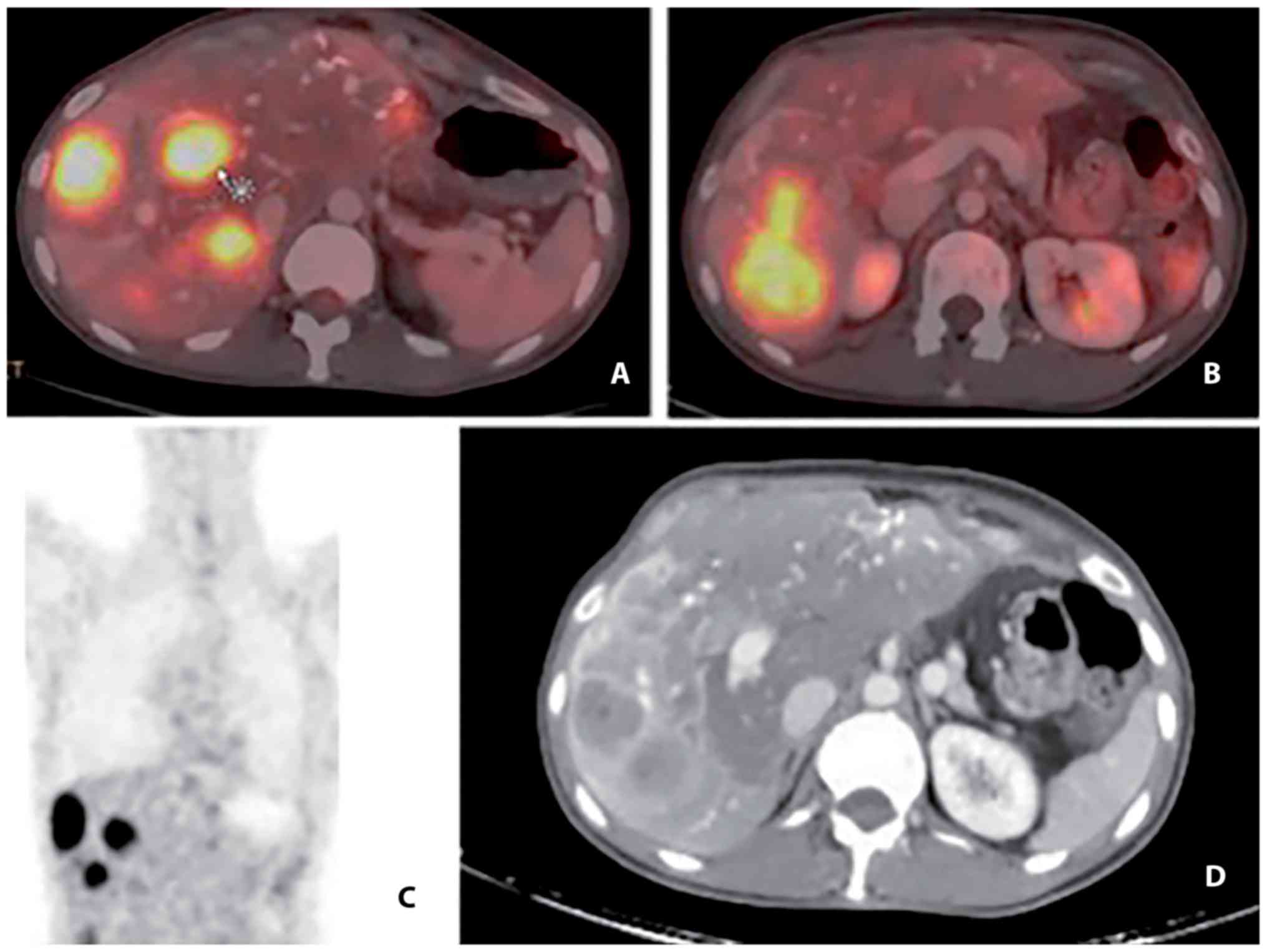
reaction. During treatment with sunitinib a 68Ga-Dotatoc
PET-CT was performed in April 2012, which demonstrated uptake, and
the patient was then referred for PRRT; however, treatment was
denied due to the high proliferation index.
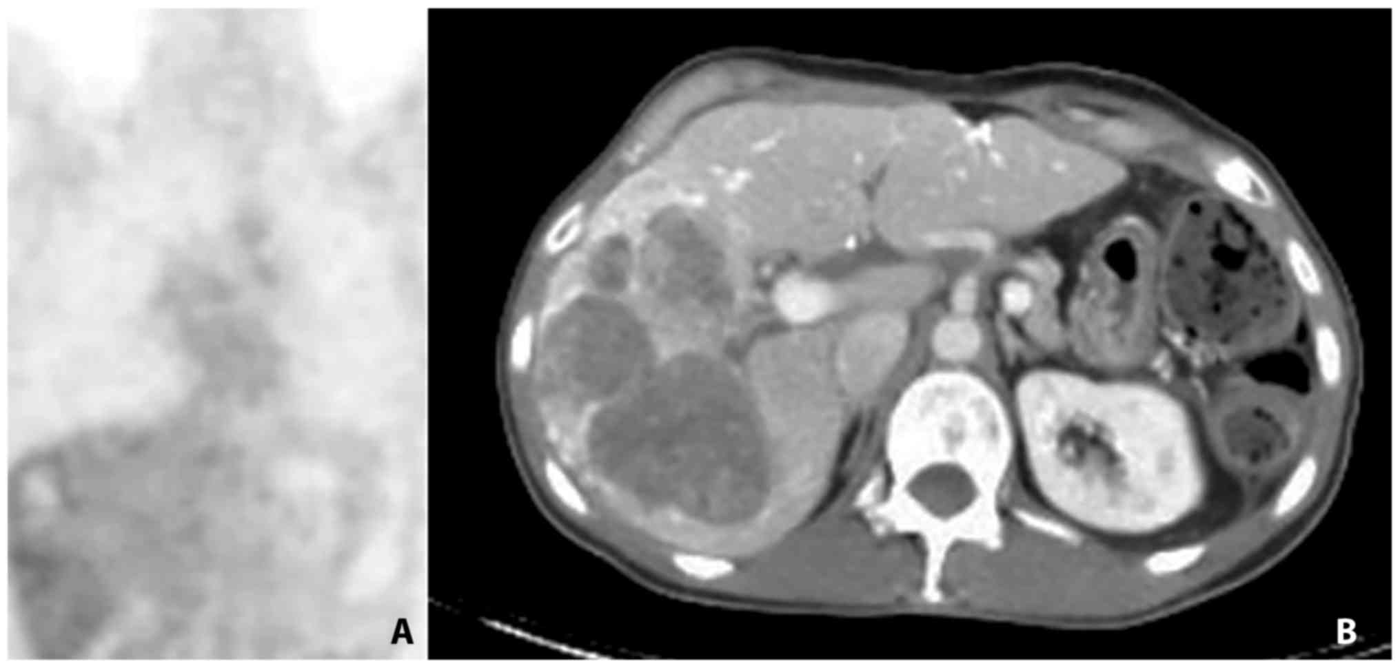
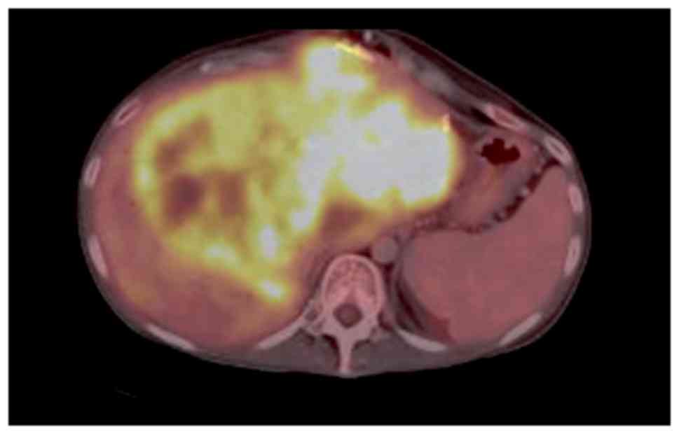
In June 2012, chemotherapy supplemented with
cisplatin and etoposide was initiated due to progression of the
disease (Fig. 3). The chemotherapy
was moderately tolerated by the patient and stopped in February
2013 when the disease was stable following three cycles of
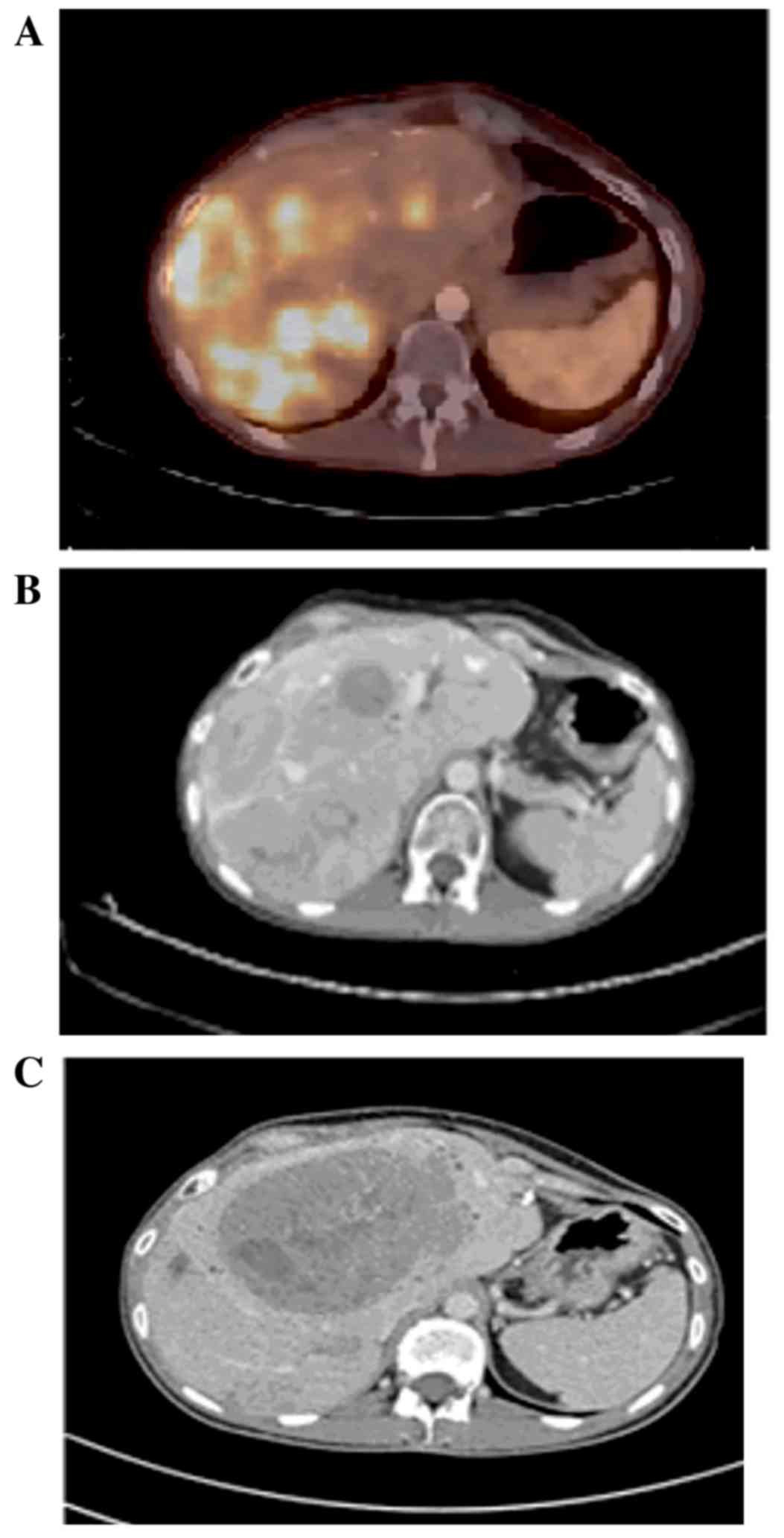
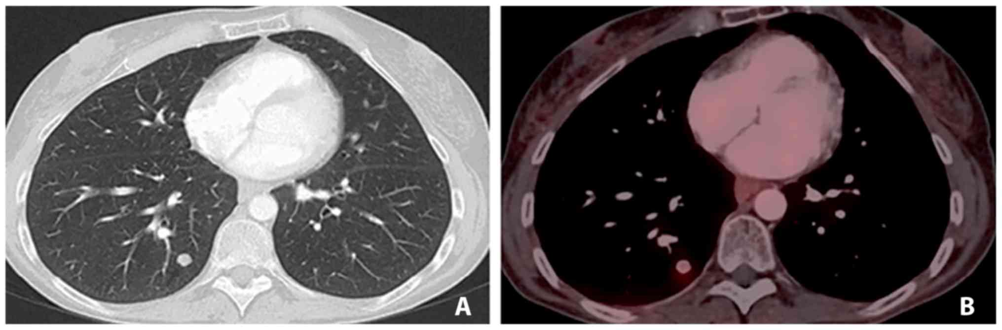
treatment. In September 2013, the patient was referred and accepted
for PRRT (Fig. 4A and B). The patient
underwent 3/4 planned treatment sessions; delays were often
experienced due to bone marrow toxicity. Therapy with PRRT was
completed in January 2014 and a good response in all but one lesion
was observed (Fig. 4C). This lesion
demonstrated a high metabolic activity on
18Fludeoxyglucose (18F-FDG) PET/CT and fine
needle aspiration revealed a Ki-67 index of 40%.
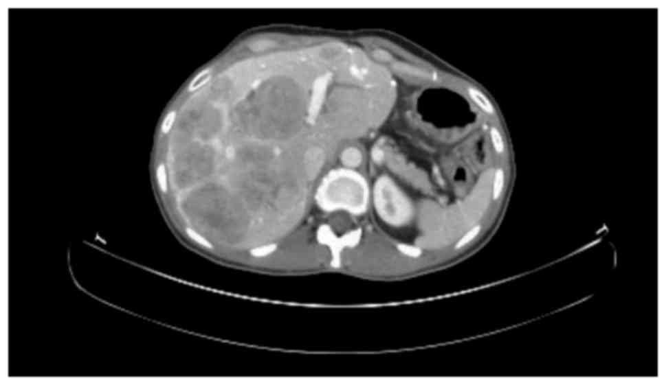
In April 2014, therapy with
temozolomide-capecitabine was initiated, and fast progression was
observed. The patient developed jaundice due to biliary obstruction
at the hilus of the liver (Fig. 5).
It was technically impossible to perform adequate biliary internal
drainage. External drainage was performed, leading to amelioration,
but no resolution of the jaundice was achieved, limiting the
treatment options.
At this time, the patient had had a diagnosis of
metastatic G3 NET for four years, and the disease had been well
controlled for long periods previously. Alternative treatment
options could have been discussed again if the level of bilirubin
was improved. During the clinical course and disease evolution,
despite the high Ki-67 index, it was agreed to perform a liver
transplantation in the patient using a marginal graft. A suitable
graft from a 68-year old brain-dead female donor with a history of
15 years of breast cancer became available, leading to a successful
transplantation in October 2014. In the explant liver, all lesions
had similar pathological features and a diagnosis of a
well-differentiated NET with Ki-67 index ≤40%. Immunosuppression
was based on tacrolimus and mycophenolate mofetil, with a switch to
sirolimus from tacrolimus after four weeks. Restaging in March 2015
revealed no signs of tumor recurrence; however, a novel
re-evaluation in October 2015, one year following transplantation,
identified numerous metastatic long nodules, demonstrating uptake
on 68Ga-Dotatoc PET-CT (Fig.
6). Therapy with octreotide analogues was initiated. The
patient is currently (January 2017) asymptomatic and has a good
quality of life.
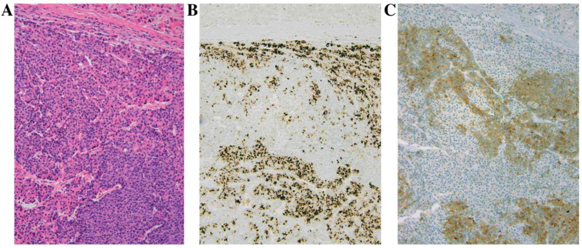
Review of the pathology specimens at this time
challenged the diagnosis. Following revision of all previous
resection specimens and the inclusion of markers for pancreatic
lipase, trypsin and chymotrypsin, a diagnosis of MAEC was
determined (Fig. 7).
Discussion
MAEC are rare tumors of the pancreas. They are
malignant epithelial neoplasms exhibiting both endocrine and acinar
differentiation. (2) By arbitrary
definition, each component must compromise ≥25% of the neoplasm for
a diagnosis of MAEC (2,4). Immunohistochemical labelling is
performed in order to diagnose the disease. This revealed regions
with acinar differentiation displaying positivity for lipase,
trypsin and/or chymotrypsin, and regions with endocrine
differentiation demonstrated positivity for synaptophysin and
chromogranin (2–4).
MAEC is challenging for pathologists to diagnose and
easily confused with ACC, solid pseudopapillary neoplasms and
predominantly with neuroendocrine neoplasms (1,4). In the
majority of cases there is a close intermingling of the acinar and
endocrine component without clear segregation of the two cell
types, thus it is not possible to identify with certainty the two
lines of differentiation by routine histology (2). Immunohistochemical labelling for
neuroendocrine and acinar cell markers provides the only evidence
for dual differentiation. In contrast to neuroendocrine
immunohistochemical markers, markers for pancreatic enzymes are not
widely available outside of large hospitals, including at the time
of diagnosis of the patient in the present case. In order to
achieve the correct diagnosis, referral to a specialized department
with access to these markers is indicated.
MAEC is thought to behave more akin to typical
pancreatic ACCs, compared with well-differentiated pancreatic
endocrine neoplasms (3,4). Pancreatic ACC is commonly treated in the
same way as adenocarcinomas, although the disease may have a more
indolent course, as suggested by increased survival rates
demonstrated in the Surveillance, Epidemiology, and End Results
Programme database for unresected (5-year survival rate, 22%) and
resected ACC (5-year survival rate, 72%) (1). In the present study analysis, it is
confirmed that the diagnosis of ACC can be difficult to make and
that incorrect classification of NETs and MAEC may favourably bias
the survival rate.
Reports on MAECs with a clear histopathological
diagnosis are scarce, in addition to evidence on disease behaviour
and treatment options. Recently, a review on ~30 presented cases in
literature was published (4). The
general consensus is that surgery is the primary treatment method
for patients with resectable tumors, and reports of patients
benefiting from tumor debulking surgery have been revealed
(4). The median overall survival time
following surgery was indicated to be ~12 months; however, this
assumption was made based on a small number of patients with
various tumor loads and lengths of follow-up (4). The patient in the present case relapsed
2.5 years following resection of the primary tumor and exhibited
local recurrence in the pancreas and liver metastases.
To the best of our knowledge, no previous studies
have been published investigating the use of functional imaging
techniques in MAEC, although in a certain case report the use of an
octreoscan following resection was discussed (6). Patients with neuroendocrine neoplasms
may have lesions with various uptakes of 18F-FDG PET/CT
and 68Ga-SMA PET/CT. Typically there is a higher uptake
of 68Ga-SMA in well differentiated NETs compared with in
poorly differentiated NECs, and a higher uptake of
18F-FDG in poorly differentiated NEC compared with in
well differentiated NETs (7,8). Particularly in patients with multiple
metastases, tracer uptake can be variable at different lesion sites
(8). Furthermore, certain lesions
within the same patient may be apparent on 68Ga-SMA
PET/CT and not on 18F-FDG PET/CT, or vice versa: this
reflects tumor heterogeneity (8).
This case study presented a patient who had distinct lesions on
68Ga-Dotatoc PET-CT, in accordance with the
histopathological diagnosis of a component of well-differentiated
NET. The patient also exhibited uptake on 18F-FDG PET/CT
in certain lesions, which may be associated with the acinar cell
component.
In NETs, an higher uptake on functional imaging
scans has been suggested to be associated with increased of overall
survival, and treatment decisions have been challenged based on the
results of functional scans (7,9–11). Treatment decisions in
tumor-node-metastasis stage IV NETs were previously determined
based on the grade of differentiation, prior to the introduction of
the Rindi grading system in 2006 (12), which was later integrated into the
World Health Organisation (WHO) 2010 classification system
(5). It is becoming more accepted
that the current WHO 2010 G3 category includes neuroendocrine
neoplasms of two distinct types: A highly proliferative group of
well-differentiated NETs (WD-NETs) and the poorly-differentiated
NECs, divided into small cell and large cell NECs (5,12–17). The pattern of uptake in functional
imaging provided evidence supporting the decision to administer the
patient in the present study, who was thought to have a diagnosis
of a well-differentiated G3 neuroendocrine neoplasm with a Ki-67
index of 40% maximum, chemotherapy (cisplatin-etoposide and
temozolomide-capecitabine), as recommended for NEC, and a tyrosine
kinase inhibitor (TKI), sunitinib, PRRT and liver transplantation
(5,12–17).
For a rare disease such as MAEC, it is not possible
to conduct trials for robust evidence of therapeutic effects and
case reports are important sources of information. The patient in
the present study had stable disease with treatment of sunitinib
for 17 months, and no objective response to cisplatin-etoposide,
but control of the disease was obtained for 15 months. Conversely,
the response to PRRT was clearly documented in all but one lesion;
however, time to tumor progression (TTP) was only four months due
to one rapidly progressive lesion. It was observed that the
combination of temozolomide-capecitabine in the fourth line of
treatment did not demonstrate any therapeutic benefit to the
patient; however, the subsequent therapeutic interventions may have
altered the properties of the tumor. Selection pressure may have
exhibited an impact on slower growing parts of the tumor, leaving
more aggressive cells behind. However, the tumor lesions in the
explant liver were extensively sampled and were not considered to
differ histopathologically. More specifically, the rapidly
progressive lesion was not revealed to have a higher Ki-67 index or
a differing cell population.
Upon revision at one year, performed for academic
reasons, and the application of immunohistochemical markers for
pancreatic enzymes, the diagnosis was changed to an MAEC. Revision
of older tissue specimens concurred with this finding. Liver
transplantation is not a standard treatment in neuroendocrine
neoplasms with a high Ki-67 index, and was performed exceptionally
as the only lifesaving option at the time of progression of a
centrally located liver lesion that had induced obstructive
jaundice in a young patient treated for metastatic disease for
>4 years. Relapse occurred 13 months following this and the
novel lesions again demonstrated uptake on 68Ga-Dotatoc
PET-CT, suggesting a component of well-differentiated NET. The
absence of fast recurrence following this surgical intervention in
spite of therapy with immune suppression may add to the suggestion
that surgery should be performed when possible in patients with
MAEC (1,4).
The response to the active sunitinib and PRRT
treatments suggested that treatment of the endocrine component
within the MAEC in addition to surgery may be beneficial. In
conclusion, a case of MAEC of the pancreas that demonstrated uptake
on 68Ga-Dotatoc PET-CT was presented, challenging the
suggestion of using these active treatments for the endocrine
component of the tumor. The majority of the lesions responded to
PRRT and a durable disease control was suggested with TKIs and
chemotherapy. However, the present case report also revealed that
surgical options must be considered in MAEC.
References
|
1
|
Wisnoski NC, Townsend CM Jr, Nealon WH,
Freeman JL and Riall TS: 672 Patients with acinar cell carcinoma of
the pancreas: A population-based comparison to pancreatic
adenocarcinoma. Surgery. 144:141–148. 2008. View Article : Google Scholar : PubMed/NCBI
|
|
2
|
Klimstra DS, Rosai J and Heffess CS: Mixed
acinar-endocrine carcinomas of the pancreas. Am J Surg Pathol.
18:765–778. 1994. View Article : Google Scholar : PubMed/NCBI
|
|
3
|
Ohike N, Kosmahl M and Klöppel G: Mixed
acinar-endocrine carcinoma of the pancreas. A clinicopathological
study and comparison with acinar-cell carcinoma. Virchows Arch.
445:231–235. 2004. View Article : Google Scholar : PubMed/NCBI
|
|
4
|
Liu Z, Dong C, Wang C, Liu Q, Sun D and
Wang L: Mixed acinar-endocrine carcinoma of pancreas: A case report
and brief review of the literature. Onco Targets Ther. 8:1633–1642.
2015. View Article : Google Scholar : PubMed/NCBI
|
|
5
|
Pasaoglu E, Dursun N, Ozyalvacli G,
Hacihasanoglu E, Behzatoglu K and Calay O: Comparison of world
health organization 2000/2004 and world health organization 2010
classifications for gastrointestinal and pancreatic neuroendocrine
tumors. Ann Diagn Pathol. 19:81–87. 2015. View Article : Google Scholar : PubMed/NCBI
|
|
6
|
Ogbonna OH, Garcon MC, Syrigos KN and Saif
MW: Mixed acinar-neuroendocrine carcinoma of the pancreas with
neuroendocrine predominance. Case Rep Med.
2013.7050922013.PubMed/NCBI
|
|
7
|
Has Simsek D, Kuyumcu S, Turkmen C, Sanlı
Y, Aykan F, Unal S and Adalet I: Can complementary 68 Ga-DOTATATE
and 18F-FDG PET/CT establish the missing link between
histopathology and therapeutic approach in gastroentero-pancreatic
neuroendocrine tumors? J Nucl Med. 55:1811–1817. 2014. View Article : Google Scholar : PubMed/NCBI
|
|
8
|
Kayani I, Bomanji JB, Groves A, Conway G,
Gacinovic S, Win T, Dickson J, Caplin M and Ell PJ: Functional
imaging of neuroendocrine tumors with combined PET/CT using
68Ga-DOTATATE (DOTA-DPhe1, Tyr3-octreotate) and 18F-FDG. Cancer.
112:2447–2455. 2008. View Article : Google Scholar : PubMed/NCBI
|
|
9
|
Panagiotidis E and Bomanji J: Role of
18F-fluorodeoxyglucose PET in the study of neuroendocrine tumors.
PET Clin. 9:43–55. 2014. View Article : Google Scholar : PubMed/NCBI
|
|
10
|
Kartalis N, Mucelli RM and Sundin A:
Recent developments in imaging of pancreatic neuroendocrine tumors.
Ann Gastroenterol. 28:193–202. 2015.PubMed/NCBI
|
|
11
|
Basu S, Ranade R and Thapa P: Correlation
and discordance of tumour proliferation index and molecular imaging
characteristics and their implications for treatment decisions and
outcome pertaining to peptide receptor radionuclide therapy in
patients with advanced neuroendocrine tumour: Developing a
personalized model? Nucl Med Commun. 36:766–774. 2015. View Article : Google Scholar : PubMed/NCBI
|
|
12
|
Rindi G, Klöppel G, Ahlman H, Caplin M,
Couvelard A, De Herder WW, Erikssson B, Falchetti A, Falconi M,
Komminoth P, et al: TNM staging of foregut (neuro)endocrine tumors:
A consensus proposal including a grading system. Virchows Arch.
449:395–401. 2006. View Article : Google Scholar : PubMed/NCBI
|
|
13
|
Garcia-Carbonero R, Sorbye H, Baudin E,
Raymond E, Wiedenmann B, Niederle B, Sedlackova E, Toumpanakis C,
Anlauf M, Cwikla JB, et al: ENETS consensus guidelines for
high-grade gastroenteropancreatic neuroendocrine tumors and
neuroendocrine carcinomas. Neuroendocrinology. 103:186–194. 2016.
View Article : Google Scholar : PubMed/NCBI
|
|
14
|
Vélayoudom-Céphise FL, Duvillard P, Foucan
L, Hadoux J, Chougnet CN, Leboulleux S, Malka D, Guigay J, Goere D,
Debaere T, et al: Are G3 ENETS neuroendocrine neoplasms
heterogeneous? Endocr Relat Cancer. 20:649–657. 2013. View Article : Google Scholar : PubMed/NCBI
|
|
15
|
Basturk O, Yang Z, Tang LH, Hruban RH,
Adsay V, McCall CM, Krasinskas AM, Jang KT, Frankel WL, Balci S, et
al: The high-grade (WHO G3) pancreatic neuroendocrine tumor
category is morphologically and biologically heterogenous and
includes both well differentiated and poorly differentiated
neoplasms. Am J Surg Pathol. 39:683–690. 2015. View Article : Google Scholar : PubMed/NCBI
|
|
16
|
Tang LH, Untch BR, Reidy DL, O'Reilly E,
Dhall D, Jih L, Basturk O, Allen PJ and Klimstra DS: Well
differentiated Neuroendocrine tumors with a morphologically
apparent high-grade component: A pathway distinct from poorly
differentiated neuroendocrine Carcinomas. Clin Cancer Res.
22:1011–1017. 2016. View Article : Google Scholar : PubMed/NCBI
|
|
17
|
Sorbye H, Welin S, Langer SW, Vestermark
LW, Holt N, Osterlund P, Dueland S, Hofsli E, Guren MG, Ohrling K,
et al: Predictive and prognostic factors for treatment and survival
in 305 patients with advanced gastrointestinal neuroendocrine
carcinoma (WHO G3): The NORDIC NEC study. Ann Oncol. 24:152–160.
2013. View Article : Google Scholar : PubMed/NCBI
|