Introduction
Gastric cancer (GC), one of the most common types of
malignant tumor worldwide, is the second most common cause of
cancer-related mortality globally (1). It is estimated that annually, there are
~1,000,000 new cases and >700,000 mortalities due to GC
worldwide (2). As a result of higher
Helicobacter pylori prevalence rates, >70% of patients
with GC are in developing countries, particularly in China
(3,4).
Despite development in comprehensive treatment including surgery,
radiotherapy and chemotherapy, the 5-year overall survival rate for
GC remains poor (5). This poor
survival rate is mainly due to recurrence and metastasis, even
following subtotal gastrectomy (6).
The tumorigenesis, development and metastasis of GC is
multifactorial, and numerous genetic and epigenetic changes
involving oncogenes, tumor suppressor genes and growth factors
changes have been demonstrated to be involved in GC (7). However, the molecular mechanism
underlying the tumorigenesis and development of GC remains unclear.
Therefore, it is of great significance to investigate the molecular
mechanisms underlying the initiation and progression of GC to
explore new therapeutic treatments for GC.
MicroRNAs (miRNAs) are a group of
non-protein-coding, highly conserved, single strand RNA molecules,
which are 21–25 nucleotides in length (8). miRNAs primarily regulate target gene
expression at the transcriptional or posttranscriptional level by
binding to the 3′ untranslated region (3′UTR) of target genes
(8,9).
Previous studies have reported that miRNAs play important functions
in various physiological and pathological processes, including cell
proliferation, cell cycle, apoptosis, differentiation and
metastasis; thereby affecting normal cell growth and development
and leading to a variety of disorders including malignancies
(10–12). In addition, the abnormal expression of
miRNAs has been identified in various types of human malignant
tumors, and their expression was significantly correlated with the
carcinogenesis, progression and metastasis of these cancer types
(13,14). Abnormally expressed miRNAs in human
cancer are able to function as tumor suppressors or oncogenes
depending on their target mRNAs (15,16).
Therefore, it is important to further examine the functions of
miRNAs in GC, in order to develop novel and efficient therapeutic
strategies for GC.
The present study aimed to evaluate the expression,
functions and mechanisms of microRNA-154 (miR-154) in GC. Firstly,
the expression of miR-154 was measured in GC tissues and cell lines
using reverse transcription-quantitative PCR (RT-qPCR). GC cells
were then transfected with miR-154 mimics or negative control (NC)
to evaluate the effects of miR-154 on the biological behavior of GC
cells. Following transfection, bioinformatics analysis, Dual
Luciferase reporter assay, RT-qPCR and western blot analysis were
adopted to explore the molecular mechanisms underlying
miR-154-inhibited growth and metastasis of GC cells.
Materials and methods
Tissue specimens
The present study was approved by the Research
Ethics Committee of The First Hospital of Lanzhou University
(Gansu, China). Full written informed consent was obtained from all
patients with GC prior to the collection of tissue specimens. A
total of 36 paired GC tissues and matched non-neoplastic gastric
tissues (normal) were obtained from patients with GC who had
undergone radical gastrectomy at The First Hospital of Lanzhou
University. None of the patients with GC had been treated with any
radiotherapy or chemotherapy prior to surgery.
Cell culture and transfection
The four human GC SGC-7901, AGS, MKN-1 and BGC-823
cell lines and the normal gastric epithelium GES-1 cell line were
all ordered from Shanghai Institute of Biochemistry and Cell
Biology (Shanghai, China). All cells were cultured in RPMI-1640
medium containing 10% fetal bovine serum (FBS; Gibco, Thermo Fisher
Scientific, Inc., Waltham, MA, USA), 100 U/ml penicillin and 100
mg/ml streptomycin. All cell lines were incubated at 37°C in a
humidified atmosphere of 5% CO2 and 95% air.
miR-154 mimics, NC miRNA mimics, metadherin (MTDH)
small interfering RNA (siRNA) and siRNA control were all purchased
from Shanghai GenePharma Co., Ltd. (Shanghai, China). Cells were
transfected with mimics or siRNA using Lipofectamine 2000
(Invitrogen; Thermo Fisher Scientific, Inc.) according to the
manufacturer's protocol.
RT-qPCR
Total RNA was isolated from tissues and cells using
TRIzol® (Invitrogen; Thermo Fisher Scientific, Inc.),
according to the manufacturer's protocol. For mature miR-154
expression, the Taqman microRNA reverse transcription kit (Applied
Biosystems; Thermo Fisher Scientific, Inc.) was used to synthesize
cDNA, followed by RT-qPCR with a Taqman microRNA assay kit (Applied
Biosystems; Thermo Fisher Scientific, Inc.). RNU6B was used as an
internal control for miR-154 expression analysis. The thermocycling
conditions were as follows: 95°C for 10 min, 40 cycles of
denaturation at 95°C for 15 sec, annealing at 60°C for 1 min, and a
final elongation step at 72°C for 10 min. To quantify MTDH mRNA
expression, total RNA was reverse transcribed into cDNA using the
PrimeScript RT reagent kit (Takara Biotechnology Co., Ltd., Dalian,
China). SYBR Green PCR Master mix (Applied Biosystems, Thermo
Fisher Scientific, Inc.) was adopted to measure MTDH mRNA
expression levels. The thermocycling conditions were as follows:
95°C for 10 min; 40 cycles at 95°C for 15 sec and 60°C for 1 min.
The data were normalized to glyceraldehyde-3-phosphate
dehydrogenase (GAPDH). RT-qPCR was performed in triplicate on an
AB7300 thermocycler (Applied Biosystems, Thermo Fisher Scientific,
Inc.). The primer sequences were as follows: miR-154, forward,
5′-TGCGCTAGGTTATCCGTGTTG-3′ and reverse,
5′-CTCAAGTGTCGTGGAGTCGGCAA-3′; U6 forward, 5′-CTCGCTTCGGCAGCACA-3′
and reverse, 5′-AACGCTTCACGAATTTGCGT-3′; MTDH forward,
5′-CTCGCTTCGGCAGCACA-3′ and reverse, 5′-AACGCTTCACGAATTTGCGT-3′;
GAPDH forward, 5′-CGTCTTCACCACCATGGAGA-3′ and reverse primer,
5′-CGCCCATCACGCCACAGTTT-3′. Relative expression level was
calculated using the 2−ΔΔCT method (17).
Cell proliferation assay
The effects of miR-154 on cell proliferation were
assessed with Cell Counting kit 8 assay (CCK8; Dojindo Molecular
Technologies, Inc., Kumamoto, Japan). GC cells were seeded into
96-well plates at a density of 3,000 cells per well. Following
transfection with mimics or siRNA, cells were incubated at 37°C in
a humidified incubator for 24, 48, 72 and 96 h. Subsequently, 10 µl
of CCK8 assay solution was added to each well of the 96 well plates
and incubated at 37°C for an additional 2 h. The absorbance of each
well at 450 nm was detected using a microplate reader (Bio-Rad,
Laboratories, Inc., Hercules, CA, USA). All experiments were
performed in triplicate.
Transwell migration and Matrigel
invasion assay
The effects of miR-154 on cell migration and
invasion were evaluated using Transwell chambers with an 8 µm pore
size (BD Biosciences, Bedford, MA, USA). For the Transwell
migration assay, cells were collected, and 5×10⁴ cells in 200 µl
serum-free medium were seeded into the upper Transwell chambers 24
h subsequent to transfection with mimics or siRNA. The lower
Transwell chambers were loaded with 500 µl medium supplemented with
20% FBS as a chemoattractant. For the Matrigel invasion assay, the
Transwell chambers were pre-coated with 2% Matrigel (BD
Biosciences, San Jose, CA, USA). Following incubation at 37°C for
24 h (migration assay) or 36 h (invasion assay), the cells on the
upper surface of the Transwell chambers were removed carefully by
cotton wool. The migrated and invaded cells were fixed and stained
with 0.5% crystal violet. Subsequent to washing with PBS three
times, the chambers were visualized with an IX71 inverted
microscope (Olympus Corporation, Tokyo, Japan).
Bioinformatic analysis
Target genes of miR-154 were searched using
TargetScan (http://www.targetscan.org).
Western blot analysis
In the present study, MTDH (1:1,000; catalog no.
sc-517220) and β-actin (1:1,000; catalog no. sc-130301) primary
antibodies were purchased from Santa Cruz Biotechnology, Inc.
(Dallas, TX, USA). At 72 h subsequent to transfection, transfected
cells (mimics/siRNA) were lysed using a radioimmunoprecipitation
assay buffer in the presence of a protease inhibitor cocktail
(Sigma-Aldrich; Merck KGaA, Darmstadt, Germany). The concentration
of total cellular protein was determined using a BCA protein assay
kit (Pierce; Thermo Fisher Scientific, Inc.). Equal amounts of
protein (20 µg) were separated by 10% SDS-PAGE and transferred onto
a polyvinylidene fluoride membrane (PVDF, EMD Millipore, Billerica,
MA, USA). Subsequent to blocking with 5% non-fat milk in TBS, the
membranes were probed with primary antibodies overnight at 4°C,
followed by incubation with goat anti-mouse horseradish peroxidase
(HRP)-conjugated secondary antibodies (1:3,000; catalog no.
sc-2005; Santa Cruz Biotechnology, Inc.) for 1 h at room
temperature. The bands were detected with enhanced
chemiluminescence solution (ECL; Pierce; Thermo Fisher Scientific,
Inc.). The protein intensities were quantified using AlphaEase FC
software. (version 4.1.0; Alpha Innotech, San Leandro, USA). This
experiment was repeated three times.
Dual Luciferase reporter assay
For the luciferase reporter assay, the luciferase
reporter vectors PGL3-MTDH-3′UTR wild type (Wt) and PGL3-MTDH-3′UTR
mutant (Mut) were purchased from Shanghai GenePharma Co., Ltd.
Cells were transfected with miR-154 mimics or NC, and
PGL3-MTDH-3′UTR Wt or PGL3-MTDH-3′UTR Mut using Lipofectamine 2000,
according to the manufacturer's protocol. The luciferase activities
were measured using Dual Luciferase Reporter Assay system (Promega
Corporation, Manheim, Germany) 48 h following transfection.
Experiments were performed in triplicate and replicated 3
times.
Statistical analysis
Data are presented as the mean ± standard deviation,
and compared using StataCorp LP 10.0 (College Station, TX, USA).
Two-tailed P<0.05 was considered to indicate a statistically
significant difference.
Results
miR-154 expression is downregulated in
GC tissues and cell lines
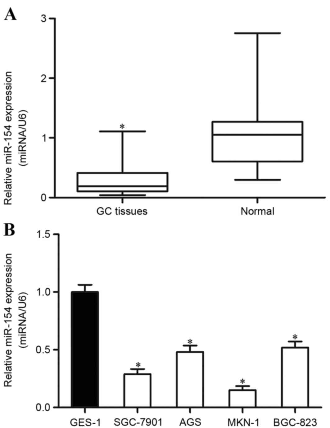
To determine whether miR-154 was involved in the
tumorigenesis and development of GC, its expression was measured
using RT-qPCR in GC tissues and matched non-neoplastic gastric
tissues. The results demonstrated that miR-154 was significantly
downregulated in GC tissues in comparison with matched
non-neoplastic gastric tissues (Fig.
1A). miR-154 expression levels were also detected in the four
GC cell lines and the normal gastric epithelium cell line. The
results revealed that all GC cell lines expressed lower levels of
miR-154 compared with the expression levels in the normal gastric
epithelium GES-1 cell line (Fig. 1B).
Together, these results indicate that miR-154 is downregulated in
GC.
Overexpression of miR-154 inhibits
proliferation, migration and invasion of GC cells
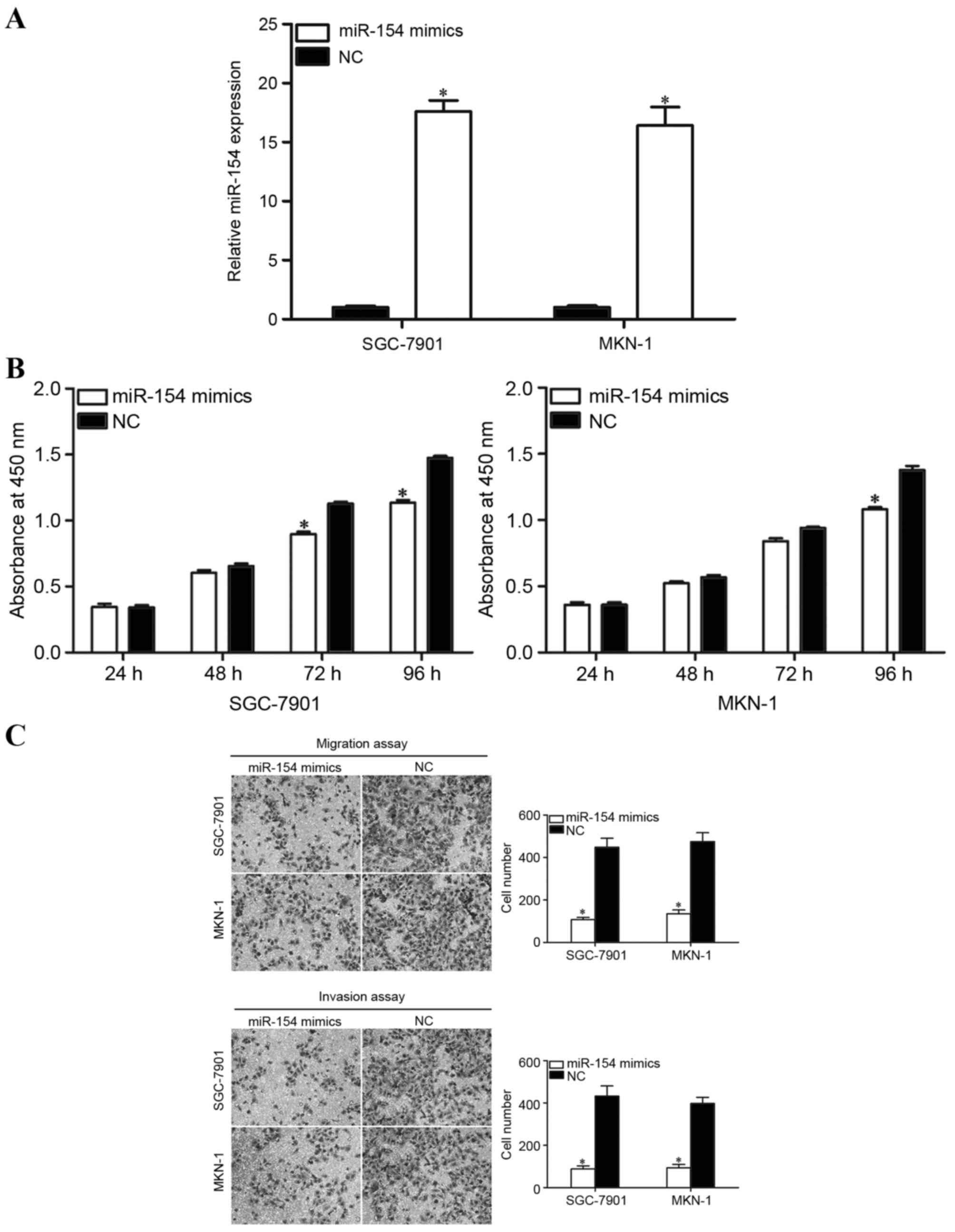
To investigate whether miR-154 affects the
proliferation of GC cells, miR-154 mimics or NC was introduced into
GC cells. SGC-7901 and MKN-1 were selected for the present
functional study due to their lower miR-154 expression levels.
Following transfection, RT-qPCR was performed to measure relative
miR-154 expression. As presented in Fig.
2A, miR-154 was significantly upregulated in SGC-7901 and MKN-1
cell lines. Cell proliferation was assessed using a CCK8 assay. As
presented in Fig. 2B, overexpression
of miR-154 inhibited the proliferation of SGC-7901 at 72 and 96 h
following transfection. The overexpression of miR-154 also
inhibited proliferation of MKN-1 cells at 96 h following
transfection.
Transwell migration and Matrigel invasion assays
were performed to explore whether miR-154 affected the migration
and invasion capacity of GC cells. As expected, the cell migration
and invasion capacity in SGC-7901 and MKN-1 cells was significantly
reduced following transfection with miR-154 mimics in comparison
with NC (Fig. 2C). These findings
indicate that miR-154 may function as a tumor suppressor in GC.
miR-154 directly targeted MTDH by
interaction with the binding site in the 3′UTR
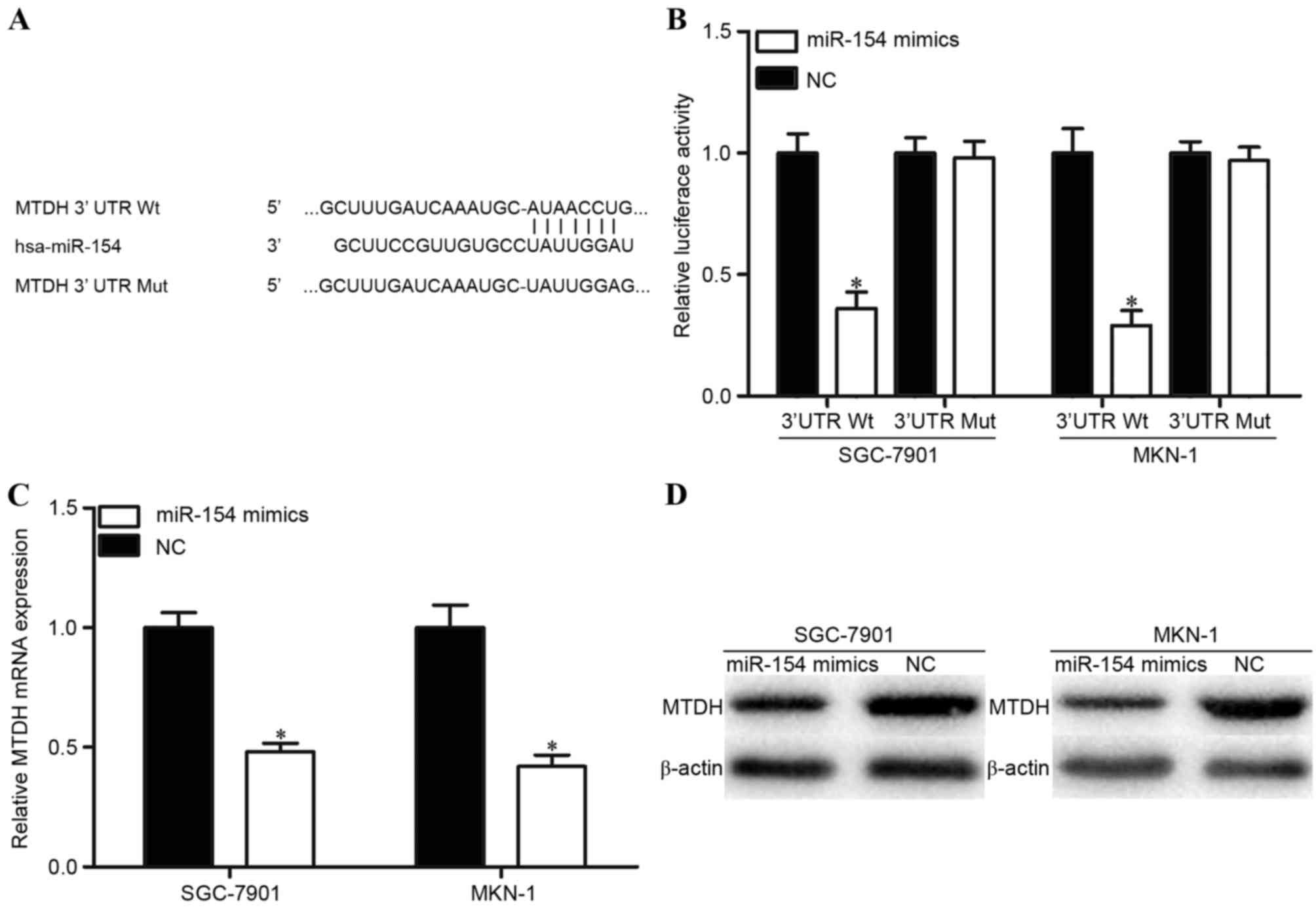
TargetScan was used to predicate potential target
genes with complementary sites of miR-154 in their 3′UTR. The
results revealed that MTDH contained a miR-154 seed match at 3′UTR
of MTDH (Fig. 3A). To evaluate this
possibility, luciferase reporter assays were performed. As
presented in Fig. 3B, the relative
luciferase activities of the PGL3-MTDH-3′UTR Wt were significantly
decreased when miR-154 mimics were co-transfected. However, the
luciferase activities of PGL3-MTDH-3′UTR Mut were unaffected by
co-transfection with miR-154 mimics. Furthermore, RT-qPCR and
western blot analysis were performed to explore whether miR-154
affects MTDH expression at transcriptional and translational
levels. The results indicated that the levels of MTDH mRNA and
protein expression in miR-154 transfected SGC-7901 and MKN-1 cells
were significantly inhibited compared with those in NC-transfected
cells (Fig. 3C and D). These results
suggest that miR-154 directly targets MTDH in GC by interacting
with the binding site in the 3′UTR of the MTDH gene.
MTDH siRNA inhibited proliferation,
migration and invasion of GC cells
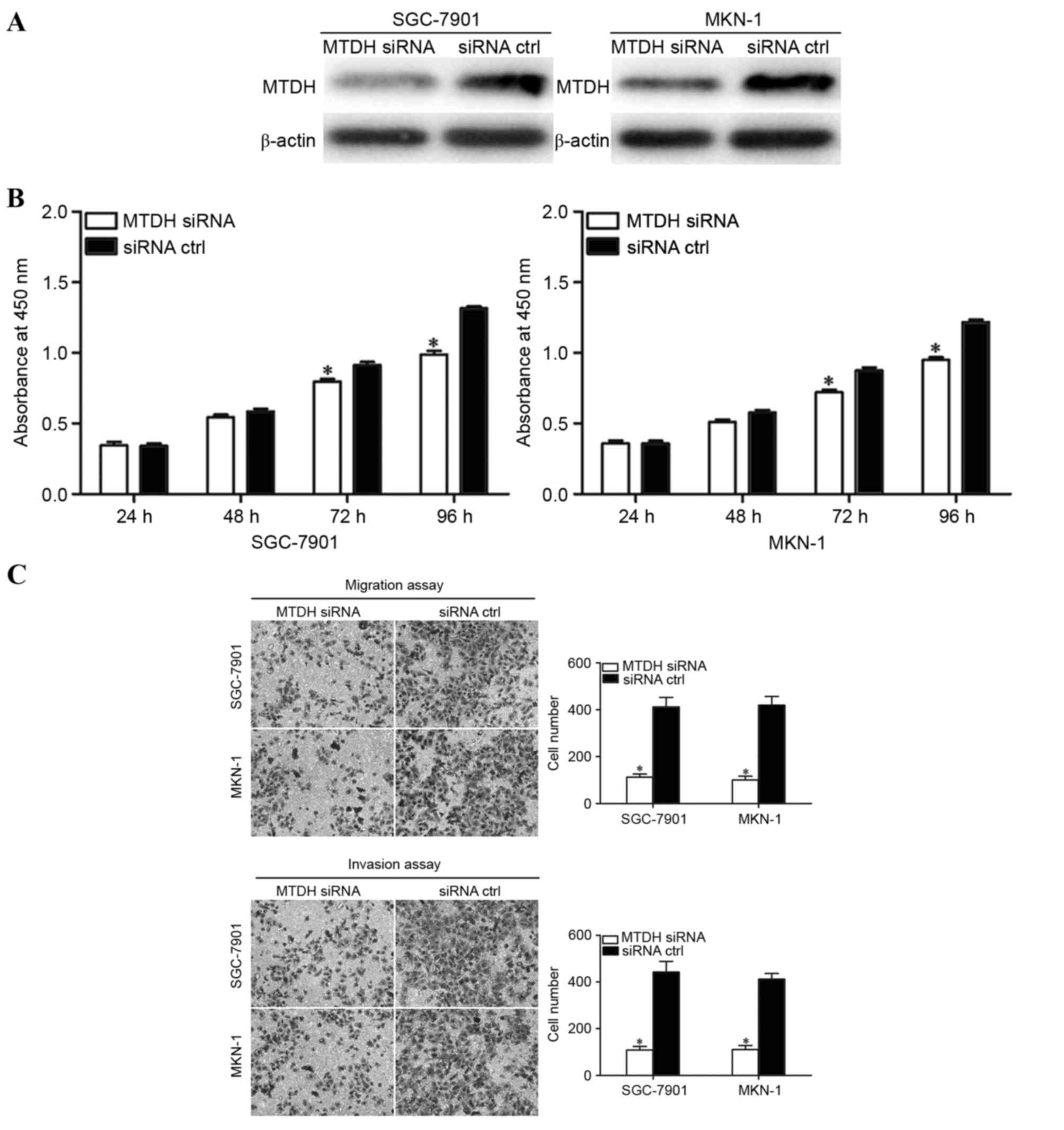
To explore the roles of MTDH in GC, GC SGC-7901 and
MKN-1 cells were transfected with MTDH siRNA or siRNA as a control.
Western blot analysis revealed that MTDH siRNA decreased MTDH
expression in SGC-7901 and MKN-1 cells compared with that in siRNA
control transfected cells (Fig. 4A).
Cell proliferation assays, Transwell migration and Matrigel
invasion assays were performed to investigate the effects of MTDH
on cellular proliferation, migration and invasion of GC cells. As
presented in Fig. 4B and C, MTDH
siRNA significantly inhibited the proliferation (following
incubation for 72 and 96 h) and motility of GC cells. These results
indicate that the inhibition of MTDH performed similar functions to
miR-154 overexpression in GC cells; therefore, MTDH may be a
functional target of miR-154 in GC.
Discussion
GC is one of the most common types of malignant
tumor and occurs as a result of genetic alterations and multiple
environmental factors (18). The main
therapy for GC has improved in recent decades. However, the
prognosis of patients with advanced GC remains poor. Therefore, it
is of great significance to understand the molecular mechanism
underlying GC carcinogenesis and development. In addition, an
increasing number of studies have indicated that abnormal
expression of miRNAs plays an important function in the initiation
and progression of GC, and that miRNAs may be investigated as a
potential novel therapeutic target for the treatment of GC
(19,20). The present study revealed that miR-154
was significantly downregulated in GC tissues and cell lines.
Overexpression of miR-154 in GC cells resulted in suppression of
cellular proliferation, migration and invasion. Furthermore, MTDH
was validated as a potential functional target gene of miR-154 in
GC. These findings suggest that miR-154 functions as a tumor
suppressor in GC, and may have the potential to be investigated as
an anticancer drug for GC.
miR-154 has been revealed to be downregulated in
non-small cell lung (NSCLC) (21),
colorectal (22,23) and prostate cancer (24). In NSCLC, the expression levels of
miR-154 were significantly decreased in the tumor tissues compared
with those in the matched non-tumorous lung tissues. Low miR-154
expression was significantly correlated with metastasis, larger
tumor size and advanced tumor node metastasis (TNM) stage of
patients with NSCLC (21). Kai et
al (22) revealed that miR-154
levels in colorectal cancer tissues were significantly lower than
those in non-cancerous tissues. Decreased expression levels of
miR-154 were markedly associated with large tumor size, positive
lymph node metastasis and advanced clinical stage. Univariate
analysis also demonstrated that patients with colorectal cancer
with low miR-154 expression levels had a poorer prognosis in this
previous study. In addition, multivariate analysis confirmed that
low miR-154 expression was an independent predictor of poor
survival rate.
miR-154 has been demonstrated to be a tumor
suppressor. In NSCLC, cells overexpression of miR-154 inhibits cell
growth, colony formation, migration and invasion, and enhances
cellular apoptosis and G0/G1 cell cycle arrest (21). Upregulation of miR-154 also suppresses
the growth of NSCLC cell xenografts in vivo (21). Zhu et al (24,25)
reported that enforced expression of miR-154 decreases the
proliferation, colony formation, migration and invasion of prostate
cancer cells via blockade of cyclin D2 and high mobility group
AT-hook 2. Xin et al (23)
indicated that miR-154 suppresses growth, colony formation and
motility by directly targeting toll-like receptor 2 (TLR2). These
findings indicate that upregulating miR-154 or providing analogous
pharmaceutical compounds exogenously may be effective therapeutic
strategies for these types of cancer.
Previous studies have demonstrated that miRNAs
negatively regulate target gene expression by binding to the 3′UTR
of target genes (26–28). In the present study, MTDH was
identified as a novel target gene of miR-154 in GC. Firstly, MTDH
was predicted as a target gene of miR-154 by using TargetScan.
Secondly, Dual Luciferase reporter assays demonstrated that miR-154
significantly decreased the luciferase activity in GC cells
transfected with MTDH-3′UTR Wt compared with MTDH-3′UTR Mut.
Thirdly, over-expression of miR-154 suppressed MTDH mRNA and
protein expression of GC cells. Finally, the functions of MTDH
siRNA were similar to those induced by miR-154 in GC cells, which
indicated that MTDH may be a functional target of miR-154 in GC.
The identification of miR-154 target genes is essential for
elucidating the functions of miR-154 in the carcinogenesis and
progression of GC, and may provide promising therapeutic targets
for patients with GC.
MTDH, located at chromosome 8q22, is a
multifunctional oncogene that has been reported to be overexpressed
in a variety of human cancers including glioma (29), hepatocellular carcinoma (30), colorectal (31) and breast cancer (32). Subsequent investigations have revealed
that MTDH contributes to multiple biological processes in the
course of cancer carcinogenesis and progression, including cellular
growth, apoptosis, metastasis, invasion, chemoresistance and
angiogenesis (33,34). The expression of MTDH mRNA and protein
levels were also upregulated in GC tissues (35,36). In
addition, high expression levels of MTDH were significantly
associated with differentiation status, TNM stage, invasive depth
and lymph node metastasis in GC (36). These studies all indicate that MTDH
may be a novel and promising target for therapeutic intervention in
GC. The present study demonstrated that MTDH was downregulated in
GC cells following transfection with miR-154 mimics. Additionally,
knockdown of MTDH inhibited growth and metastasis of GC cells.
These results suggest that miR-154 may be investigated as a
targeted therapy against MTDH and to block the growth and
metastasis of GC.
In conclusion, the present study identified that
miR-154 was downregulated in GC tissues and cells. Overexpression
of miR-154 effectively inhibited the proliferation, migration and
invasion of GC cells. Additionally, MTDH was demonstrated as a
direct functional target gene of miR-154 in GC. The present study
provides new insights into the tumorigenesis and development of GC.
It also suggests that the miR-154/MTDH axis may act as a
therapeutic target for patients with GC.
References
|
1
|
Jemal A, Bray F, Center MM, Ferlay J, Ward
E and Forman D: Global cancer statistics. CA Cancer J Clin.
61:69–90. 2011. View Article : Google Scholar : PubMed/NCBI
|
|
2
|
Liu J, Xue H, Zhang J, Suo T, Xiang Y,
Zhang W, Ma J, Cai D and Gu X: MicroRNA-144 inhibits the metastasis
of gastric cancer by targeting MET expression. J Exp Clin Cancer
Res. 34:352015. View Article : Google Scholar : PubMed/NCBI
|
|
3
|
Wang CS, Hsieh CC, Chao TC, Jan YY, Jeng
LB, Hwang TL, Chen MF, Chen PC, Chen JS and Hsueh S: Resectable
gastric cancer: Operative mortality and survival analysis. Chang
Gung Med J. 25:216–227. 2002.PubMed/NCBI
|
|
4
|
Ye YW, Dong RZ, Zhou Y, Du CY, Wang CM, Fu
H and Shi YQ: Prognostic analysis of familial gastric cancer in
Chinese population. J Surg Oncol. 104:76–82. 2011. View Article : Google Scholar : PubMed/NCBI
|
|
5
|
Meyer HJ and Wilke H: Treatment strategies
in gastric cancer. Dtsch Arztebl Int. 108:698–705. 2011.PubMed/NCBI
|
|
6
|
Kim SJ, Wang YG, Lee HW, Kang HG, La SH,
Choi IJ, Irimura T, Ro JY, Bresalier RS and Chun KH: Up-regulation
of neogenin-1 increases cell proliferation and motility in gastric
cancer. Oncotarget. 5:3386–3398. 2014. View Article : Google Scholar : PubMed/NCBI
|
|
7
|
Otani K, Li X, Arakawa T, Chan FK and Yu
J: Epigenetic-mediated tumor suppressor genes as diagnostic or
prognostic biomarkers in gastric cancer. Expert Rev Mol Diagn.
13:445–455. 2013. View Article : Google Scholar : PubMed/NCBI
|
|
8
|
Hutvágner G and Zamore PD: A microRNA in a
multiple-turnover RNAi enzyme complex. Science. 297:2056–2060.
2002. View Article : Google Scholar : PubMed/NCBI
|
|
9
|
Cortés-Sempere M and de Ibáñez Cáceres I:
microRNAs as novel epigenetic biomarkers for human cancer. Clin
Transl Oncol. 13:357–362. 2011. View Article : Google Scholar : PubMed/NCBI
|
|
10
|
Croce CM and Calin GA: miRNAs, cancer, and
stem cell division. Cell. 122:6–7. 2005. View Article : Google Scholar : PubMed/NCBI
|
|
11
|
Hwang HW and Mendell JT: MicroRNAs in cell
proliferation, cell death and tumorigenesis. Br J Cancer.
96:(Suppl). R40–R44. 2007.PubMed/NCBI
|
|
12
|
Gregory RI and Shiekhattar R: MicroRNA
biogenesis and cancer. Cancer Res. 65:3509–3512. 2005. View Article : Google Scholar : PubMed/NCBI
|
|
13
|
Stenvang J, Petri A, Lindow M, Obad S and
Kauppinen S: Inhibition of microRNA function by antimiR
oligonucleotides. Silence. 3:12012. View Article : Google Scholar : PubMed/NCBI
|
|
14
|
Bou Kheir T, Futoma-Kazmierczak E,
Jacobsen A, Krogh A, Bardram L, Hother C, Grønbæk K, Federspiel B,
Lund AH and Friis-Hansen L: miR-449 inhibits cell proliferation and
is down-regulated in gastric cancer. Mol Cancer. 10:292011.
View Article : Google Scholar : PubMed/NCBI
|
|
15
|
Lu J, Getz G, Miska EA, Alvarez-Saavedra
E, Lamb J, Peck D, Sweet-Cordero A, Ebert BL, Mak RH, Ferrando AA,
et al: MicroRNA expression profiles classify human cancers. Nature.
435:834–838. 2005. View Article : Google Scholar : PubMed/NCBI
|
|
16
|
Volinia S, Calin GA, Liu CG, Ambs S,
Cimmino A, Petrocca F, Visone R, Iorio M, Roldo C, Ferracin M, et
al: A microRNA expression signature of human solid tumors defines
cancer gene targets. Proc Natl Acad Sci USA. 103:2257–2261. 2006.
View Article : Google Scholar : PubMed/NCBI
|
|
17
|
Livak KJ and Schmittgen TD: Analysis of
relative gene expression data using real-time quantitative PCR and
the 2(−Delta Delta C(T)) Method. Methods. 25:402–408. 2001.
View Article : Google Scholar : PubMed/NCBI
|
|
18
|
Ma G, Dai W, Sang A, Yang X and Gao C:
Upregulation of microRNA-23a/b promotes tumor progression and
confers poor prognosis in patients with gastric cancer. Int J Clin
Exp Pathol. 7:8833–8840. 2014.PubMed/NCBI
|
|
19
|
Chang L, Guo F, Huo B, Lv Y, Wang Y and
Liu W: Expression and clinical significance of the microRNA-200
family in gastric cancer. Oncol Lett. 9:2317–2324. 2015.PubMed/NCBI
|
|
20
|
Qiu T, Zhou X, Wang J, Du Y, Xu J, Huang
Z, Zhu W, Shu Y and Liu P: MiR-145, miR-133a and miR-133b inhibit
proliferation, migration, invasion and cell cycle progression via
targeting transcription factor Sp1 in gastric cancer. FEBS Lett.
588:1168–1177. 2014. View Article : Google Scholar : PubMed/NCBI
|
|
21
|
Lin X, Yang Z, Zhang P and Shao G: miR-154
suppresses non-small cell lung cancer growth in vitro and in vivo.
Oncol Rep. 33:3053–3060. 2015.PubMed/NCBI
|
|
22
|
Kai Y, Qiang C, Xinxin P, Miaomiao Z and
Kuailu L: Decreased miR-154 expression and its clinical
significance in human colorectal cancer. World J Surg Oncol.
13:1952015. View Article : Google Scholar : PubMed/NCBI
|
|
23
|
Xin C, Zhang H and Liu Z: miR-154
suppresses colorectal cancer cell growth and motility by targeting
TLR2. Mol Cell Biochem. 387:271–277. 2014. View Article : Google Scholar : PubMed/NCBI
|
|
24
|
Zhu C, Shao P, Bao M, Li P, Zhou H, Cai H,
Cao Q, Tao L, Meng X, Ju X, et al: miR-154 inhibits prostate cancer
cell proliferation by targeting CCND2. Urol Oncol. 32(31): e9–e16.
2014.
|
|
25
|
Zhu C, Li J, Cheng G, Zhou H, Tao L, Cai
H, Li P, Cao Q, Ju X, Meng X, et al: miR-154 inhibits EMT by
targeting HMGA2 in prostate cancer cells. Mol Cell Biochem.
379:69–75. 2013. View Article : Google Scholar : PubMed/NCBI
|
|
26
|
Liu X, Wang S, Yuan A, Yuan X and Liu B:
MicroRNA-140 represses glioma growth and metastasis by directly
targeting ADAM9. Oncol Rep. 36:2329–2338. 2016.PubMed/NCBI
|
|
27
|
Shi C and Zhang Z: MicroRNA-362 is
downregulated in cervical cancer and inhibits cell proliferation,
migration and invasion by directly targeting SIX1. Oncol Rep.
37:501–509. 2017.PubMed/NCBI
|
|
28
|
Wang LL, Wang L, Wang XY, Shang D, Yin SJ,
Sun LL and Ji HB: MicroRNA-218 inhibits the proliferation,
migration, and invasion and promotes apoptosis of gastric cancer
cells by targeting LASP1. Tumour Biol. 37:15241–15252. 2016.
View Article : Google Scholar : PubMed/NCBI
|
|
29
|
Kim DH, Mohapatra G, Bollen A, Waldman FM
and Feuerstein BG: Chromosomal abnormalities in glioblastoma
multiforme tumors and glioma cell lines detected by comparative
genomic hybridization. Int J Cancer. 60:812–819. 1995. View Article : Google Scholar : PubMed/NCBI
|
|
30
|
Poon TC, Wong N, Lai PB, Rattray M,
Johnson PJ and Sung JJ: A tumor progression model for
hepatocellular carcinoma: Bioinformatic analysis of genomic data.
Gastroenterology. 131:1262–1270. 2006. View Article : Google Scholar : PubMed/NCBI
|
|
31
|
Gnosa S, Shen YM, Wang CJ, Zhang H,
Stratmann J, Arbman G and Sun XF: Expression of AEG-1 mRNA and
protein in colorectal cancer patients and colon cancer cell lines.
J Transl Med. 10:1092012. View Article : Google Scholar : PubMed/NCBI
|
|
32
|
Wang Y, Klijn JG, Zhang Y, Sieuwerts AM,
Look MP, Yang F, Talantov D, Timmermans M, Meijer-van Gelder ME, Yu
J, et al: Gene-expression profiles to predict distant metastasis of
lymph-node-negative primary breast cancer. Lancet. 365:671–679.
2005. View Article : Google Scholar
|
|
33
|
Hu G, Wei Y and Kang Y: The multifaceted
role of MTDH/AEG-1 in cancer progression. Clin Cancer Res.
15:5615–5620. 2009. View Article : Google Scholar : PubMed/NCBI
|
|
34
|
Sarkar D, Emdad L, Lee SG, Yoo BK, Su ZZ
and Fisher PB: Astrocyte elevated gene-1: Far more than just a gene
regulated in astrocytes. Cancer Res. 69:8529–8535. 2009. View Article : Google Scholar : PubMed/NCBI
|
|
35
|
Baygi ME and Nikpour P: Deregulation of
MTDH gene expression in gastric cancer. Asian Pac J Cancer Prev.
13:2833–2836. 2012. View Article : Google Scholar : PubMed/NCBI
|
|
36
|
Dong L, Qin S, Li Y, Zhao L, Dong S, Wang
Y, Zhang C and Han S: High expression of astrocyte elevated gene-1
is associated with clinical staging, metastasis, and unfavorable
prognosis in gastric carcinoma. Tumour Biol. 36:2169–2178. 2015.
View Article : Google Scholar : PubMed/NCBI
|