Introduction
Despite substantial improvement in the survival
rates of patients with cancer, cancer remains the most common
mortality-associated disease in the United States in 2015 (1). Renal cell carcinoma (RCC) is a lethal
malignant tumor type in the urinary system, accounting for 3% of
all malignancies globally in 2015 (2). In China, the incidence of this
malignancy is notably increasing. Nephrectomy remains the
first-line therapy in patients with a solid tumor (3). However, the frequent metastasis greatly
impedes the curative effect following kidney resection. Nearly 30%
of patients with RCC progress to metastatic disease subsequent to
radical nephrectomy surgery (4).
Numerous independent risk factors, including nodule number, tumor
stage [American Joint Committee on Cancer (AJCC, 2010)] (5) and grade, were identified to have a close
correlation with the recurrence and metastasis of patients with RCC
(6). Nevertheless, these parameters
are not absolutely accurate. Hence, the introduction of novelty
predictive and prognostic biomarkers will aid oncologists in
performing preventative treatment and individual therapy in
patients with RCC.
Cytokeratin 19 (CK19) is an epithelial cytoskeleton
marker and is highly expressed in epithelial tumor types (7). One previous study has revealed that CK19
may be an appropriate marker for the detection of cancer cells
(8). Furthermore, it may function as
a prognostic indicator for patients with cancer (9). Endoglin (CD105) was first reported over
two decades ago and identified as an accessory receptor for
transforming growth factor-β (TGF-β) (10,11). It is
notably upregulated in endothelial cells and has been considered to
be a biomarker for tumor angiogenesis (12). The role of CD105 in the early
metastasis of a tumor has gained attention, including in squamous
cell carcinoma and pancreatic ductal adenocarcinoma (13,14). In
RCC, the function of CD105 remains controversial (15,16).
Cluster of differentiation 146 (CD146) is a 113 kD membrane
glycoprotein which was first reported in malignant melanoma
(17). One previous study has
reported that CD146 is only expressed in blood vessels and smooth
muscle cells (18). Subsequent
research has demonstrated that CD146 has multiple functions in
development, immunity and angiogenesis (17). CD146 is additionally implicated in the
development of the kidney and the nervous system (19,20).
However, to the best of our knowledge, there are no previous
reports on the association between CD146 expression and RCC
metastasis following surgical treatment.
Circulating tumor cells (CTCs) are cells that exist
in the blood that have antigenic and/or genetic traits of a
specific tumor type (21). They have
been used to indicate a poor prognosis in a variety of cancer
types, including breast cancer, prostate cancer, colorectal cancer
and lung cancer (22–25). The detection of CTCs is easy to
implement via peripheral blood samples and may be conducive to
improving cancer prognostication and treatment (26).
In the present study, the aim was to explore the
association between CK19, CD105 and CD146 and the early metastasis
of RCC following surgery, and to evaluate whether the expression of
CK19, CD105 and CD146 is associated with the early metastasis of
patients with CTC-positive RCC.
Materials and methods
Ethics statement
The present study was approved by the Ethical
Committee of Second Affiliated Hospital of Medical School, Xi'an
Jiaotong University (Xi'an, China). The procedures in the present
study followed the principles of the Declaration of Helsinki.
Written informed consent was obtained from all patients prior to
the study.
Patient selection
The present retrospective study recruited 200 RCC
cases between January 2015 and October 2015. Of these, there were
100 male and 100 female patients with RCC at stage I, II and III
(AJCC, 2010) (5) of the disease (from
70, 122 and 8 patients, respectively) who were accepted for
surgical treatment at the Department of Urology, Second Affiliated
Hospital of Medical School, Xi'an Jiaotong University (Xi'an,
China). Inclusion criteria were as follows: i) Pathological
diagnosis confirmed RCC; ii) clinical stage cT1-3 (AJCC, 2010)
(5), ready for nephrectomy; iii)
distant metastasis were not identified prior kidney resection.
Exclusion criteria were as follows: i) Patients with RCC with other
malignancies; ii) patients who had undergone chemotherapy or
radiotherapy prior to surgery. A total of 100 patients with no sign
of tumor in pathology were additionally included as the control
group. The mean age of the patients with RCC and without were 60
years (age range from 40 to 79 years) and 61.36 years (age range
from 39 to 75 years), respectively.
Blood sampling and tumor tissues
collecting
Once informed consent was obtained from all
patients, 10 ml peripheral venous blood samples were collected at
four different times (Q1, 1 day pre-operation; Q2, 1 day
post-operation; Q3, 1 week post-operation; and Q4, 1 month
post-operation), transferred to the laboratory and used for further
experiments. Tumor tissues and adjacent normal tissues were
resected during surgery, collected and stored at −80°C for
subsequent experiments.
Hematoxylin-eosin (H&E)
staining
All surgical specimens were collected and
transferred to the laboratory. Subsequently, the samples were fixed
using 4% paraformaldehyde for 48 h at room temperature. Then, the
samples were embedded in paraffin and submitted to H&E staining
at room temperature for 48 h.
Immunohistochemistry
Tumor samples (4 µm thick) were fixed with 4%
paraformaldehyde at room temperature for 48 h and embedded in
paraffin. Following deparaffinization and rehydration as follows:
Ethanol for 15 min 3 times, 95% ethanol for 2 h twice, 90% ethanol
for 1 h, 80% ethanol for 1 h and 70% ethanol for 30 min, the CD105
antigen was retrieved using a citric acid buffer (pH 6.0; Boster
Biological Technology, Pleasanton, CA, USA). Subsequently, the
samples were blocked using endogenous peroxidase for 30 min at room
temperature. Following washing with potassium-free
phosphate-buffered saline (PBS) buffer 3 times, the samples were
incubated with monoclonal antibodies against CK19 (cat no. 13092),
CD105 (cat no. 14606) and CD146 (cat no. 13475) (Cell Signaling
Technology, Inc., Danvers, MA, USA; dilution 1:100) overnight at
4°C. Subsequent to rewarming, the samples were incubated with the
corresponding secondary antibodies [anti-mouse IgG (cat no. BA1051)
and anti-rabbit IgG (cat no. BA1055) horseradish
peroxidase-conjugated secondary antibodies; Boster Biological
Technology; dilution 1:1,000] for 1 h at room temperature. Then the
sections were stained using Mayer's hematoxylin for 5 min at room
temperature, followed by dehydration (70% ethanol for 30 min, 80%
ethanol for 1 h, 90% ethanol for 1 h, 95% ethanol for 2 h twice and
ethanol for 15 min 3 times) and transparency. Finally, the sections
were mounted with neutral resins and visualized using a light
microscope (×20 and ×40).
Western blotting
Briefly, total proteins were washed using PBS buffer
3 times and extracted using RIPA lysis buffer (50 mM Tris-HCl pH
7.4, 150 mM NaCl, 0.1% Triton X-100, 1% PMSF) for 10 min at 4°C.
Following centrifugation for 15 min at 14,000 × g at 4°C. Protein
concentration was determined using a Bicinchoninic Acid Assay kit
(Sigma-Aldrich; Merck KGaA, Darmstadt, Germany). Proteins were
separated using sodium dodecyl sulfate-polyacrylamide gel
electrophoresis (10%) and transferred to polyvinylidene difluoride
membranes. Then, the membranes were blocked with 5% nonfat milk for
1 h at room temperature and incubated with primary antibodies
against CK19 (cat no. 13092), CD105 (cat no. 14606) and CD146 (cat
no. 13475) (Cell Signaling Technology, Inc.; dilution 1:1,000)
overnight at 4°C. The membranes were washed 3 times using
tris-buffered saline with Tween-20 buffer (10 mM Tris-HCl pH 7.4,
150 mM NaCl and 0.1% Tween-20) and incubated with horseradish
peroxidase-conjugated secondary antibodies [anti-mouse IgG (cat no.
BA1051) and anti-rabbit IgG (cat no. BA1055); Boster Biological
Technology; dilution 1:1,000] at room temperature for 1 h. Finally,
the protein bands were visualized using an enhanced
chemiluminescence substrate.
Enzyme-linked immunosorbent assay
(ELISA)
Blood samples (5 ml) were collected from patients
and controls. Following centrifugation at 4°C at 1,200 × g for 10
min, the serum levels of CK19, CD105 and CD146 were determined
using an ELISA kit (R&D Systems, Inc., Minneapolis, MN, USA)
according to the manufacturer's protocol. Each sample was detected
in duplicate.
CTC analysis
The CellSearch system (Janssen Diagnostics LLC,
Raritan, NJ, USA) was used for the detection and evaluation of CTCs
according to the manufacturer's protocol. A blood volume of 7.5 ml
was prepared for each sample analysis.
Statistical analysis
All analyses were calculated using SPSS 19.0
software (IBM Corp., Armonk, NY, USA). The results are represented
as the mean ± standard deviation. A paired Student's t-test was
used for the analysis of numerical data. P<0.05 was considered
to indicate a statistically significant difference.
Results
Patient characteristics
The present study analyzed 200 patients with RCC who
underwent nephrectomy at the Second Affiliated Hospital of Medical
School, Xi'an Jiaotong University between January 2015 and October
2015. An additional 100 patients without RCC were recruited as
controls. The mean age of the patient and control groups were 60
years (ranging from 40 to 79 years) and 61.36 years (ranging from
39 to 75 years), respectively. All patients had a non-metastatic
form of the disease (stage I–III) (Table
I).
 | Table I.Clinical characteristics of patients
with RCC. |
Table I.
Clinical characteristics of patients
with RCC.
| Variables | Frequency | Percent (%) |
|---|
| Age (years) |
|
|
| Median age
(range) | 60 (40–79) |
|
| Sex |
|
|
|
Female | 100 | 50.0 |
| Male | 100 | 50.0 |
| Tumor grade |
|
|
| I | 70 | 35.0 |
| II | 122 | 61.0 |
| III |
8 | 4.0 |
| TNM stage |
|
|
|
T1aN0M0 | 72 | 36.0 |
|
T1bN0M0 | 98 | 49.0 |
|
T2aN0M0 | 23 | 11.5 |
|
T3aN0M0 |
7 |
3.5 |
| Pathological
type |
|
|
| Clear
cell RCC | 166 | 83.0 |
|
Papillary RCC | 34 | 17.0 |
| Surgical |
|
|
|
NSS | 135 | 67.5 |
| RN | 65 | 32.5 |
| CTC |
|
|
|
Positive | 90 | 45.0 |
|
Negative | 110 | 55.0 |
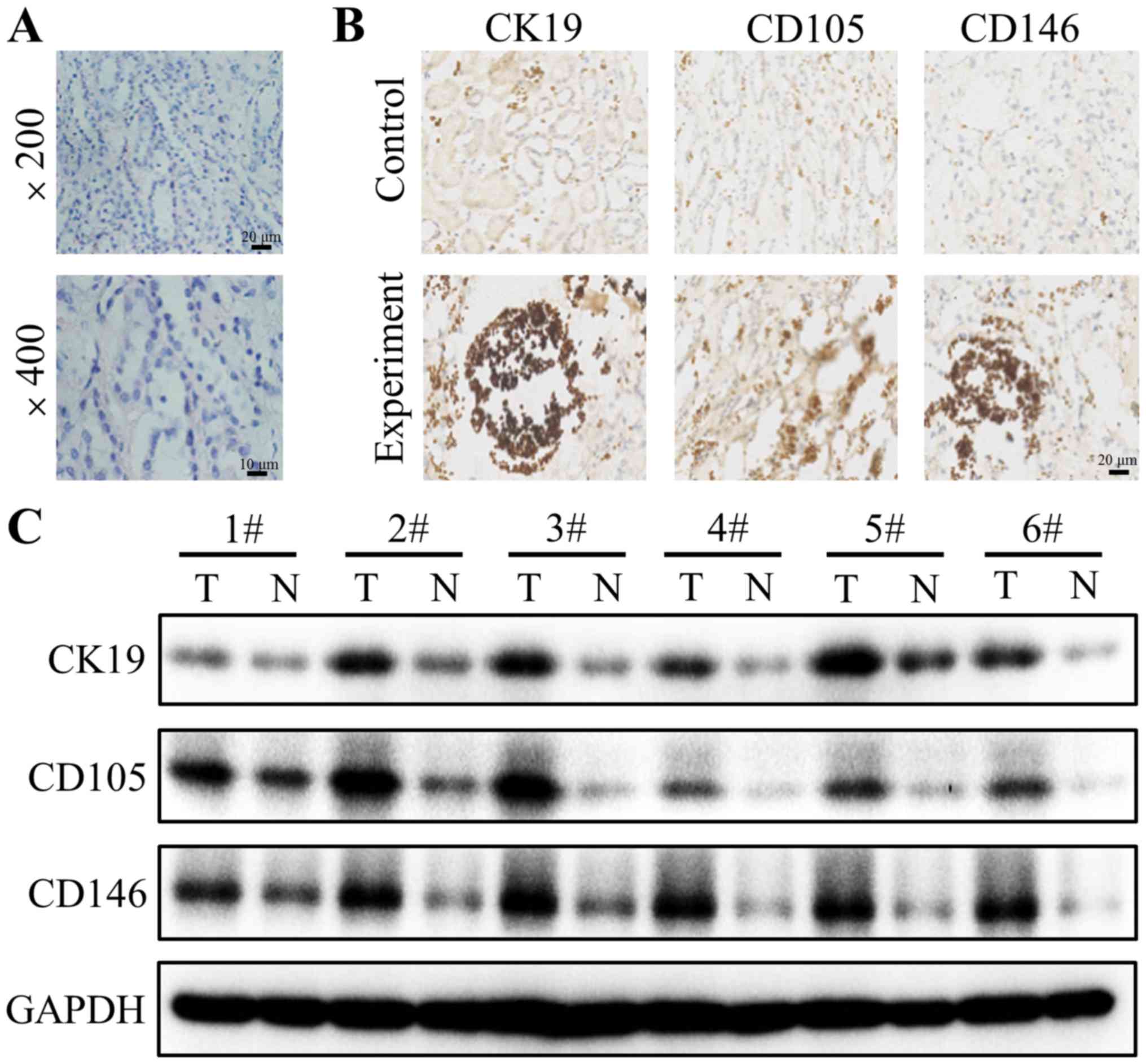
CK19, CD105 and CD146 expression
Firstly, H&E staining was used to confirm
whether the resected tissues were from the solid tumor. The results
revealed notable tumor cell clusters different from the adjacent
epithelial cells (Fig. 1A).
Subsequently, the expression of CK19, CD105 and CD146 in tumor
tissues and adjacent normal tissues were detected using
immunohistochemistry staining. Positive CK19 staining was observed
in the cytoplasm of the tumor cells of 200 patients (Fig. 1B). The staining of the tumor cells of
all patients was strong and cytoplasmic. The distribution of CD105
and CD146 staining were also located in the cytoplasm of tumor
cells (Fig. 1B). To further explore
the differential expression of CK19, CD105 and CD146 between tumor
and normal tissues, western blotting was performed. The results
demonstrated that CK19, CD105 and CD146 are highly expressed in
tumor tissues compared with their corresponding adjacent normal
tissues (Fig. 1C).
Association between the early
metastasis of patients with RCC and the expression of CK19, CD105
and CD146
The application of ELISA to analyze biomarker
expression in the peripheral blood of patients with a tumor is an
emerging concept in the management of cancer (25). Thus, the expression of CK19, CD105 and
CD146 were detected at four different time points in patient and
control groups using ELISA (Q1, 1 day pre-operation; Q2, 1 day
post-operation; Q3, 1 week post-operation; and Q4, 1 month
post-operation). The results revealed that there is a significant
difference between the expression of CK19, CD105 between the
patient and control groups (P<0.05), but not for CD146 (Table II). Then, the differential
expressions of CK19, CD105 and CD146 between all four time points
were investigated through pairwise comparison. The results
indicated no statistically significant differences (data not
shown).
| Table II.CK19, CD105 and CD146 expression
between patient and control groups. |
Table II.
CK19, CD105 and CD146 expression
between patient and control groups.
| Variables | Patients group | Control group | P-value |
|---|
| CK19 |
3.29±1.89 |
2.73±1.01 | 0.038 |
| CD105 |
6.07±3.18 |
4.90±1.91 | 0.014 |
| CD146 |
204.90±100.63 |
217.18±80.26 | 0.471 |
CTCs have been identified as a predictor of early
metastasis in patients with cancer (26). Based on whether patients were
CTC-positive or CTC-negative, subgroup analysis was performed. The
results demonstrated that the expression of CK19, CD105 and CD146
was not significantly different between CTCs-positive and
CTC-negative groups or between four time points using pairwise
comparison (data not shown). These results are in accordance with
previous results in the present study. Under a CTC-positive
condition, the expression of CK19, CD105 and CD146 in patient and
control groups was detected and compared according to the
difference between the time point Q1 and the other three time
points (Q2-4). The results of the present study suggested that the
three biomarkers had no statistical difference in the patient group
compared with the control group based on the difference between the
Q2 and Q1 time points (data not shown). A significant difference
between the difference of expression of CK19 and CD105 between the
Q1 and Q3 time points of CTC-positive patient and non-RCC control
groups was identified (P<0.05; Table
III). However, no significant difference was identified with
CD146. On the basis of the difference between Q1 and Q4, the
expressions of all three markers exhibited a statistically
significant difference (P<0.05; Table
IV). Nevertheless, no significant difference was identified in
a CTC-negative condition (P>0.05; Table V).
| Table III.Expression of CK19, CD105 and CD146
in patients with a CTC-positive condition according to the
difference between the time points of Q1 and Q3. |
Table III.
Expression of CK19, CD105 and CD146
in patients with a CTC-positive condition according to the
difference between the time points of Q1 and Q3.
| Variables | CTC-positive
group | Control group | P-value |
|---|
| CK19 |
0.667±0.877 |
−0.176±0.266 | 0.010 |
| CD105 |
0.760±0.849 |
−0.446±0.346 | 0.001 |
| CD146 |
19.495±34.640 |
−19.390±113.124 | 0.337 |
| Table IV.Expression of CK19, CD105 and CD146
in patients with a CTC-positive condition according to the
difference between the time points of Q1 and Q4. |
Table IV.
Expression of CK19, CD105 and CD146
in patients with a CTC-positive condition according to the
difference between the time points of Q1 and Q4.
| Variables | CTC positive
group | Control group | P-value |
|---|
| CK19 |
1.082±1.555 |
−0.026±0.163 | 0.038 |
| CD105 |
1.403±2.395 |
−0.201±0.120 | 0.049 |
| CD146 |
57.943±56.742 |
−28.270±79.113 | 0.015 |
| Table V.Expression of CK19, CD105 and CD146
in patients with a CTC-negative condition according to the
difference between the time points of Q1 and Q4. |
Table V.
Expression of CK19, CD105 and CD146
in patients with a CTC-negative condition according to the
difference between the time points of Q1 and Q4.
| Variables | CTC positive
group | Control group | P-value |
|---|
| CK19 |
0.100±1.448 |
−0.026±0.163 | 0.788 |
| CD105 |
−0.471±0.566 |
−0.201±0.120 | 0.156 |
| CD146 |
−28.790±44.910 |
−28.270±79.113 | 0.98 |
Discussion
Tendency to invade and transfer is one of the
obstacles in the treatment of RCC. Clinical parameters including
Fuhrman stage may not entirely reflect prognosis and early
metastasis (27). Patients may have a
recurrence or metastasis with a low-grade and early-stage tumor
following surgery. Thus, oncologists and clinicians produced
insight into alternative or complementary indicators, expecting to
predict disease progression and early prognosis more accurately
(28). The aim of the present study
was to explore peripheral blood biomarkers (CK19, CD105 and CD146)
for the early metastasis of RCC following surgery.
CK19 is a characteristic intermediate filament of
epithelial cells, which has gained attention as a marker for
micro-metastasis (29). It has been
reported that CK 19 may be detected in the tumor cells of patients
with breast cancer (30). Although
additional validation studies are essential, the present study
demonstrated that CK19 is highly expressed in tumor tissues
compared with that in normal tissues, which were confirmed using
immunohistochemical staining and western blotting. CD105, a
co-receptor of TGF-β, is implicated in tumor proliferation,
migration and differentiation (31).
It has been reported to be a marker of tumor neovascularization,
and to provide prognostic data (31).
It has been demonstrated that the high expression of CD105 is
closely correlated with a more favorable prognosis compared with
lower expression (32). However,
other studies have demonstrated that the high CD105 expression is
positively associated with a poorer progression-free survival
(33). In the present study, CD105
expression in tumor tissues was greater compared with that in
normal tissues, which was consistent with the latter. CD146 was
revealed to participate in cancer progression. It may enhance
cancer migration and invasion in melanoma, gallbladder
adenocarcinoma and breast cancer (34–36).
Furthermore, CD105 had been considered to be an indicator of early
metastasis including in gastric cancer and lung cancer (37). However, the expression of CD146 was
greater in the intermediate/high grade of oral mucoepidermoid
carcinoma compared with that in patients with local recurrence and
distant metastasis (38). To the best
of our knowledge, the role of CD146 in RCC cells had not yet been
explored. In the present study, CD146 expression in tumor tissues
was revealed to be higher compared with that in normal tissues,
which corresponded with the results of a previous study (37).
In the subsequent experiment, the results indicated
that the expressions of CK19 and CD105 had statistically
significant differences between two groups (P<0.05), but no such
significant difference was identified for CD146. Further
experiments revealed no significant difference between four time
points (data not shown). Furthermore, CTCs were examined, a crucial
blood biomarker in cancer (26). CTC
clusters have been reported to be present in patients with
metastatic colorectal cancer and prostate cancer, and have a
positive correlation with decreased survival in lung cancer and
breast cancer (39,40). Furthermore, the CTC clusters may
indicate a poor outcome in breast cancer via detection using the
CellSearch system (41). In view of
the indicated role of CTCs on tumor prognosis and early metastasis,
subgroup analysis was performed based on whether the patients were
CTC positive or not, and the difference between the point of Q1 and
other three points (Q2-4). Similar to the results presented before,
the results revealed no statistically significant difference
between the CTC-positive or -negative condition, and in the
difference between the time points Q1 and Q2 (data not shown).
Nevertheless, the expression of CK19 and CD105 exhibited a
significant difference with a CTC-positive condition according to
the difference between the time points Q1 and Q3 (P<0.05).
Furthermore, on the basis of the difference between Q1 and Q4, the
expression of CK19, CD105 and CD146 were significantly different
(P<0.05). The results concluded that CK19, CD105 and CD146 could
not be used individually to predict the early metastasis of
patients with RCC subsequent to surgery. The expressions of CK19,
CD105, and CD146 at Q2 (1 day post-operation) may not accurately
reflect the levels of biomarkers following surgery, due to the
state of stress caused by surgical trauma, which resulted in no
statistical difference between the CTC-positive condition in the
difference between the point of Q1 and Q2. But at the Q3 time point
(1 week post-operation), patients had recovered from the surgery.
The expression of CK19 and CD105 exhibited a significant difference
with a CTC-positive condition based on the difference between Q1
and Q3 (P<0.05), although CD146 did not. Furthermore, a
statistically significant difference was present in the expression
of CK19, CD105 and CD146 on the basis of the difference between Q1
and Q4 (P<0.05), indicating that CK19, CD105 and CD146 may be
novel predictors for the early metastasis of CTC-positive condition
in the difference between Q1 and Q3/Q4. Additionally, CK19, CD105
and CD146 may be auxiliary indicators for CTCs to estimate the
prognosis of patients with RCC.
Altogether, the results suggest that CK19, CD105 and
CD146 markers of peripheral blood may be considered to be effective
tools to evaluate the early metastasis of patients with a
CTC-positive condition between Q1 and Q3/Q4. CK19, CD105 and CD146
may be useful for CTCs to evaluate the prognosis of patients with
RCC, although more widespread studies with larger scale samples are
required in order to verify the results of the present study. As a
nonsurgical strategy, CK19, CD105 and CD146 may be the potential
targets for treatment of RCC.
Acknowledgements
The present study was supported by the Key Science
and Technology Program of Shaanxi Province (grant no.
2011K12-34).
Competing interests
The authors declare that they have no competing
interests.
References
|
1
|
Siegel RL, Miller KD and Jemal A: Cancer
statistics, 2016. CA Cancer J Clin. 66:7–30. 2016. View Article : Google Scholar : PubMed/NCBI
|
|
2
|
Siegel RL, Miller KD and Jemal A: Cancer
Statistics, 2015. CA Cancer J Clin. 65:5–29. 2015. View Article : Google Scholar : PubMed/NCBI
|
|
3
|
Heuer R, Gill IS, Guazzoni G, Kirkali Z,
Marberger M, Richie JP and de la Rosette JJ: A critical analysis of
the actual role of minimally invasive surgery and active
surveillance for kidney cancer. Eur Urol. 57:223–232. 2010.
View Article : Google Scholar : PubMed/NCBI
|
|
4
|
Ritchie AW and Chisholm GD: The natural
history of renal carcinoma. Semin Oncol. 10:390–400.
1983.PubMed/NCBI
|
|
5
|
Edge SB, Byrd DR, Compton CC, Fritz AG,
Greene FL and Trotti A: AJCC cancer staging manual. 7th.
Springer-Verlag; New York: pp. 547–560. 2009
|
|
6
|
Chin AI, Lam JS, Figlin RA and Belldegrun
AS: Surveillance strategies for renal cell carcinoma patients
following nephrectomy. Rev Urol. 8:1–7. 2006.PubMed/NCBI
|
|
7
|
Sun DW, Zhang YY, Sun XD, Chen YG, Qiu W,
Ji M and Lv GY: Prognostic value of cytokeratin 19 in
hepatocellular carcinoma: A meta-analysis. Clin Chim Acta.
448:161–169. 2015. View Article : Google Scholar : PubMed/NCBI
|
|
8
|
Schmitt AM, Anlauf M, Rousson V, Schmid S,
Kofler A, Riniker F, Bauersfeld J, Barghorn A, Probst-Hensch NM,
Moch H, et al: WHO 2004 criteria and CK19 are reliable prognostic
markers in pancreatic endocrine tumors. Am J Surg Pathol.
31:1677–1682. 2007. View Article : Google Scholar : PubMed/NCBI
|
|
9
|
Stathopoulos EN, Sanidas E, Kafousi M,
Mavroudis D, Askoxylakis J, Bozionelou V, Perraki M, Tsiftsis D and
Georgoulias V: Detection of CK-19 mRNA-positive cells in the
peripheral blood of breast cancer patients with histologically and
immunohistochemically negative axillary lymph nodes. Ann Oncol.
16:240–246. 2005. View Article : Google Scholar : PubMed/NCBI
|
|
10
|
Quackenbush EJ and Letarte M:
Identification of several cell surface proteins of non-T, non-B
acute lymphoblastic leukemia by using monoclonal antibodies. J
Immunol. 134:1276–1285. 1985.PubMed/NCBI
|
|
11
|
Wong SH, Hamel L, Chevalier S and Philip
A: Endoglin expression on human microvascular endothelial cells
association with betaglycan and formation of higher order complexes
with TGF-beta signalling receptors. Eur J Biochem. 267:5550–5560.
2000. View Article : Google Scholar : PubMed/NCBI
|
|
12
|
Nassiri F, Cusimano MD, Scheithauer BW,
Rotondo F, Fazio A, Yousef GM, Syro LV, Kovacs K and Lioyd RV:
Endoglin (CD105): A review of its role in angiogenesis and tumor
diagnosis, progression and therapy. Anticancer Res. 31:2283–2290.
2011.PubMed/NCBI
|
|
13
|
Nair S, Nayak R, Bhat K, Kotrashetti VS
and Babji D: Immunohistochemical expression of CD105 and TGF-beta1
in oral squamous cell carcinoma and adjacent apparently normal oral
mucosa and its correlation with clinicopathologic features. Appl
Immunohistochem Mol Morphol. 24:35–41. 2016. View Article : Google Scholar : PubMed/NCBI
|
|
14
|
Lytras D, Leontara V, Kefala M, Foukas PG,
Giannakou N, Pouliakis A, Dervenis C, Panayiotides IG and
Karakitsos P: Microvessel landscape assessment in pancreatic ductal
adenocarcinoma: Unclear value of targeting endoglin (CD105) as
prognostic factor of clinical outcome. Pancreas. 44:87–92. 2015.
View Article : Google Scholar : PubMed/NCBI
|
|
15
|
Sandlund J, Hedberg Y, Bergh A, Grankvist
K, Ljungberg B and Rasmuson T: Endoglin (CD105) expression in human
renal cell carcinoma. BJU Int. 97:706–710. 2006. View Article : Google Scholar : PubMed/NCBI
|
|
16
|
Dubinski W, Gabril M, Iakovlev VV,
Scorilas A, Youssef YM, Faragalla H, Kovacs K, Rotondo F, Metias S,
Arsanious A, et al: Assessment of the prognostic significance of
endoglin (CD105) in clear cell renal cell carcinoma using automated
image analysis. Hum Pathol. 43:1037–1043. 2012. View Article : Google Scholar : PubMed/NCBI
|
|
17
|
Wang Z and Yan X: CD146, a
multi-functional molecule beyond adhesion. Cancer Lett.
330:150–162. 2013. View Article : Google Scholar : PubMed/NCBI
|
|
18
|
Sers C, Riethmuller G and Johnson JP:
MUC18, a melanoma-progression associated molecule, and its
potential role in tumor vascularization and hematogenous spread.
Cancer Res. 54:5689–5694. 1994.PubMed/NCBI
|
|
19
|
Takaha N, Taira E, Taniura H, Nagino T,
Tsukamoto Y, Matsumoto T, Kotani T, Sakuma S and Miki N: Expression
of gicerin in development, oncogenesis and regeneration of the
chick kidney. Differentiation. 58:313–320. 1995. View Article : Google Scholar : PubMed/NCBI
|
|
20
|
Taira E, Kohama K, Tsukamoto Y, Okumura S
and Miki N: Gicerin/CD146 is involved in neurite extension of
NGF-treated PC12 cells. J Cell Physiol. 204:632–637. 2005.
View Article : Google Scholar : PubMed/NCBI
|
|
21
|
Harris L, Fritsche H, Mennel R, Norton L,
Ravdin P, Taube S, Somerfield MR, Hayes DF and Bast RC Jr; American
Society of Clinical Oncology, : American Society of Clinical
Oncology 2007 update of recommendations for the use of tumor
markers in breast cancer. J Clin Oncol. 25:5287–5312. 2007.
View Article : Google Scholar : PubMed/NCBI
|
|
22
|
Saloustros E and Mavroudis D: Cytokeratin
19-positive circulating tumor cells in early breast cancer
prognosis. Future Oncol. 6:209–219. 2010. View Article : Google Scholar : PubMed/NCBI
|
|
23
|
de Bono JS, Scher HI, Montgomery RB,
Parker C, Miller MC, Tissing H, Doyle GV, Terstappen LW, Pienta KJ
and Raghavan D: Circulating tumor cells predict survival benefit
from treatment in metastatic castration-resistant prostate cancer.
Clin Cancer Res. 14:6302–6309. 2008. View Article : Google Scholar : PubMed/NCBI
|
|
24
|
Cohen SJ, Punt CJ, Iannotti N, Saidman BH,
Sabbath KD, Gabrail NY, Picus J, Morse MA, Mitchell E, Miller MC,
et al: Prognostic significance of circulating tumor cells in
patients with metastatic colorectal cancer. Ann Oncol.
20:1223–1229. 2009. View Article : Google Scholar : PubMed/NCBI
|
|
25
|
Hou JM, Krebs MG, Lancashire L, Sloane R,
Backen A, Swain RK, Priest LJ, Greystoke A, Zhou C, Morris K, et
al: Clinical significance and molecular characteristics of
circulating tumor cells and circulating tumor microemboli in
patients with small-cell lung cancer. J Clin Oncol. 30:525–532.
2012. View Article : Google Scholar : PubMed/NCBI
|
|
26
|
Lianidou ES, Markou A and Strati A: The
role of CTCs as tumor biomarkers. Adv Exp Med Biol. 867:341–367.
2015. View Article : Google Scholar : PubMed/NCBI
|
|
27
|
Volpe A and Patard JJ: Prognostic factors
in renal cell carcinoma. World J Urol. 28:319–327. 2010. View Article : Google Scholar : PubMed/NCBI
|
|
28
|
Aceto N, Bardia A, Miyamoto DT, Donaldson
MC, Wittner BS, Spencer JA, Yu M, Pely A, Engstrom A, Zhu H, et al:
Circulating tumor cell clusters are oligoclonal precursors of
breast cancer metastasis. Cell. 158:1110–1122. 2014. View Article : Google Scholar : PubMed/NCBI
|
|
29
|
Yu XF, Yang HJ, Lei L, Wang C and Huang J:
CK19 mRNA in blood can predict non-sentinel lymph node metastasis
in breast cancer. Oncotarget. 7:30504–30510. 2016.PubMed/NCBI
|
|
30
|
Alix-Panabières C, Vendrell JP, Slijper M,
Pellé O, Barbotte E, Mercier G, Jacot W, Fabbro M and Pantel K:
Full-length cytokeratin-19 is released by human tumor cells: A
potential role in metastatic progression of breast cancer. Breast
Cancer Res. 11:R392009. View
Article : Google Scholar : PubMed/NCBI
|
|
31
|
Smith SJ, Tilly H, Ward JH, Macarthur DC,
Lowe J, Coyle B and Grundy RG: CD105 (Endoglin) exerts prognostic
effects via its role in the microvascular niche of paediatric high
grade glioma. Acta Neuropathol. 124:99–110. 2012. View Article : Google Scholar : PubMed/NCBI
|
|
32
|
Tanaka F, Otake Y, Yanagihara K, Kawano Y,
Miyahara R, Li M, Ishikawa S and Wada H: Correlation between
apoptotic index and angiogenesis in non-small cell lung cancer:
Comparison between CD105 and CD34 as a marker of angiogenesis. Lung
Cancer. 39:289–296. 2003. View Article : Google Scholar : PubMed/NCBI
|
|
33
|
Nikiteas NI, Tzanakis N, Theodoropoulos G,
Atsaves V, Christoni Z, Karakitsos P, Lazaris AC, Papachristodoulou
A, Klonaris C and Gazouli M: Vascular endothelial growth factor and
endoglin (CD-105) in gastric cancer. Gastric Cancer. 10:12–17.
2007. View Article : Google Scholar : PubMed/NCBI
|
|
34
|
Lei X, Guan CW, Song Y and Wang H: The
multifaceted role of CD146/MCAM in the promotion of melanoma
progression. Cancer Cell Int. 15:32015. View Article : Google Scholar : PubMed/NCBI
|
|
35
|
Wang W, Yang ZL, Liu JQ, Jiang S and Miao
XY: Identification of CD146 expression, angiogenesis, and
lymphangiogenesis as progression, metastasis, and poor-prognosis
related markers for gallbladder adenocarcinoma. Tumour Biol.
33:173–182. 2012. View Article : Google Scholar : PubMed/NCBI
|
|
36
|
Zabouo G, Imbert AM, Jacquemier J, Finetti
P, Moreau T, Esterni B, Birnbaum D, Bertucci F and Chabannon C:
CD146 expression is associated with a poor prognosis in human
breast tumors and with enhanced motility in breast cancer cell
lines. Breast Cancer Res. 11:R12009. View
Article : Google Scholar : PubMed/NCBI
|
|
37
|
Koyama Y, Okayama H, Kumamoto K, Saito K,
Nakamura I, Ohki S and Takenoshita S: Overexpression of endoglin
(CD105) is associated with recurrence in radically resected gastric
cancer. Exp Ther Med. 1:627–633. 2010. View Article : Google Scholar : PubMed/NCBI
|
|
38
|
Pires FR, Shih IeM, da Cruz Perez DE, de
Almeida OP and Kowalski LP: Mel-CAM (CD146) expression in parotid
mucoepidermoid carcinoma. Oral Oncol. 39:277–281. 2003. View Article : Google Scholar : PubMed/NCBI
|
|
39
|
Molnar B, Ladanyi A, Tanko L, Sréter L and
Tulassay Z: Circulating tumor cell clusters in the peripheral blood
of colorectal cancer patients. Clin Cancer Res. 7:4080–4085.
2001.PubMed/NCBI
|
|
40
|
Brandt B, Junker R, Griwatz C, Heidi S,
Brinkmann O, Semjonow A, Assmann G and Zanker KS: Isolation of
prostate-derived single cells and cell clusters from human
peripheral blood. Cancer Res. 56:4556–4561. 1996.PubMed/NCBI
|
|
41
|
Méhes G, Witt A, Kubista E and Ambros PF:
Circulating breast cancer cells are frequently apoptotic. Am J
Pathol. 159:17–20. 2001. View Article : Google Scholar : PubMed/NCBI
|