Introduction
As one of the major types of esophageal cancer,
esophageal squamous cell carcinoma (ESCC) is one of the most
aggressive malignancies of the digestive tract, with high
cancer-associated morbidity and mortality worldwide, due to the
late diagnosis at advanced stages and the lack of effective
treatment (1,2). ESCC occurs mostly in developing
countries (3). ESCC is characterized
by early lymphatic and hematogenous metastasis, which is the main
cause of the poor prognosis in patients with this malignancy
(4). Among the available prognostic
factors for ESCC, the International Union Against Cancer (UICC)
tumor-node-metastasis stage has been recognized as the most
important factor, since staging is determined by the involvement of
lymph nodes, depth of invasion and presence of distant metastasis
(5). However, the prognostic value of
the stage is often affected by the heterogeneity of ESCC patients.
Therefore, there is an urgent requirement to clarify the molecular
mechanisms that drive the aggressive tumor progression and to
identify more specific and sensitive biomarkers for the diagnosis
and targeted therapy of ESCC.
Nectin-2, also termed cluster of differentiation 112
and poliovirus receptor-related protein 2, in addition to Nectin-1,
−3 and −4, belongs to the Nectin family of cell adhesion molecules,
which exert similar functions to immunoglobulin in the formation
and continuation of tight junctions and adherence connections
(6). Four members in this family have
the same structure, containing extracellular loops, one
transmembrane segment and a short cytoplasmic domain (7). Studies have reported the aberrant
expression of Nectins and revealed their important roles in
numerous cancer types (8–10). Notably, strong Nectin-2 expression was
observed in leukemic blasts, myeloma, breast cancer, squamous cell
and adenosquamous carcinomas, adenocarcinoma of gallbladder,
colorectal carcinoma, pancreatic ductal adenocarcinomas and ovarian
cancer, but its expression is reduced or absent in hepatocellular
carcinoma (11–17). However, to the best of our knowledge,
the involvement of Nestin-2 in ESCC remains unknown. The present
study aimed to investigate the expression pattern of Nectin-2, its
clinical significance and roles in the malignant phenotypes of
ESCC.
Materials and methods
Patients and tissue samples
The Ethics Committees of People's Hospital of
Lianshui County, People's Hospital of Xuyi County and Huai'an First
People's Hospital (Huai'an, Jiangsu, China) approved the present
study. All patients agreed to the procedure and signed consent
forms.
A total of 106 patients with ESCC were diagnosed by
endoscopic biopsy pathology between February 2006 and March 2012 at
People's Hospital of Lianshui County, People's Hospital of Xuyi
County and Huai'an First People's Hospital (Huai'an, Jiangsu,
China). These patients were enrolled in the present study based on
the following criteria: Newly diagnosed cancer of the esophagus
without preoperative adjuvant chemotherapy or radiotherapy, and
histologically-confirmed primary ESCC. There were 78 men and 28
women, ranging in age between 38 and 82 years (mean age, 50.6
years; median age, 50 years). The histological grade and clinical
stage of the tumors were defined based on the UICC 2009 criteria
(18). Of the 106 patients with ESCC,
16, 30 and 60 cases were classified as grades I–II, III and IV,
respectively. In addition, 26, 30 and 50 cases were well,
moderately and poorly differentiated, respectively. The
clinicopathological characteristics of the 106 EOC patients are
summarized in Table I. In total, 20
samples of normal esophageal mucosa were used as a control group.
All specimens were collected from endoscopic biopsy, immediately
snap-frozen in liquid nitrogen and maintained at −80°C until
use.
 | Table I.Association between Nectin-2 and
various clinicopathological features of patients with esophageal
squamous cell carcinoma. |
Table I.
Association between Nectin-2 and
various clinicopathological features of patients with esophageal
squamous cell carcinoma.
|
|
| Nectin-2 expression,
n (%) |
|
|---|
|
|
|
|
|
|---|
| Clinicopathological
features | Cases, n | High | Low | P-value |
|---|
| Age |
|
|
| NS |
| <60
years | 40 | 20 (50.0) | 20 (50.0) |
|
| ≥60
years | 66 | 35 (53.0) | 31 (47.0) |
|
| Gender |
|
|
| NS |
| Male | 70 | 37 (52.9) | 33 (47.1) |
|
|
Female | 36 | 18 (50.0) | 18 (50.0) |
|
| Differentiation |
|
|
| 0.02 |
|
Well-moderate | 56 | 20 (35.7) | 36 (64.3) |
|
| Poor | 50 | 35 (70.0) | 15 (30.0) |
|
| Tumor size |
|
|
| NS |
| <5
cm | 46 | 25 (54.3) | 21 (45.7) |
|
| ≥5
cm | 60 | 30 (50.0) | 30 (50.0) |
|
| TNM stage |
|
|
| 0.006 |
| I–II | 16 | 1 (6.3) | 15 (93.7) |
|
| III | 30 | 9 (30.0) | 21 (70.0) |
|
| IV | 60 | 45 (75.0) | 15 (75.0) |
|
Cell culture and transfection
The human ESCC ECA109 and KYSE510 cell lines were
purchased from the Chinese Academy of Sciences (Shanghai, China)
and were cultured in RPMI-1640 medium (Gibco; Thermo Fisher
Scientific, Inc., Waltham, MA, USA) supplemented with 10% fetal
bovine serum (HyClone; GE Healthcare Life Sciences, Logan, UT, USA)
and 100 U/ml penicillin/streptomycin (Gibco; Thermo Fisher
Scientific, Inc.) and was maintained in a humidified 5%
CO2 atmosphere at 37°C.
The Nectin-2-targeted small interfering (si)RNA
(si-Nectin-2; catalog no. sc-43169; sequence: GCCUUUUCUCAGACUCGATT)
and negative control siRNA (si-con; catalog no. sc-44236; sequence:
GACCAATCTCAGCAGACAGTGCCATCATG) were purchased from Santa Cruz
Biotechnology, Inc. (Dallas, TX, USA) and transfected into cells
using Lipofectamine 2000 (Invitrogen; Thermo Fisher Scientific,
Inc.), according to the manufacturer's protocol. After 48 h, the
subsequent experiments were performed.
Reverse transcription-quantitative
polymerase chain reaction (RT-qPCR)
RT-qPCR was performed to detect the expression level
of Nectin-2 mRNA in ESCC tissues and normal esophageal mucosa,
according to the protocol described in a previous study (19). GAPDH was used as an internal control.
The qPCR primers were as follows: Nectin-2 forward,
5′-CGCCTCGATACCATGAGTT-3′ and reverse, 5′-ACATCCCTGGCAGAAAGC-3′;
and GAPDH forward5′-AGGTCGGTGTGAACGGATTTG-3′ and reverse,
5′-GGGGTCGTTGATGGCAACA-3′. The quantification cycle (Cq) is defined
as the fractional cycle number at which the fluorescence passes the
fixed threshold. The relative expression of Nectin-2 mRNA was
normalized to the expression of GAPDH mRNA using the
2−ΔΔCq method (20).
Western blot analysis
Western blot analysis was performed to detect the
expression level of Nectin-2 protein in ESCC tissues and normal
esophageal mucosa. Proteins in tissues and cell lysates were
extracted using cell lysis buffer (Sigma-Aldrich; Merck KGaA,
Darmstadt, Germany) and separated by 10% SDS-PAGE followed by
electrotransference onto nitrocellulose membranes, which were then
blocked with nonfat dry milk at room temperature for 1 h.
Subsequently, the membranes were incubated overnight at 4°C, with
the primary antibodies against Nectin-2 (dilution, 1:500; rabbit
anti-human Nectin-2 antibody; cat. no. AF1166c; Abgent, Inc., San
Diego, CA, USA) and GAPDH (dilution, 1:2,000; cat. no. SAB168068;
Sigma-Aldrich; Merck KGaA). Subsequently, the membranes were washed
with TBST several times and then incubated with horseradish
peroxidase-conjugated secondary antibodies (dilution, 1:3,000; cat.
no. SAB186192; Sigma-Aldrich; Merck KGaA) at room temperature for 1
h. Immunoblots were visualized by the enhanced chemiluminescence
detection system (Pierce; Thermo Fisher Scientific, Inc.).
Immunohistochemistry
Immunohistochemistry was performed to detect the
expression pattern and subcellular localization of Nectin-2 protein
in 106 ESCC tissues and 20 normal esophageal mucosa. All tissue
samples were fixed with 4% formaldehyde, embedded in paraffin, and
sectioned into 4-µm slices. The sections were deparaffinized in
xylene and rehydrated in graded ethanol. Endogenous peroxidase
activity was blocked for 15 min with 3% hydrogen peroxide in
phosphate-buffered saline (PBS). The sections were incubated with
primary rabbit anti-human Nectin-2 antibody (dilution, 1:500; cat.
no. AF1166c; Abgent, Inc.) overnight at 4°C and then incubated with
IgG-horseradish peroxidase (dilution, 1:1,000; cat. no. ZS6061;
Zhongshan Biotechnology, Co., Ltd., Zhongshan, China) for 1 h at
37°C. Immunostaining was performed using the avidin biotin
peroxidase complex method and antigen-antibody reactions were
visualized with the chromogen diaminobenzidine.
To evaluate the results of immunohistochemistry, the
sections were observed under optical microscopy (×10 and ×200) by
two pathologists who were blinded to the clinicopathological
characteristics of the 106 patients with ESCC. The immunoreactive
score (IRS) was calculated by multiplying the intensity score and
the percentage score of immunostaining. The former was defined as
follows: 3, strong; 2, moderate; 1, weak; and 0, negative. The
latter was defined as follows: 0, <5% of cells; 1, 5–25%; 2,
26–50%; 3, 50–75%; and 4, >75%. The median IRS value (3.9) of
Nectin-2 protein was used as a cutoff point to divide all 106
patients with ESCC into Nectin-2-high (n=55) and Nectin-2-low
(n=51) groups.
Cell migration and invasion
assays
Transwell assay was performed to assess the
migration and invasion abilities of ESCC cells transfected with
si-Nectin-2 or si-con using a modified two-chamber migration assay
with a pore size of 8 µm (Merck KGaA).
For the migration assay, 1×10⁵ cells were
suspended with serum-free RPMI-1640 (Gibco; Thermo Fisher
Scientific, Inc.) and seeded in the upper chambers of the 24-well
Transwell plates. The lower well of the chamber was supplemented
with RPMI-1640 containing 10% serum (Gibco; Thermo Fisher
Scientific, Inc.). Following incubation for 24 h, any cells
remaining on top of the insert were removed by scraping and the
migrating cells on the underside of the membrane were fixed with 4%
formaldehyde, stained with 0.5% crystal violet, and examined under
an optical microscope (magnification, ×10 and ×200; Olympus
Corporation, Tokyo, Japan) in 10 randomly selected fields.
For the invasion assay, 1×10⁵ cells were
suspended with serum-free RPMI-1640 (Gibco; Thermo Fisher
Scientific, Inc.) and seeded in the upper chambers of the 24-well
Transwell chambers coated with Matrigel (1 mg/ml; BD Biosciences,
Franklin Lakes, NJ, USA). The lower chamber was supplemented with
RPMI-1640 containing 10% serum (Gibco; Thermo Fisher Scientific,
Inc.) as a chemoattractant. Subsequent to incubation for 24 h, the
invading cells on the underside of the membrane were fixed with 4%
formaldehyde, stained with 0.5% crystal violet, and examined under
an optical microscope (magnification, ×10 and ×200; Olympus
Corporation) in 10 randomly selected fields.
Statistical analysis
All statistical analyses in the current study were
conducted by using SPSS version 11.0 for Windows (SPSS Inc.,
Chicago, IL, USA). Each experiment was performed in triplicate.
Data were expressed as the mean ± standard error of three repeated
assays. Differences between various groups were assessed using
Students t-test. Fishers exact test and χ2 were
performed to assess the associations between Nectin-2 expression
and different clinicopathological characteristics. P<0.05 was
considered to indicate a statistically significant difference.
Results
Elevated Nectin-2 expression in human
ESCC tissues
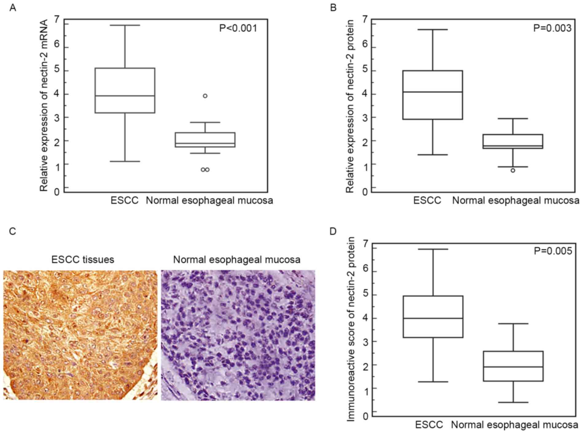
RT-qPCR showed that the relative expression of
Nectin-2 mRNA in ESCC tissues was significantly elevated compared
with that in the normal esophageal mucosa (ESCC vs. normal,
4.09±1.37 vs. 2.00±0.67; P<0.001; Fig.
1A). Similarly, western blot analysis also found an increased
expression level of Nectin-2 protein in ESCC tissues compared with
normal esophageal mucosa (ESCC vs. normal, 3.93±1.40 vs. 1.76±0.57;
P=0.003; Fig. 1B).
In addition, the expression pattern and subcellular
localization of Nectin-2 protein in ESCC and normal esophageal
mucosa was further examined by immunohistochemistry. As shown in
Fig. 1C, positive staining of
Nectin-2 protein was localized in the cytoplasm of cancer cells in
ESCC tissues. Of the 106 ESCC tissues, 38 (35.8%) and 42 (39.6%),
respectively, demonstrated strong and moderate positive
immunoreactivity for the Nectin-2 protein, whereas 16 (15.1%)
showed a low Nectin-2 expression and 10 (9.4%) were
Nectin-2-negative. By contrast, all normal esophageal mucosa were
weakly or not immunostained for Nectin-2 expression. Additionally,
the statistical analysis demonstrated that the IRS of Nectin-2
protein in the ESCC tissues was significantly increased compared
with normal esophageal mucosa (ESCC vs. normal, 3.96±1.41 vs.
1.92±1.87; P=0.005; Fig. 1D), which
was consistent with the results of RT-qPCR and western blot
analysis.
Elevated expression of Nectin-2
protein is associated with aggressive progression of ESCC
patients
Table I summarized the
association of Nectin-2 protein expression with the
clinicopathological parameters of ESCC patients. There were
significant associations between Nectin-2 protein expression and
clinicopathological parameters, such as age, gender and tumor size
(P>0.05). Conversely, significant associations were observed
between Nectin-2 protein expression, and tumor stage and
differentiation in ESCC patients. The average IRS of Nectin-2
protein in ESCC tissues obtained from patients with advanced tumor
stage (P=0.006; Table I) and poor
differentiation (P=0.02; Table I) was
increased compared with tissues obtained from patients with early
tumor stage and well-moderate differentiation.
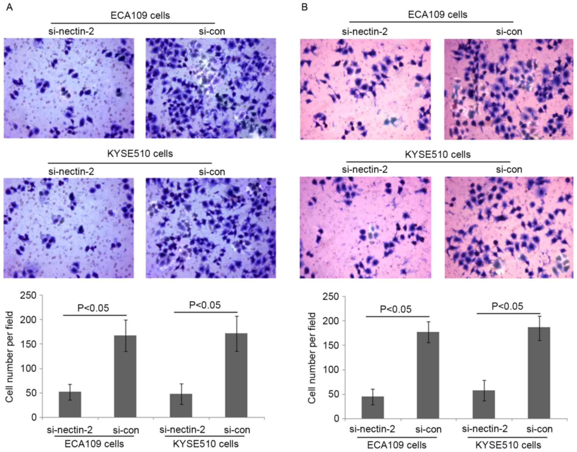
Loss of Nectin-2 protein expression
inhibited ESCC cell migration and invasion in vitro
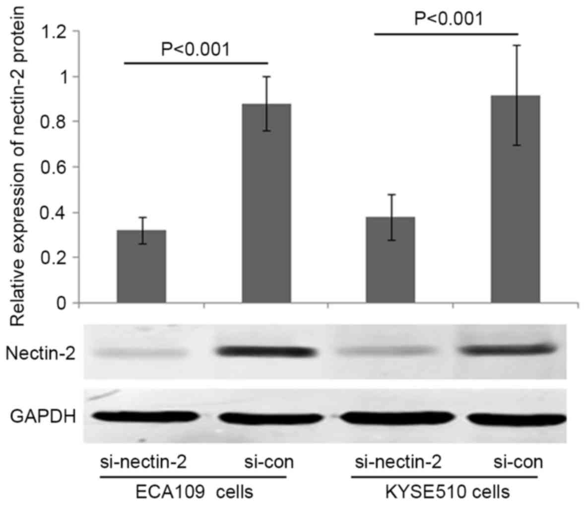
To elucidate the involvement of Nectin-2 in ESCC
cell migration and invasion abilities, Nectin-2 siRNA was used to
knockdown the expression of Nectin-2 in the human ESCC ECA109 and
KYSE510 cell lines. As shown in Fig.
2, the relative protein expression of Nectin-2 in ECA109 and
KYSE510 cells was markedly decreased by transfection with Nectin-2
siRNA (P<0.001). The migration and invasion abilities of ECA109
and KYSE510 cells transfected with Nectin-2 siRNA were suppressed
compared with the two control groups (P<0.05; Fig. 3).
Discussion
Metastasis is a complex process of cancer cell
migration and invasion to new tissues, and has been recognized as
the principal cause of mortality in ESCC patients (21,22). Thus,
it is important to better understand the mechanisms underlying the
carcinogenesis and to identify an indicator capable of predicting
the malignant potential of ESCC. In the current study, the data
illustrated the expression patterns of Nectin-2 in ESCC tissues,
followed by demonstrating the association between the Nectin-2
expression and clinicopathological parameters based on 106 clinical
samples, and identified the role of Nectin-2 in malignant
phenotypes of ESCC cells. Differential expression of Nectin-2 at
mRNA and protein levels was measured among primary ESCC tissues and
normal esophageal mucosa by RT-qPCR, western blot analysis and
immunohistochemistry, which all showed the upregulation of Nectin-2
in ESCC tissues compared with normal esophageal mucosa. Nectin-2
upregulation was then found to be closely correlated with advanced
tumor stage and poor differentiation. Additional in vitro
experiments presented here showed that the two human ESCC cell
lines transfected with Nectin-2 siRNA had reduced migration and
invasion abilities. These findings suggested that Nectin-2 could
play a vital role in the malignant progression of ESCC.
Nectin-2 belongs to the Nectin family of
Ca2+-independent immunoglobulin-like cell adhesion
molecules and functions as a junction molecule regulating the cell
adhesion between epithelial cells by creating trans-dimmers
with neighbor cells of Nectin-2 member (23). It is ubiquitously expressed in cells
of various tissues, including neuronal, hematopoietic, endothelial
and epithelial cells (24). Nectin-2
has been concerned in different diseases in humans, and has
currently been defined as a marker, and potential therapeutic
target in numerous types of cancers. Nectin-2 is often
overexpressed in cancer cells, and is associated with a poor
prognosis. For example, Karabulut et al (13) performed ELISA and determined that the
serum Nectin-2 levels in 140 colorectal cancer patients were
dramatically compared with patients in the healthy control group.
The diagnostic and prognostic roles of serum levels of Nectin-2 in
patients with colorectal cancer were also reported. Miao et
al (14) identified Nectin-2 as a
biomarker for metastasis and poor prognosis of squamous
cell/adenosquamous carcinomas and adenocarcinoma of the
gallbladder. Oshima et al (15) observed the overexpression of Nectin-2
in breast and ovarian cancers and anti-tumor activity of
anti-Nectin-2 monoclonal antibodies via strong antibody-dependent
cellular cytotoxicity, indicating its potential as a target for
antibody therapy against breast and ovarian cancers. Liang et
al (16) reported that positive
Nectin-2 expression was associated with progression and poor
prognosis in pancreatic ductal adenocarcinoma patients. By
contrast, Huang et al (17)
found that Nectin-2 expression was significantly reduced in
hepatocellular carcinoma tissues, as well as the majority of
hepatocellular carcinoma cell lines, as determined by western blot
analysis and immunofluorescence analyses. This study also confirmed
that the downregulation of Nectin-2 may be an important mechanism
through which hepatocellular carcinoma cells evade the natural
killer cell-mediated immunosurveillance, and may function as a
marker for poor prognosis in patients with this malignancy
(17). These data suggest that
Nectin-2 may function as either an oncogene or a tumor suppressor
in various cancers.
The present study evaluated the expression of
Nectin-2 in 106 ESCC tissues at the mRNA and protein levels. The
results showed that Nectin-2 expression at the mRNA and protein
levels was evidently increased in human ESCC tissues compared with
adjacent normal esophageal tissues. In addition, consistent with
the results of studies in other types of cancers (14,17), the
immunohistochemistry analysis demonstrated that staining for
Nectin-2 was present in the cytoplasm of the cancer cells.
Consequently, it was hypothesized that overexpression of Nectin-2
plays an important role in the progression of ESCC. Since tumor
cell motility is a sign of invasiveness and an essential step in
metastasis, the function of Nectin-2 in ECA109 and KYSE510 cells
was investigated using a gene knockdown technique. The present
study showed that the migration and invasion of the ESCC ECA109 and
KYSE510 cell lines subsequent to transfection with siRNA specific
to Nectin-2 were evidently reduced compared to the negative control
cells, indicating that Nectin-2 may act as an oncogene for
promoting cell migratory ability and invasiveness in ESCC
progression.
In conclusion, to the best of our knowledge, the
present data revealed for the first time that Nectin-2 is
overexpressed in ESCC and associated with aggressive cancer
progression. The present data also indicated that the silencing of
Nectin-2 with siRNA in ESCC cells may inhibit malignant biological
properties in cells, indicating potential as a possible marker or
therapeutic target for ESCC. Additional studies are required to
elucidate the mechanisms underlying the functions of Nectin-2 as a
useful marker for ESCC.
References
|
1
|
Baba Y, Watanabe M, Yoshida N and Baba H:
Neoadjuvant treatment for esophageal squamous cell carcinoma. World
J Gastrointest Oncol. 6:121–128. 2014. View Article : Google Scholar : PubMed/NCBI
|
|
2
|
Shang L and Wang M: Molecular alterations
and clinical relevance in esophageal squamous cell carcinoma. Front
Med. 7:401–410. 2013. View Article : Google Scholar : PubMed/NCBI
|
|
3
|
Yang CS, Chen X and Tu S: Etiology and
prevention of esophageal cancer. Gastrointest Tumors. 3:3–16. 2016.
View Article : Google Scholar : PubMed/NCBI
|
|
4
|
Li JS, Ying JM, Wang XW, Wang ZH, Tao Q
and Li LL: Promoter methylation of tumor suppressor genes in
esophageal squamous cell carcinoma. Chin J Cancer. 32:3–11. 2013.
View Article : Google Scholar : PubMed/NCBI
|
|
5
|
Lehrbach DM, Nita ME and Cecconello I:
Molecular aspects of esophageal squamous cell carcinoma
carcinogenesis. Arq Gastroenterol. 40:256–261. 2003. View Article : Google Scholar : PubMed/NCBI
|
|
6
|
Devilard E, Xerri L, Dubreuil P, Lopez M
and Reymond N: Nectin-3 (CD113) interacts with Nectin-2 (CD112) to
promote lymphocyte transendothelial migration. PLoS One.
8:e774242013. View Article : Google Scholar : PubMed/NCBI
|
|
7
|
Zhang X and Lui WY: Dysregulation of
nectin-2 in the testicular cells: An explanation of cadmium-induced
male infertility. Biochim Biophys Acta. 1839:873–884. 2014.
View Article : Google Scholar : PubMed/NCBI
|
|
8
|
Liu J, Qian X, Chen Z, Xu X, Gao F, Zhang
S, Zhang R, Qi J, Gao GF and Yan J: Crystal structure of cell
adhesion molecule nectin-2/CD112 and its binding to immune receptor
DNAM-1/CD226. J Immunol. 188:5511–5520. 2012. View Article : Google Scholar : PubMed/NCBI
|
|
9
|
Mandai K, Rikitake Y, Mori M and Takai Y:
Nectins and nectin-like molecules in development and disease. Curr
Top Dev Biol. 112:197–231. 2015. View Article : Google Scholar : PubMed/NCBI
|
|
10
|
Samanta D and Almo SC: Nectin family of
cell-adhesion molecules: Structural and molecular aspects of
function and specificity. Cell Mol Life Sci. 72:645–658. 2015.
View Article : Google Scholar : PubMed/NCBI
|
|
11
|
Pende D, Spaggiari GM, Marcenaro S,
Martini S, Rivera P, Capobianco A, Falco M, Lanino E, Pierri I,
Zambello R, et al: Analysis of the receptor-ligand interactions in
the natural killer-mediated lysis of freshly isolated myeloid or
lymphoblastic leukemias: Evidence for the involvement of the
Poliovirus receptor (CD155) and Nectin-2 (CD112). Blood.
105:2066–2073. 2005. View Article : Google Scholar : PubMed/NCBI
|
|
12
|
Wu MR, Zhang T, Alcon A and Sentman CL:
DNAM-1-based chimeric antigen receptors enhance T cell effector
function and exhibit in vivo efficacy against melanoma. Cancer
Immunol Immunother. 64:409–418. 2015. View Article : Google Scholar : PubMed/NCBI
|
|
13
|
Karabulut M, Gunaldi M, Alis H, Afsar CU,
Karabulut S, Serilmez M, Akarsu C, Seyit H and Aykan NF: Serum
nectin-2 levels are diagnostic and prognostic in patients with
colorectal carcinoma. Clin Transl Oncol. 18:160–171. 2016.
View Article : Google Scholar : PubMed/NCBI
|
|
14
|
Miao X, Yang ZL, Xiong L, Zou Q, Yuan Y,
Li J, Liang L, Chen M and Chen S: Nectin-2 and DDX3 are biomarkers
for metastasis and poor prognosis of squamous cell/adenosquamous
carcinomas and adenocarcinoma of gallbladder. Int J Clin Exp
Pathol. 6:179–190. 2013.PubMed/NCBI
|
|
15
|
Oshima T, Sato S, Kato J, Ito Y, Watanabe
T, Tsuji I, Hori A, Kurokawa T and Kokubo T: Nectin-2 is a
potential target for antibody therapy of breast and ovarian
cancers. Mol Cancer. 12:602013. View Article : Google Scholar : PubMed/NCBI
|
|
16
|
Liang S, Yang Z, Li D, Miao X, Yang L, Zou
Q and Yuan Y: The clinical and pathological significance of
Nectin-2 and DDX3 expression in pancreatic ductal adenocarcinomas.
Dis Markers. 2015:3795682015. View Article : Google Scholar : PubMed/NCBI
|
|
17
|
Huang X, Qu P, Chen Y, Zhou X, Wu Y, Liu
F, Wang D, Zhang J and An J: Low expression of CD112 is associated
with poor overall survival in patients with hepatocellular
carcinoma. Hum Pathol. 45:1944–1950. 2014. View Article : Google Scholar : PubMed/NCBI
|
|
18
|
Yamasaki M, Miyata H, Miyazaki Y,
Takahashi T, Kurokawa Y, Nakajima K, Takiguchi S, Mori M and Doki
Y: Evaluation of the nodal status in the 7th edition of the
UICC-TNM classification for esophageal squamous cell carcinoma:
Proposed modifications for improved survival stratification: Impact
of lymph node metastases on overall survival after esophagectomy.
Ann Surg Oncol. 21:2850–2856. 2014. View Article : Google Scholar : PubMed/NCBI
|
|
19
|
Chen G, Peng J, Zhu W, Tao G, Song Y, Zhou
X and Wang W: Combined downregulation of microRNA-133a and
microRNA-133b predicts chemosensitivity of patients with esophageal
squamous cell carcinoma undergoing paclitaxel-based chemotherapy.
Med Oncol. 31:2632014. View Article : Google Scholar : PubMed/NCBI
|
|
20
|
Livak KJ and Schmittgen TD: Analysis of
relative gene expression data using real-time quantitative PCR and
the 2(-Delta Delta C(T)) method. Methods. 25:402–408. 2001.
View Article : Google Scholar : PubMed/NCBI
|
|
21
|
Chen MQ, Xu BH and Zhang YY: Analysis of
prognostic factors for esophageal squamous cell carcinoma with
distant organ metastasis at initial diagnosis. J Chin Med Assoc.
77:562–566. 2014. View Article : Google Scholar : PubMed/NCBI
|
|
22
|
Salian V, Dinakar C, Shetty P and Ajila V:
Etiological trends in oral squamous cell carcinoma: A retrospective
institutional study. Cancer Transl Med. 2:33–36. 2016. View Article : Google Scholar
|
|
23
|
Sakisaka T and Takai Y: Biology and
pathology of nectins and nectin-like molecules. Curr Opin Cell
Biol. 16:513–521. 2004. View Article : Google Scholar : PubMed/NCBI
|
|
24
|
Sakisaka T, Ikeda W, Ogita H, Fujita N and
Takai Y: The roles of nectins in cell adhesions: Cooperation with
other cell adhesion molecules and growth factor receptors. Curr
Opin Cell Biol. 19:593–602. 2007. View Article : Google Scholar : PubMed/NCBI
|